
You have no items in your shopping cart.
Fluorescent Protein Research
Superfolder GFP (sfGFP): Robust Folding for Challenging Fusion Proteins
Explore the folding advantages of superfolder GFP, compare sfGFP and EGFP, understand GFP1–10/GFP11 complementation, and select Biorbyt reagents for detection and assay development.
1. What Is Superfolder GFP?
Superfolder green fluorescent protein, commonly abbreviated as sfGFP, is an engineered variant of green fluorescent protein originally derived from Aequorea victoria. It was developed to fold efficiently under experimental conditions that can prevent conventional GFP variants from maturing correctly.
The defining advantage of sfGFP is folding robustness. It can retain productive folding and chromophore formation when attached to poorly folded, aggregation-prone or structurally challenging fusion partners.
These properties make sfGFP useful for fusion-protein design, recombinant expression screening, protein-folding studies, live-cell imaging, split-fluorescent-protein tagging and biosensor development.
Key point: sfGFP is primarily selected for reliable folding. It should not automatically be described as brighter than every other green fluorescent protein under all experimental conditions.
Superfolder GFP at a Glance
Property | Superfolder GFP |
|---|---|
Common abbreviation | sfGFP |
Protein family | Engineered Aequorea victoria GFP |
Approximate molecular weight | 27.7 kDa for the original corrected sfGFP construct |
Fluorescence colour | Green |
Principal advantage | Robust and efficient protein folding |
Common applications | Fusion proteins, folding reporters, live-cell imaging, split-GFP tagging and biosensors |
Detection methods | Direct fluorescence, GFP11-tag antibodies, broad GFP antibodies and GFP-binding nanobodies |
Imaging compatibility | Conventional GFP fluorescence channels |
2. Superfolder GFP vs EGFP
Both EGFP and sfGFP are engineered green fluorescent proteins, but they were optimized for different experimental priorities. EGFP is widely used as a general-purpose fluorescent reporter, whereas sfGFP is commonly selected when poor folding or a difficult fusion partner reduces reporter performance.
Feature | EGFP | Superfolder GFP |
|---|---|---|
Main design priority | Improved fluorescence and expression | Improved folding robustness |
Routine cellular imaging | Widely used | Suitable |
Difficult fusion partners | Performance may be construct-dependent | Designed for challenging fusion contexts |
Folding-reporter applications | Possible | Particularly well suited |
Split-protein systems | Available in selected engineered systems | Commonly used as the basis of GFP1–10/GFP11 complementation |
Best selected when | Routine green fluorescence is required | Poor folding or fusion instability limits the experiment |
Selection guidance: Use EGFP for routine green fluorescence imaging when a fusion performs reliably. Consider sfGFP when a conventional GFP construct shows weak fluorescence, aggregation or poor maturation.
Explore broader GFP biology and detection options on the Biorbyt GFP and EGFP resource page .
3. Recommended sfGFP-Related and GFP-Family Reagents
Biorbyt offers a directly relevant GFP11-tag antibody for split-superfolder GFP workflows, supporting GFP-family antibodies for detection and capture, and a recombinant GFP protein for control and assay-development applications.
Featured Product for Split sfGFP Workflows
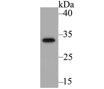
GFP11-tag Antibody validation image for orb621407 .
Directly Relevant to Split sfGFP
GFP11-tag Antibody — orb621407
GFP11 corresponds to the eleventh beta-strand of the superfolder GFP beta-barrel. When GFP11 and the complementary GFP1–10 fragment are expressed in the same cell, they can assemble to restore green fluorescence.
orb621407 is a rabbit polyclonal antibody raised against the short GFP11 peptide sequence. It is intended for Western blot detection of GFP11-tagged fusion proteins , allowing expression and molecular weight to be verified independently of fluorescence complementation.
Host: Rabbit
Clonality: Polyclonal
Application: WB
WB dilution: 1:1000
Conjugation: Unconjugated
Concentration: 1 mg/ml
Available sizes: 50/100 µl
Customer samples: Yeast, human and mouse
Application scope: orb621407 is tested for detection of the GFP11 tag by Western blot. It should not be presented as a universal antibody for all intact, folded full-length sfGFP constructs or as validated for IF, ICC or IP unless additional validation becomes available.
Supporting GFP-Family Antibodies
The products below support broader GFP-family detection, imaging, immunoprecipitation and capture workflows. Their public product information does not specifically claim validation against full-length sfGFP, so compatibility should be confirmed using the exact construct and application.
SKU | Product | Applications | Available Size |
|---|---|---|---|
GFP Antibody Goat polyclonal IgG | ELISA, IF, IHC, IP, WB | 100 µg | |
Rabbit GFP Recombinant Monoclonal Antibody Rabbit recombinant monoclonal · Clone BLRE04L | ICC, IP, WB | 100 µl | |
Green Fluorescent Protein Antibody Chicken polyclonal IgY | ELISA, ICC, IHC, WB | 400 µl | |
GFP and Other Derivatives of GFP Antibody Camelid VHH monoclonal · Clone GE10 | ELISA, IP, WB | 50/100/250/1000/2000 µg |
Recombinant GFP Protein Control
Positive Control and Assay Development
Recombinant GFP Protein, His — orb1516874
orb1516874 is a recombinant His-tagged GFP protein expressed in E. coli. It can be used as a positive control, antibody-testing antigen or reference material during ELISA and other GFP assay-development workflows.
This product is a recombinant conventional GFP protein and should not be described as a recombinant sfGFP protein.
4. Split sfGFP: How It Works and Where It Is Used
Split superfolder GFP separates the fluorescent protein into two complementary components: the larger GFP1–10 fragment and the short GFP11 peptide. When both fragments are present in the same cell and brought into a compatible molecular context, they can assemble to restore green fluorescence.
GFP1–10 Larger non-fluorescent fragment containing the first ten beta-strands
+
GFP11 Tag Short peptide corresponding to the eleventh beta-strand
→
Complemented GFP Reconstituted fluorescent complex producing a green signal
GFP11 contains approximately 16 amino acids, making it substantially smaller than a complete fluorescent-protein tag. Its compact size can reduce interference with the target protein and enables insertion into endogenous genomic loci using CRISPR-based homology-directed repair and synthetic ssDNA donor oligonucleotides.
Key Applications
Endogenous Protein Tagging
The compact GFP11 sequence can be inserted into endogenous genes while minimizing the size of the introduced tag. Complementation with GFP1–10 then enables visualization of the tagged protein.
Challenging Fusion Proteins
Full-length sfGFP offers robust folding when conventional GFP variants perform poorly. Split sfGFP provides an additional option when a substantially smaller tag is preferred.
Imaging and Biosensors
sfGFP and split-sfGFP systems support live-cell localization, protein-interaction studies and fluorescent biosensor development.
Independent expression confirmation: GFP1–10/GFP11 fluorescence confirms successful complementation, but does not independently confirm expression or molecular weight of the GFP11-tagged protein. orb621407 can support Western blot verification.
5. Detecting and Validating sfGFP Constructs
sfGFP constructs can be analysed through direct fluorescence, broad anti-GFP antibody detection or GFP11-tag antibody detection. The most appropriate method depends on whether the experiment uses full-length sfGFP or a split GFP1–10/GFP11 system.
Direct or Complemented Fluorescence
Direct fluorescence is suitable for live-cell imaging, localization studies and time-course experiments. Full-length sfGFP fluoresces after folding and chromophore maturation, while split sfGFP requires successful complementation.
- Suitable for living cells
- No primary or secondary antibody required
- Preserves spatial and temporal information
- Does not confirm fusion-protein molecular weight
Antibody-Based Detection
Antibody detection is useful when direct fluorescence is weak or when protein expression, molecular weight or fusion integrity must be confirmed.
- Use orb621407 for WB detection of GFP11-tagged proteins.
- Use broad GFP-family antibodies for full-length GFP or related constructs.
- Use IP-validated GFP antibodies or nanobodies for enrichment and capture.
Compatibility note: orb621407 is raised against the GFP11 peptide and is tested for Western blot. Broader GFP-family antibodies may recognize full-length sfGFP because much of the GFP framework is retained, but compatibility should be confirmed using the exact construct and application.
Essential Experimental Controls
- Untagged or untransfected sample: measures autofluorescence and non-specific antibody background.
- Known sfGFP- or GFP11-positive sample: confirms that the fluorescence or antibody-detection system is functioning.
- GFP1–10-only or non-complemented control: measures background fluorescence in split-sfGFP experiments.
- Fusion-partner detection or molecular-weight confirmation: helps determine whether the complete fusion protein remains intact.
Interpretation: Fluorescence confirms maturation or complementation of the fluorescent component, but does not by itself prove that the attached protein is correctly folded, correctly localized or biologically active.
6. Companion Reagents and Workflow Tools
sfGFP and GFP11 workflows may also require secondary antibodies, capture beads, Western blot substrates and protein-handling resources.
Detection Guide Secondary Antibody Selection Guide Select a compatible secondary antibody according to the primary-antibody host, experimental application and required conjugate. Immunoprecipitation Protein A/G Magnetic Beads — orb3148406 Magnetic Protein A/G beads for antibody capture and immunoprecipitation using compatible conventional IgG antibodies. Western Blot Detection ECL Plus Western Blotting Substrate — orb527071 Chemiluminescent HRP substrate for sensitive Western blot detection of GFP-family and GFP11-tagged proteins. Technical Resource Protein Handling and Storage Guide General guidance for recombinant-protein reconstitution, storage, aliquoting and freeze-thaw management.
7. Why Choose Biorbyt for sfGFP-Related Workflows?
Biorbyt provides a GFP11-tag-specific Western blot antibody alongside several reagent formats for broader GFP-family detection, capture and assay development.
Direct GFP11 Detection
orb621407 supports Western blot detection of GFP11-tagged fusion proteins.
Multiple Antibody Formats
Polyclonal, recombinant monoclonal, chicken IgY and camelid VHH formats are available.
Application Coverage
Supporting reagents are available for WB, ICC, IHC, IF, IP and ELISA workflows.
Multiplex Flexibility
Different antibody hosts help support experiments involving multiple primary antibodies.
Capture and Enrichment
GFP-binding VHH reagents support compact capture, enrichment and pull-down systems.
Recombinant Controls
Recombinant GFP protein is available for controls and assay-development studies.
8. Frequently Asked Questions
What is the main difference between sfGFP and EGFP?
EGFP is widely used as a general-purpose fluorescent reporter. sfGFP was engineered for improved folding, particularly when attached to difficult or poorly folded fusion partners.
What is GFP11?
GFP11 is the short eleventh beta-strand of the split-superfolder GFP system. It can complement the larger GFP1–10 fragment to reconstitute a fluorescent GFP complex.
Is orb621407 a full-length sfGFP antibody?
No. orb621407 is raised against the short GFP11 tag sequence and is tested for Western blot. It should be described as a GFP11-tag antibody for split-sfGFP workflows, not as a universal full-length sfGFP antibody.
Can a standard GFP antibody detect full-length sfGFP?
Many antibodies raised against full-length GFP or multiple GFP derivatives are reasonable candidates because much of the GFP framework is retained. Recognition depends on the antibody epitope and exact sfGFP construct.
Can sfGFP be detected without an antibody?
Yes. Full-length sfGFP can normally be detected directly through green fluorescence. GFP1–10/GFP11 complementation can also restore fluorescence in split-sfGFP systems.
Does fluorescence confirm that the complete fusion protein is functional?
No. Fluorescence confirms maturation or complementation of the fluorescent component. It does not independently prove that the fusion partner is intact, correctly localized or biologically active.
10. Scientific References
- Pédelacq JD, Cabantous S, Tran T, Terwilliger TC, Waldo GS. Engineering and characterization of a superfolder green fluorescent protein. Nature Biotechnology. 2006;24:79–88. doi:10.1038/nbt1172
- Pédelacq JD, Cabantous S, Tran T, et al. Corrigendum: Engineering and characterization of a superfolder green fluorescent protein. Nature Biotechnology. 2006;24:1170. doi:10.1038/nbt0906-1170d
- Cabantous S, Terwilliger TC, Waldo GS. Protein tagging and detection with engineered self-assembling fragments of green fluorescent protein. Nature Biotechnology. 2005;23:102–107. doi:10.1038/nbt1044
- Cabantous S, Waldo GS. In vivo and in vitro protein solubility assays using split GFP. Nature Methods. 2006;3:845–854. doi:10.1038/nmeth932
- Kamiyama D, Sekine S, Barsi-Rhyne B, et al. Versatile protein tagging in cells with split fluorescent protein. Nature Communications. 2016;7:11046. doi:10.1038/ncomms11046
- Leonetti MD, Sekine S, Kamiyama D, Weissman JS, Huang B. A scalable strategy for high-throughput GFP tagging of endogenous human proteins. Proceedings of the National Academy of Sciences . 2016;113:E3501–E3508. doi:10.1073/pnas.1606731113
- Aronson DE, Costantini LM, Snapp EL. Superfolder GFP is fluorescent in oxidizing environments when targeted via the Sec translocon. Traffic. 2011;12:543–548. doi:10.1111/j.1600-0854.2011.01168.x