
You have no items in your shopping cart.
mCherry Red Fluorescent Protein | Antibodies & Live-Cell Imaging
mCherry is a widely used monomeric red fluorescent protein (RFP) valued for its stable red fluorescence, photostability, low cytotoxicity, and compatibility with fusion protein applications. As a DsRed-derived fluorescent protein, mCherry is commonly used in live-cell imaging, reporter gene studies, protein localization, tumor imaging, and immune cell tracking.
Overview
mCherry is a widely used monomeric red fluorescent protein (RFP) valued for its stable red fluorescence, photostability, low cytotoxicity, and compatibility with fusion protein applications. As a DsRed-derived fluorescent protein, mCherry is commonly used in live-cell imaging, reporter gene studies, protein localization, tumor imaging, and immune cell tracking.
Biorbyt provides high-quality anti-mCherry antibodies to support reliable detection of mCherry and mCherry-tagged proteins in WB, IF, IHC, ICC, and flow cytometry applications.
What is mCherry?
mCherry is an engineered monomeric red fluorescent protein widely used as a fluorescent tag and reporter in molecular and cell biology research. It was developed from DsRed, an early red fluorescent protein originally isolated from Discosoma coral, but was optimized to overcome DsRed's tetramer formation, slow maturation, and potential interference with fusion protein applications.
Key Properties of mCherry
The major advantage of mCherry is its stable monomeric structure, which reduces protein aggregation and minimizes disruption to target protein localization or function. This makes mCherry highly suitable for fusion protein studies, intracellular localization analysis, live-cell imaging, reporter gene assays, and stable cell line generation.
mCherry has an excitation maximum of approximately 587 nm and an emission maximum of approximately 610 nm, producing a stable red fluorescence signal compatible with GFP and other fluorophores for multicolor imaging and co-localization studies.
The fluorescence performance of mCherry is also associated with engineered mutations around the chromophore region. For example, substitutions at residues 163 and 83 may alter the local chromophore environment, helping improve fluorescence stability and brightness while maintaining the monomeric properties required for protein tagging.
Applications of mCherry
1. Live-Cell Imaging and Multicolor Fluorescence Labeling
mCherry is widely used for live-cell imaging because of its stable red fluorescence and good photostability. It allows researchers to monitor cell migration, protein trafficking, cell morphology, and intracellular dynamics in real time.
Its red fluorescence channel is compatible with GFP, EGFP, CFP, DAPI, and other fluorophores, making mCherry useful for multicolor fluorescence imaging and co-localization analysis. Common applications include live-cell imaging, confocal microscopy, time-lapse imaging, protein co-localization, and subcellular tracking.
2. Fusion Protein and Reporter Gene Studies
The monomeric structure of mCherry makes it highly suitable for fusion protein studies. It can be fused to proteins of interest to examine protein localization, intracellular transport, organelle distribution, and membrane dynamics.
mCherry is also commonly used as a reporter gene in plasmid vectors, lentiviral systems, CRISPR workflows, and stable cell line generation. Its visible red fluorescence allows rapid screening of successfully transfected or transduced cells.
3. Tumor Research and In Vivo Imaging
mCherry is frequently used in cancer biology to generate fluorescent tumor cell models. mCherry-expressing cells allow researchers to monitor tumor growth, metastasis, tumor cell migration, drug response, and tumor microenvironment interactions.
Because red fluorescence generally shows lower tissue autofluorescence than green fluorescence, mCherry is useful for xenograft imaging, orthotopic tumor models, metastasis tracking, and in vivo fluorescence imaging.
4. Macrophage and Tumor Immune Microenvironment Research
mCherry is also used in macrophage and immune microenvironment studies, especially in research involving CD68-positive macrophages and tumor-associated macrophages (TAMs).
mCherry-labeled cells or reporter systems can help study macrophage infiltration, immune cell migration, TAM distribution, M1/M2 polarization, and cell-cell interactions within tumors. Combined with CD68 antibody staining, mCherry supports macrophage tracking and tumor immune microenvironment analysis.
mCherry Antibody and Protein
High-quality anti-mCherry antibody are used to detect mCherry fluorescent protein and mCherry-tagged fusion proteins with strong specificity and sensitivity. They support reliable detection across WB, IF, ICC, IHC, and flow cytometry workflows, making them valuable for confirming protein expression, validating reporter systems, enhancing fluorescence signals, and verifying fusion protein integrity..
Biorbyt offers a selection of mCherry antibodies designed for different research needs, including broad IF/IHC/WB detection, high-specificity monoclonal detection, low-background staining, cell-based workflows, and fusion tag validation.
Product Name | SKU | Applications | Size / Format | Price |
|---|---|---|---|---|
mCherry Goat Polyclonal Antibody --- Literature-validated(include Nature) | IEM, IF, IHC-Fr, IHC-P, WB | 100 μg | $280 | |
mCherry Mouse Monoclonal Antibody --- Literature-validated, High-specificity | ELISA, IHC-P, WB | 100 μg | $280 | |
mCherry Rabbit Polyclonal Antibody --- Supports FC, ICC and WB for cell-based workflows | FC, ICC, WB | 0.1 mg | $360 | |
mCherry Antibody Pre-Adsorbed --- Low-background option for IF/IHC/WB | ELISA, IF, IHC, WB | 100 μg | $640 | |
mCherry Antibody --- Camelid monoclonal option for ELISA, IF and WB | ELISA, IF, WB | 50 μg | $290 | |
mCherry-tag Mouse Monoclonal Antibody --- Optimized for IP and WB fusion tag detection | IP, WB | 30 μl | $110 | |
MCherry Rabbit Polyclonal Antibody (Biotin) --- Biotin-conjugated format for flexible detection | FC, ICC, IF, IHC-Fr, IHC-P, WB | 100 μl | $500 |
Image Validation of Biorbyt Featured mCherry Antibody Products
Representative validation images of Biorbyt anti-mCherry antibodies across key experimental platforms, including Western Blot (WB), Immunofluorescence (IF), and Flow Cytometry (FC). These images demonstrate the specificity, sensitivity, and application performance of Biorbyt mCherry antibodies in different research workflows.
For direct comparison across mCherry antibody products, data are organized by experimental application, including WB, IF, and FC validation results, to support reliable antibody selection in protein detection and fluorescent reporter studies.
Western Blot (WB)
Immunofluorescence (IF)
Flow Cytometry (FC)
mCherry Goat Polyclonal Antibody
SKU: orb11618
.png)
WB: 293T-mCherry cells
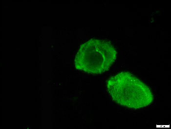
IF: 293T-mCherry cells
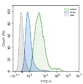
FC: 293T-mCherry cells
mCherry Chicken Polyclonal Antibody
SKU: orb611194

WB: 293T-mCherry cells

IF: 293T-mCherry cells

FC: 293T-mCherry cells
mCherry Mouse Monoclonal Antibody
SKU: orb66657

WB: 293T-mCherry cells

IF: 293T-mCherry cells

FC: 293T-mCherry cells
mCherry Rabbit Polyclonal Antibody
SKU: orb181783

WB: 293T-mCherry cells

IF: 293T-mCherry cells

FC: 293T-mCherry cells
mCherry Rabbit Polyclonal Antibody
SKU: orb704228

WB: 293T-mCherry cells

IF: 293T-mCherry cells
mCherry Recombinant Rabbit Monoclonal Antibody
SKU: orb2327512

WB: 293T-mCherry cells

IF: 293T-mCherry cells
Biorbyt also provides a range of recombinant mCherry proteins for use in antibody validation, Western blot positive controls, ELISA assay development, and fluorescence-related workflows. Recombinant mCherry proteins can help confirm antibody specificity, optimize detection conditions, and support reporter system validation in molecular and cell biology research.
- Routine mCherry controls: Recombinant mCherry Protein (orb178365), Recombinant mCherry Protein, His-tagged (orb1516870), Recombinant mCherry Protein, N-His (orb3059590).
- Apoptosis imaging and phosphatidylserine binding studies: Recombinant human Annexin V-mCherry protein (orb2821395).
- mCherry-labeled VLP controls and fluorescent particle workflows: Virus-Like Particles (VLPs) isotype control-mCherry Fluorescent (orb2985962).
Choosing the Right mCherry Antibody
Different anti-mCherry antibodies may be better suited for different experimental workflows.
- Monoclonal antibodies are generally preferred for high-specificity applications such as Western blot and immunohistochemistry.
- Polyclonal antibodies often provide stronger overall signal intensity and may perform better in immunofluorescence or low-expression systems.
- Pre-adsorbed antibodies help reduce nonspecific staining and are useful in complex tissue samples.
- Tag-specific antibodies are recommended for fusion protein pull-down, immunoprecipitation, and recombinant protein validation studies.
- Biotin-conjugated antibodies support flexible secondary detection systems and multicolor staining workflows.
Although mCherry produces intrinsic red fluorescence suitable for live-cell imaging, antibody-based detection remains important for confirming full-length fusion protein integrity, amplifying weak fluorescence signals, and validating protein expression in fixed or low-expression samples.
Because mCherry shares conserved sequence regions with other DsRed-derived fluorescent proteins such as tdTomato and DsRed, some anti-mCherry antibodies may show cross-reactivity with related RFPs. Researchers should therefore select antibodies with validated specificity and include appropriate experimental controls when multiple red fluorescent proteins are present in the same study.
mCherry and Other Fluorescent Proteins
mCherry belongs to the DsRed-derived red fluorescent protein family, but researchers often compare it with other commonly used fluorescent proteins when selecting labels for imaging, reporter assays, or fusion protein studies. Key differences include fluorescence color, protein structure, brightness, photostability, and suitability for specific applications.
Fluorescent Protein | Fluorescence Color | Structure | Excitation / Emission | Key Features | Common Applications |
|---|---|---|---|---|---|
mCherry | Red | Monomer | ~587 / 610 nm | Stable monomeric RFP; good photostability; low cytotoxicity; suitable for fusion proteins | Fusion protein labeling, live-cell imaging, reporter assays, tumor cell tracking, macrophage imaging |
Green | Monomer | ~488 / 509 nm | Classic green fluorescent protein; bright and widely compatible | Protein localization, gene expression tracking, live-cell imaging, transfection monitoring | |
Orange-red / Red | Tandem dimer | ~554 / 581 nm | Very bright; contains two chromophores; larger than monomeric tags | Reporter gene studies, in vivo imaging, lineage tracing, live-animal imaging | |
Red | Monomer | ~569 / 594 nm | Bright monomeric RFP; fast maturation; strong live-cell signal | Live-cell imaging, multicolor imaging, protein tagging | |
mOrange | Orange | Monomer | ~548 / 562 nm | Orange fluorescent protein; useful for spectral separation | Multicolor imaging, protein tagging, fluorescence microscopy |
mKate2 | Far-red | Monomer | ~588 / 633 nm | Far-red emission; good photostability; deeper tissue imaging | In vivo imaging, multicolor fluorescence imaging, cell tracking |
mPlum | Far-red | Monomer | ~590 / 649 nm | Red-shifted emission; lower brightness but useful for far-red channel | Multicolor imaging, far-red fluorescence experiments |
Compared with GFP, mCherry provides a red-shifted fluorescence channel that helps reduce spectral overlap in multicolor experiments. Compared with tdTomato, mCherry is less bright but has a smaller monomeric structure, making it more suitable for fusion protein labeling and intracellular localization studies. This balance of monomeric stability, photostability, and imaging compatibility makes mCherry a versatile fluorescent protein for cell biology and molecular biology research.
FAQ — mCherry Red Fluorescent Protein
Q1: What is mCherry used for?
mCherry is mainly used as a red fluorescent tag or reporter protein. Researchers use it for live-cell imaging, fusion protein labeling, reporter gene assays, stable cell line generation, tumor cell tracking, macrophage imaging, and multicolor fluorescence experiments. Its monomeric structure makes it especially useful when the fluorescent tag must not strongly interfere with the target protein.
Q2: Is mCherry a monomer or dimer?
mCherry is a monomeric red fluorescent protein. This is one of its key advantages over earlier DsRed-derived fluorescent proteins and tandem dimer proteins such as tdTomato. Because mCherry is monomeric, it is less likely to cause protein aggregation or alter the localization of fusion proteins.
Q3: What are the excitation and emission wavelengths of mCherry?
mCherry has an excitation maximum of approximately 587 nm and an emission maximum of approximately 610 nm. These spectral properties make it compatible with red fluorescence channels and allow it to be combined with GFP, EGFP, DAPI, CFP, and other fluorophores in multicolor imaging experiments.
Q4: What is the difference between mCherry and tdTomato?
tdTomato is generally brighter because it is a tandem dimer containing two fluorescent chromophores. mCherry is less bright but has a true monomeric structure, making it more suitable for fusion protein studies, protein localization, and intracellular tracking. In general, tdTomato is preferred when maximum brightness is required, while mCherry is preferred when tag size and monomeric behavior are more important.
Q5: What is the difference between mCherry and GFP?
GFP emits green fluorescence, while mCherry emits red fluorescence. mCherry is often used with GFP in dual-color or multicolor experiments because the two signals can be separated using different fluorescence channels. Compared with GFP, mCherry's red-shifted fluorescence may also reduce background interference in some cell and tissue imaging systems.
Q6: Why use an anti-mCherry antibody if mCherry already fluoresces?
Direct mCherry fluorescence is useful for rapid visualization and live-cell imaging, but antibody-based detection provides additional sensitivity and validation. Anti-mCherry antibodies can amplify weak signals, detect mCherry in fixed samples, confirm full-length fusion protein expression by Western blot, and help distinguish intact fusion proteins from degraded fluorescent fragments.
Q7: Can anti-mCherry antibodies cross-react with tdTomato or other RFPs?
Yes, cross-reactivity may occur because mCherry, tdTomato, DsRed, and other RFP variants share conserved sequence regions derived from DsRed. Polyclonal antibodies are generally more likely to cross-react than highly specific monoclonal antibodies. Researchers should check antibody validation data and include suitable positive and negative controls when multiple RFPs are present in the same study.
Q8: Is mCherry suitable for live-cell imaging?
Yes. mCherry is suitable for live-cell imaging because it has stable red fluorescence, good photostability, and relatively low cytotoxicity. It is commonly used for time-lapse imaging, cell migration studies, protein trafficking analysis, and long-term cellular tracking.
Q9: Can mCherry be used in tumor and immune microenvironment research?
Yes. mCherry is widely used to label tumor cells, immune cells, and reporter systems in cancer biology. It supports tumor growth monitoring, metastasis tracking, xenograft imaging, macrophage tracking, CD68-positive cell analysis, and tumor immune microenvironment studies.
Q10. How should researchers choose between direct fluorescence and anti-mCherry antibody detection?
Direct fluorescence is suitable for rapid screening, live-cell observation, and preliminary localization. Anti-mCherry antibody detection is better for confirmatory experiments, fixed samples, Western blot validation, signal amplification, and verifying fusion protein integrity. In many workflows, researchers use direct fluorescence for screening and antibody-based detection for final validation.
References
- Shaner, N. C., Campbell, R. E., Steinbach, P. A., Giepmans, B. N. G., Palmer, A. E., & Tsien, R. Y. (2004). Improved monomeric red, orange and yellow fluorescent proteins derived from Discosoma sp. red fluorescent protein. Nature Biotechnology, 22(12), 1567–1572. https://doi.org/10.1038/nbt1037
- Campbell, R. E., Tour, O., Palmer, A. E., Steinbach, P. A., Baird, G. S., Zacharias, D. A., & Tsien, R. Y. (2002). A monomeric red fluorescent protein. Proceedings of the National Academy of Sciences, 99(12), 7877–7882. https://doi.org/10.1073/pnas.082243699
- Shaner, N. C., Steinbach, P. A., & Tsien, R. Y. (2005). A guide to choosing fluorescent proteins. Nature Methods, 2(12), 905–909. https://doi.org/10.1038/nmeth819
- Rodriguez, E. A., Campbell, R. E., Lin, J. Y., Lin, M. Z., Miyawaki, A., Palmer, A. E., Shu, X., Zhang, J., & Tsien, R. Y. (2017). The growing and glowing toolbox of fluorescent and photoactive proteins. Trends in Biochemical Sciences, 42(2), 111–129. https://doi.org/10.1016/j.tibs.2016.09.010
- Bindels, D. S., Haarbosch, L., van Weeren, L., Postma, M., Wiese, K. E., Mastop, M., Aumonier, S., Gotthard, G., Royant, A., Hink, M. A., & Gadella, T. W. J. (2017). mScarlet: a bright monomeric red fluorescent protein for cellular imaging. Nature Methods, 14(1), 53–56. https://doi.org/10.1038/nmeth.4074
- Gross, L. A., Baird, G. S., Hoffman, R. C., Baldridge, K. K., & Tsien, R. Y. (2000). The structure of the chromophore within DsRed, a red fluorescent protein from coral. Proceedings of the National Academy of Sciences, 97(22), 11990–11995. https://doi.org/10.1073/pnas.97.22.11990
- Merzlyak, E. M., Goedhart, J., Shcherbo, D., Bulina, M. E., Shcheglov, A. S., Fradkov, A. F., Gaintzeva, A., Lukyanov, K. A., Lukyanov, S., Gadella, T. W. J., & Chudakov, D. M. (2007). Bright monomeric red fluorescent protein with an extended fluorescence lifetime. Nature Methods, 4(7), 555–557. https://doi.org/10.1038/nmeth1062
- Chudakov, D. M., Matz, M. V., Lukyanov, S., & Lukyanov, K. A. (2010). Fluorescent proteins and their applications in imaging living cells and tissues. Physiological Reviews, 90(3), 1103–1163. https://doi.org/10.1152/physrev.00038.2009
- Day, R. N., & Davidson, M. W. (2009). The fluorescent protein palette: tools for cellular imaging. Chemical Society Reviews, 38(10), 2887–2921. https://doi.org/10.1039/b901966a