
You have no items in your shopping cart.
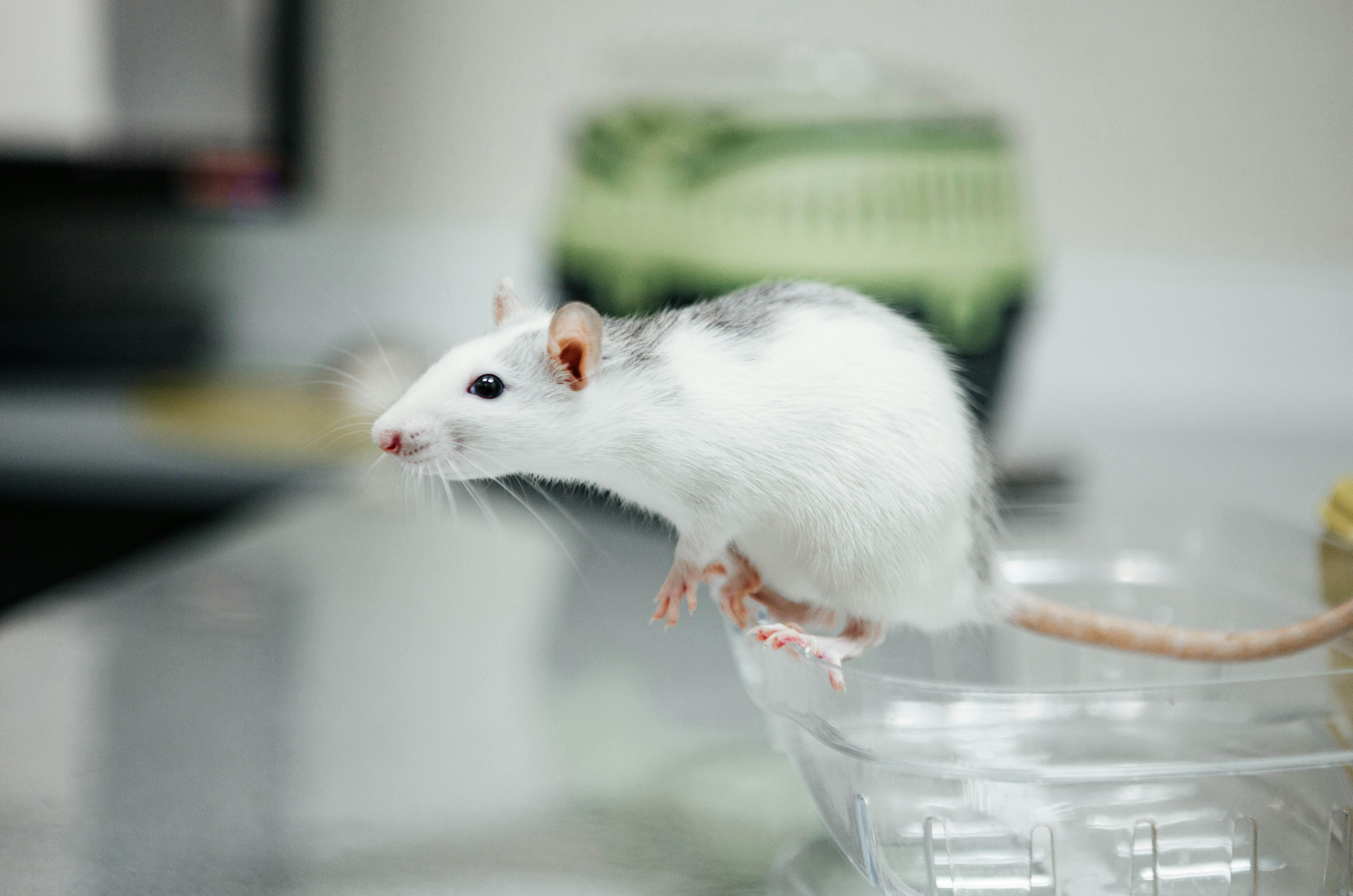
An animal model of human disease is an experimental system established in medical research that reproduces the clinical manifestations of human diseases in laboratory animals. These models serve as indispensable tools for studying disease mechanisms, evaluating therapeutic interventions, and screening drug candidates.
The complexity of human disease development makes it impractical to study disease mechanisms directly in humans due to ethical constraints and experimental limitations. Animal models allow researchers to manipulate variables that cannot be controlled under natural conditions, enabling more accurate observation of disease progression and more effective evaluation of prevention and treatment strategies.
Core Technical Service Categories
I. Basic Experimental Services
Standardized animal husbandry, precise compound dosing, and comprehensive sample collection workflows.
- Animal Housing & Maintenance
- Drug Administration & Compound Dosing
- Tissue Collection & Histopathology Preparation
II. Tumor Xenograft Models
Custom oncology modeling platforms utilizing validated tumor cell lines and patient-derived tissues.
- Common Strains & Model Characteristics
- Induced / Syngeneic / CDX / PDX Models
- Common Tumor Cell Line Screening
III. Disease Models
Broad preclinical modeling covering major physiological systems, metabolic disorders, and TCM syndromes.
- Cardiovascular Models (Atherosclerosis / HF / Ischemia etc.)
- Respiratory & Digestive (COPD / Fibrosis / Gastritis / Colitis / HCC etc.)
- Renal-Metabolic-Immune (Nephropathy / Diabetes / RA / Lupus etc.)
- Neuropsychiatric (Stroke / Parkinson’s / Alzheimer’s / Depression etc.)
- Bone-Skin-ENT (Osteoporosis / Psoriasis / Cataracts etc.)
- TCM Syndromes (Kidney / Spleen / Blood Stagnation / Liver Qi etc.)
IV. Animal Behavior
Quantitative behavioral test batteries to evaluate cognitive function, affective states, and motor coordination.
- Learning & Memory Assessments
- Anxiety / Depression Evaluation
- Social Interaction Profiling
- Motor Coordination / Sleep & Fatigue Assays
V. Small Animal Imaging
Non-invasive multimodal in vivo imaging for anatomical observation and physiological tracking.
- MRI / MicroCT Anatomical Imaging
- PET / SPECT / CT / OCT Modalities
- In Vivo Fluorescence & Ultrasound
- Laser Speckle Blood Flow Imaging
VI. Preclinical Pharmacology (Non-GLP)
Exploratory toxicology and pharmacokinetic screening to evaluate compound safety and biodistribution.
- Single / Repeated Dose Toxicity Studies
- Toxicokinetics (TK) Analysis
- Local Toxicity / Carcinogenicity Screening
- In Vivo Pharmacokinetics (PK) Profiling
VII. Other Services
Specialized functional testing and irradiation studies to support complex experimental protocols.
- Electrophysiology Recording & Analysis
- Pulmonary Function Testing
- Irradiation Studies & Model Conditioning
Comprehensive Guide to Common Modeling Methods
1. Tumor Models
Established chemical and surgical induction protocols for liver and gastric cancers.
- Hepatocellular Carcinoma — DEN Method: Diethylnitrosamine via gavage (0.25–1 mL of 0.25% solution) or drinking water (2–10 mL/kg/day) for ~6 months in rats.
- Hepatocellular Carcinoma — DBA Method: 4-Dimethylaminoazobenzene induction using 0.06% DBA diet with vitamin B₂ kept below 1.5–2 mg/kg for 4–6 months in rats.
- Hepatocellular Carcinoma — 2AAF Method: 0.03% 2-Acetylaminofluorene in feed at 2–3 mg/animal/day for 3–4 months in rats.
- Hepatocellular Carcinoma — OAAT Method: Topical 1% O-aminoazotoluene benzene solution applied to interscapular skin every other day for 7–8 weeks.
- Hepatocellular Carcinoma — Aflatoxin Method: Aflatoxin mixed into feed at 0.001–0.015 mg/kg for 6 months.
- Gastric Cancer — MC Thread Implantation: Surgical thread with ~5 mg methylcholanthrene inserted into glandular stomach mucosa of mice under sterile conditions; develops in 4–8 months.
- Gastric Cancer — Asymmetric Nitrosamine Method: 0.25 mL/kg dose; forestomach papillomas at 3 months with 85–100% progressing to forestomach carcinoma by 7–8 months (Kunming strain mice most susceptible).
2. Peptic Ulcer Models
Reliable ulcer induction techniques commonly utilized for anti-ulcer drug evaluation.
- Stress Ulcer Model (Cold Restraint): Rats fasted 24–48h, restrained, and immersed vertically in 20–23°C water up to xiphoid process for 20–24 hours.
- Histamine-Induced Ulcer Model: Subcutaneous histamine phosphate (50 mg/kg) injected twice at a 2-hour interval after 24h fast; induces ulcers in esophagus, stomach, and duodenum.
- Salicylic Acid-Induced Ulcer Model: Salicylic acid administered by gavage (100 mg/kg) after a 24-hour fast, with examination at 4 hours.
- Pyloric Ligation Model: Sterile pyloric ligation under anesthesia with fasting and examination at 19 hours post-surgery.
3. Hypertension Models
Surgical and stress-induced modeling approaches reproducing elevated blood pressure phenotypes.
- Renal Artery Stenosis Model: Silver clip placed around isolated renal artery in dogs or rabbits; contralateral kidney removed after 10–12 days for unilateral stenosis, producing long-term hypertension.
- Renal Encapsulation Model: Left kidney wrapped with X-shaped double-layer latex membrane and right kidney removed in rats (~30% develop hypertension after ~20 days).
- Stress-Induced Hypertension Model: Combined noise and electric foot shock stress administered twice daily for 2 hours per session (~20 days to hypertension).
4. Diabetes Models
Chemical, viral, and dietary induction methods for Type 1 and Type 2 diabetes research.
- Virus-Induced Model (Type 1): Subcutaneous inoculation of M variant encephalomyocarditis virus in female DBA/2 mice; hyperglycemia develops within 4–7 days.
- Alloxan Method (β-Cell Destruction): Single IV injection of alloxan (40 mg/kg) in SD rats; successful modeling defined by blood glucose >300 mg/dL maintained for 2 weeks.
- Streptozotocin (STZ) Method (Most Common): STZ in acidified saline administered intravenously at 40–100 mg/kg; successful modeling defined by blood glucose >400 mg/dL for 3 consecutive days.
- High-Sucrose Diet Model (Type 2): 54% sucrose diet in 5-week-old SHR/NLHCP rats; OGTT abnormalities appear at 1 month and insulin response abnormalities at 6.5 months.
5. Respiratory Disease Models
Validated modeling protocols for pulmonary infections, emphysema, edema, and fibrosis.
- Pneumonia Model: Intratracheal inoculation of bacterial suspension (1×10⁶ to 1×10⁹ CFU/0.25 mL) followed by 0.5 mL air and upright rotation in rats.
- Pulmonary Emphysema Model: Intratracheal or IV administration of proteolytic enzymes (papain, trypsin, or elastase), with papain producing the most typical lesions.
- Pulmonary Edema Model: Induced via NO inhalation, intratracheal 50% glucose, bilateral cervical vagotomy, IV 10% chloroform, or IP 6% ammonium chloride.
- Pulmonary Fibrosis Model (Bleomycin): Intratracheal instillation of bleomycin A5 solution (5 mg/kg) in anesthetized rats followed by upright rotation.
Quick Reference Table of Common Animal Models
| Disease Type | Common Methods | Common Species |
|---|---|---|
| Liver Cancer | DEN / DBA / 2AAF / Aflatoxin | Rat |
| Gastric Cancer | MC thread / Nitrosamine | Mouse / Rat |
| Peptic Ulcer | Stress restraint / Histamine / Pyloric ligation | Rat |
| Hypertension | Renal artery stenosis / Renal wrapping / Stress | Rat / Dog / Rabbit |
| Diabetes (T1) | STZ / Alloxan / Virus | Rat / Mouse |
| Diabetes (T2) | High-sucrose diet | Rat |
| Pneumonia | Intratracheal bacterial inoculation | Rat |
| Pulmonary Emphysema | Papain / Elastase intratracheal | Rat |
| Pulmonary Fibrosis | Bleomycin intratracheal | Rat / Mouse |
Note: The methods listed above represent commonly used modeling approaches in biomedical research. Selection of appropriate models should consider specific experimental objectives, animal welfare requirements, and institutional guidelines.