You have no items in your shopping cart.
Featured
KO/KD
Validated
Validated
Description
Research Area
Cell Biology
Images & Validation
−Item 1 of 12
| Tested Applications | IF, IHC, KO/KD Validated, WB |
|---|---|
| Reactivity | Human, Mouse, Porcine, Rat |
| Application Notes |
Key Properties
−| Antibody Type | Primary Antibody |
|---|---|
| Host | Rabbit |
| Clonality | Polyclonal |
| Isotype | IgG |
| Immunogen | A synthetic peptide of human LC3B |
| Target | MAP1LC3B |
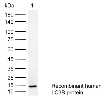
| Molecular Weight | Observed: 14kDa, 16kDa |
| Purification | Affinity purification |
| Conjugation | Unconjugated |
Storage & Handling
−| Storage | Maintain refrigerated at 2-8°C for up to 2 weeks. For long term storage store at -20°C in small aliquots to prevent freeze-thaw cycles. |
|---|---|
| Form/Appearance | Liquid |
| Buffer/Preservatives | PBS with 0.02% sodium azide, 50% glycerol, pH 7.3. |
| Concentration | batch dependent |
| Expiration Date | 12 months from date of receipt. |
| Disclaimer | For research use only |
Alternative Names
−MAP1LC3B Antibody: LC3B, ATG8F, MAP1LC3B-a, MAP1A/1BLC3, MAP1ALC3, Autophagy-related protein LC3 B
Similar Products
−- Item 1 of 15
LC3 Rabbit Polyclonal Antibody [orb33328]
ICC, IF, IHC-P, WB
Bovine
Human, Mouse, Rat
Rabbit
Polyclonal
Unconjugated
100 μg - Item 1 of 7
Cleaved LC3A Antibody [orb33336]
ICC, IF, IHC-P, WB
Rat, Zebrafish
Human, Mouse
Rabbit
Polyclonal
Unconjugated
50 μl, 100 μl - Item 1 of 4
LC3B Rabbit Polyclonal Antibody [orb6295]
IF, IHC-Fr, IHC-P
Bovine, Canine, Equine, Gallus, Human, Porcine, Zebrafish
Mouse, Rabbit, Rat
Rabbit
Polyclonal
Unconjugated
50 μl, 100 μl, 200 μl - Item 1 of 5
APG8b(MAP1LC3B) Antibody (N-term T29) [orb1937429]
FC, IHC-P, WB
Rat
Human, Mouse
Rabbit
Polyclonal
Unconjugated
50 μl, 100 μl - Item 1 of 1
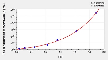
Rat Microtubule-Associated Proteins 1A/1B Light Chain 3B (MAP1LC3B) ELISA Kit [orb782694]
Rat
0.16-10 ng/mL
0.067 ng/mL
48 T, 96 T
Quality Guarantee
Explore bioreagents carefree to elevate your research. All our products are rigorously tested for performance. If a product does not perform as described on its datasheet, our scientific support team will provide expert troubleshooting, a prompt replacement, or a refund. For full details, please see our Terms & Conditions and Buying Guide. Contact us at [email protected].

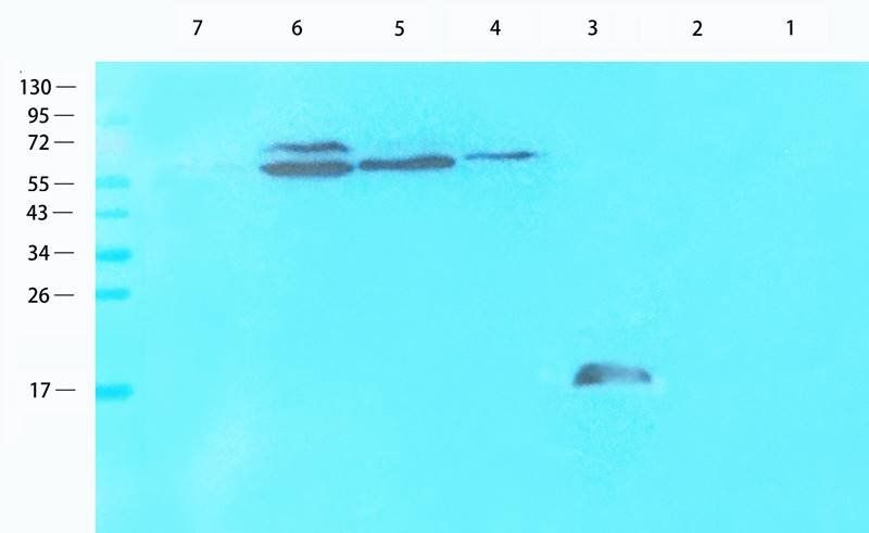
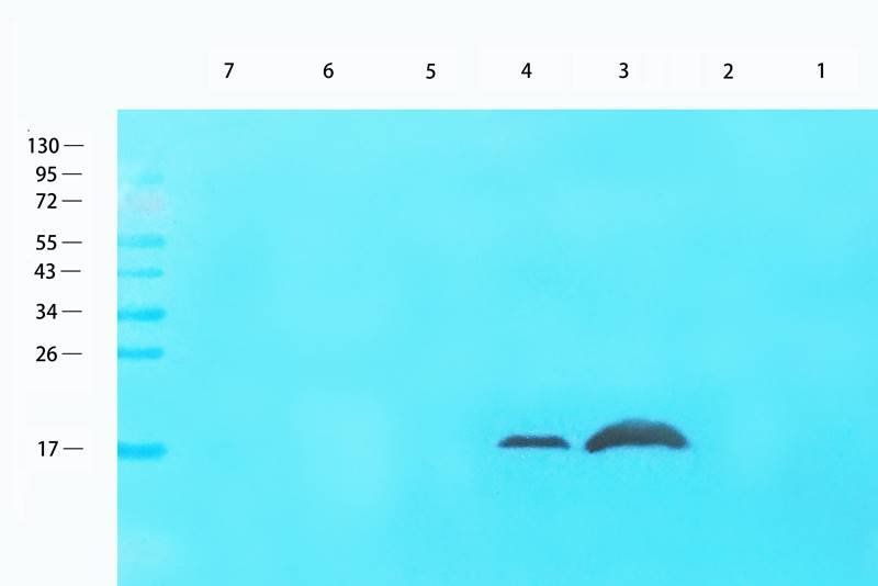
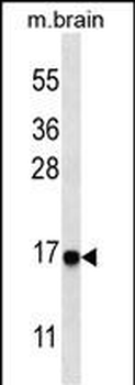
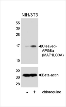
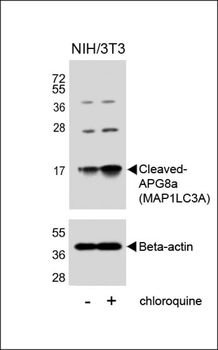
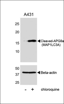
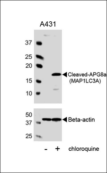
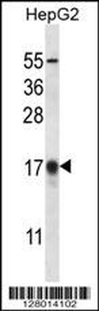
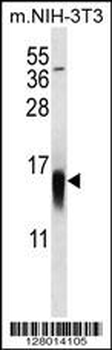
Western blot analysis of extracts of various cell lines, using LC3B antibody (orb1274430) at 1:1000 dilution. C6 cells were treated by Chloroquine (50 μM) for 20 hours. Hela cells were treated by Chloroquine (50 μM) for 20 hours. NIH/3T3 cells were treated by Chloroquine (50 μM) for 20 hours. Secondary antibody: HRP Goat Anti-Rabbit IgG (H+L) at 1:10000 dilution. Lysates/proteins: 25 ug per lane. Blocking buffer: 3% nonfat dry milk in TBST. Detection: ECL Basic Kit. Exposure time: 10s.
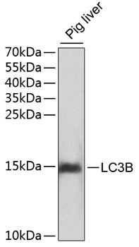
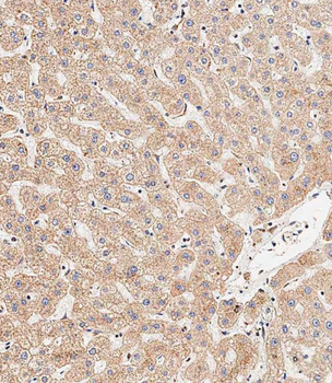
Western blot analysis of extracts of pig liver, using LC3B antibody (orb1274430). Secondary antibody: HRP Goat Anti-Rabbit IgG (H+L) at 1:10000 dilution. Lysates/proteins: 25 ug per lane. Blocking buffer: 3% nonfat dry milk in TBST.
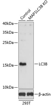
Western blot analysis of extracts from normal (control) and LC3B knockout (KO) 293T cells, using LC3B antibody (orb1274430) at 1:1000 dilution. Secondary antibody: HRP Goat Anti-Rabbit IgG (H+L) at 1:10000 dilution. Lysates/proteins: 25 ug per lane. Blocking buffer: 3% nonfat dry milk in TBST. Detection: ECL Basic Kit. Exposure time: 90s.
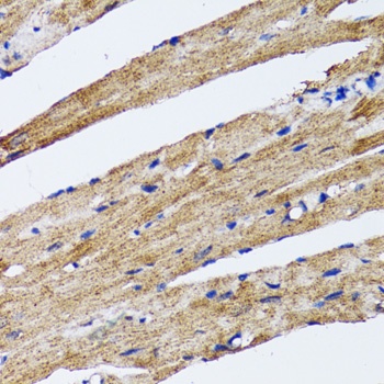
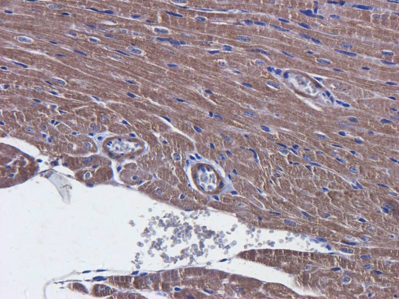
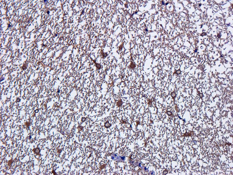
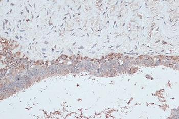
Immunohistochemistry of paraffin-embedded rat heart using LC3B antibody (orb1274430) at dilution of 1:100 (40x lens).
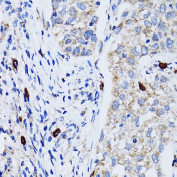
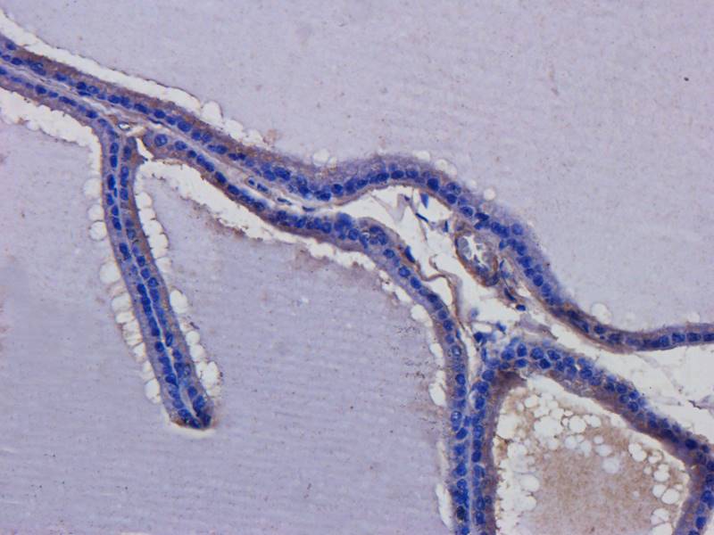
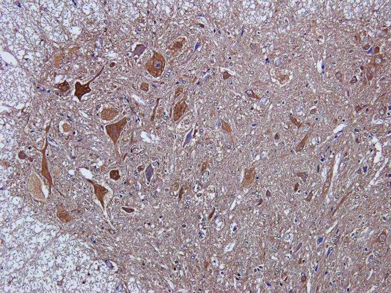
Immunohistochemistry of paraffin-embedded human lung cancer using LC3B antibody (orb1274430) at dilution of 1:100 (40x lens).
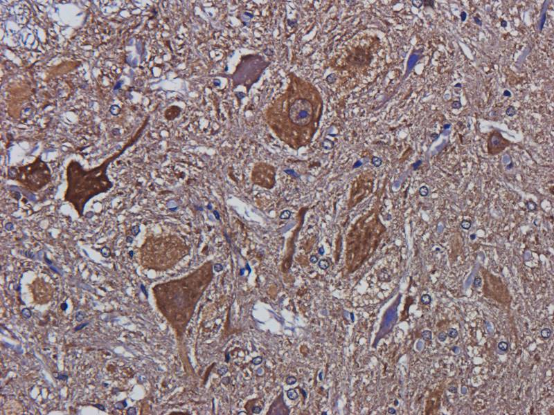
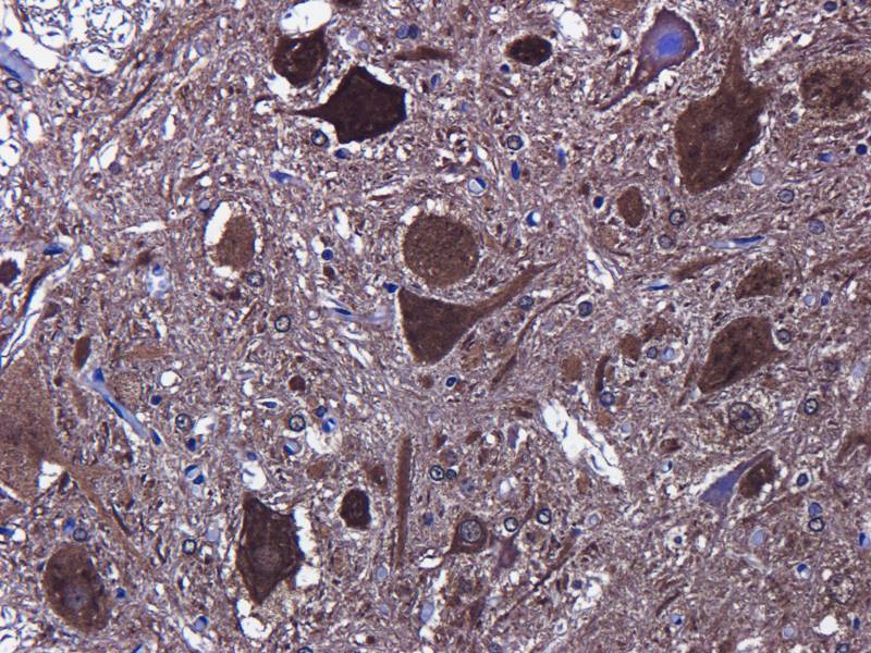
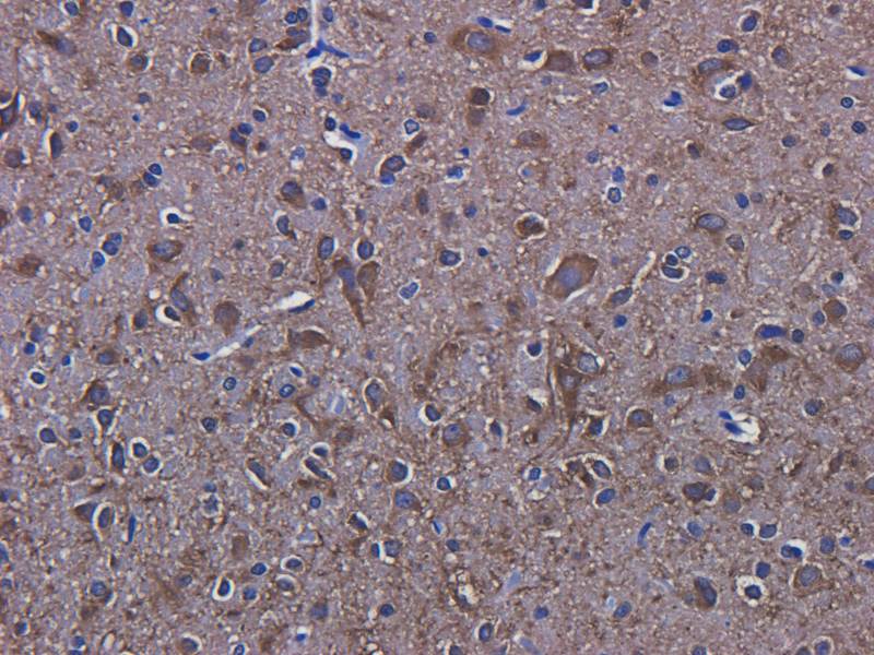
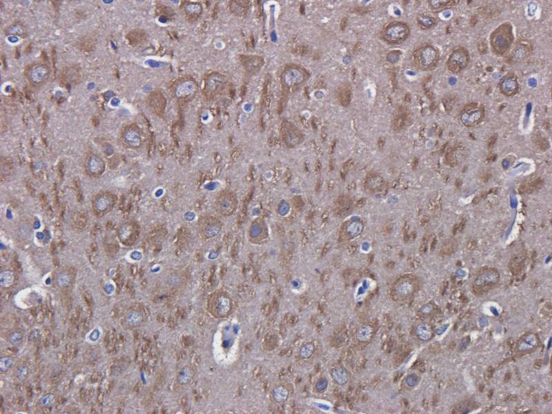
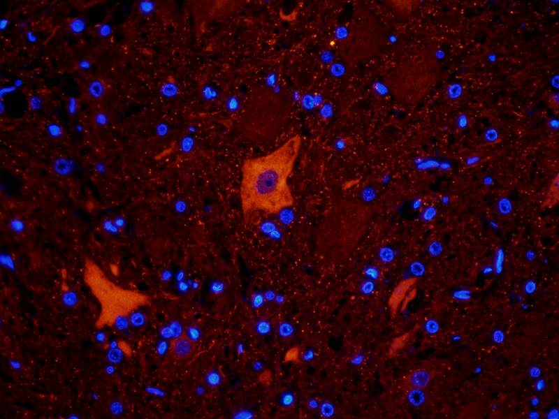
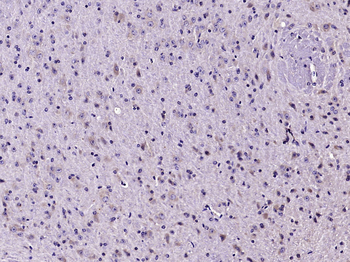
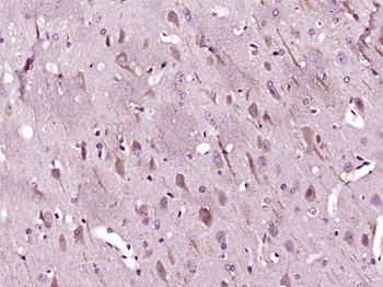
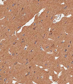
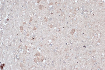
Immunohistochemistry of paraffin-embedded mouse brain using LC3B antibody (orb1274430) at dilution of 1:100 (40x lens).
Immunohistochemistry of paraffin-embedded mouse heart using LC3B antibody (orb1274430) at dilution of 1:100 (40x lens).
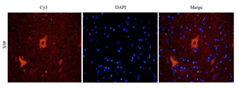
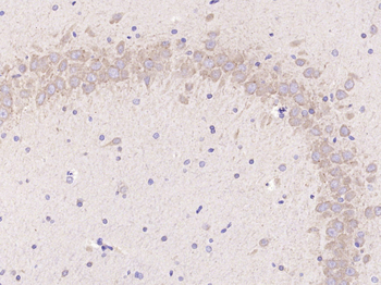
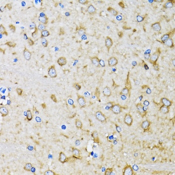
Immunohistochemistry of paraffin-embedded rat brain using LC3B antibody (orb1274430) at dilution of 1:100 (40x lens).
Immunohistochemistry of paraffin-embedded human breast cancer using LC3B antibody (orb1274430) at dilution of 1:100 (40x lens).
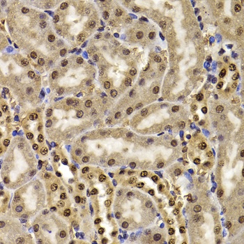
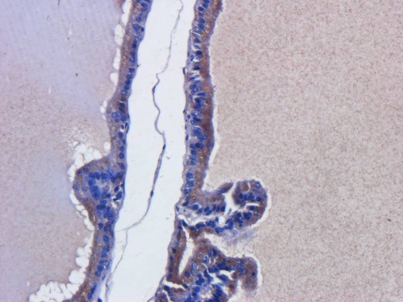
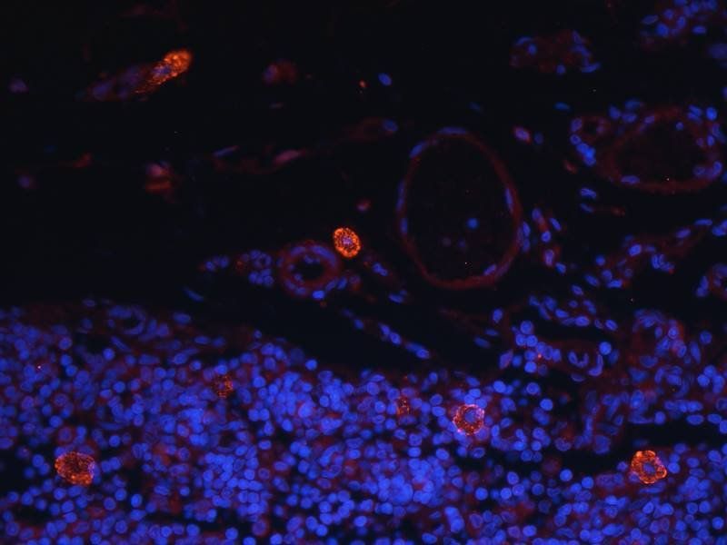
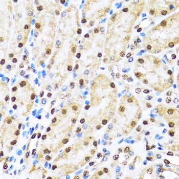
Immunohistochemistry of paraffin-embedded mouse kidney using HIRIP3 antibody (orb1256659) at dilution of 1:100 (40x lens).
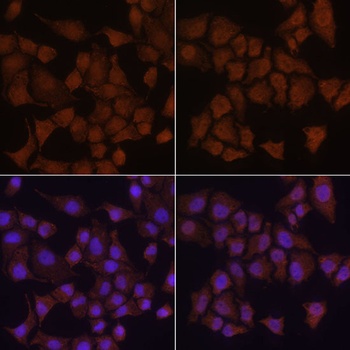
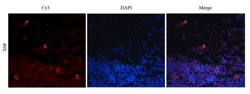
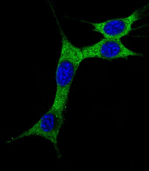
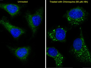
Immunofluorescence analysis of HeLa cells using LC3B antibody (orb1274430) at dilution of 1:100. Hela cells were treated by Chloroquine (50 μM) for 20 hours (left). Blue: DAPI for nuclear staining.
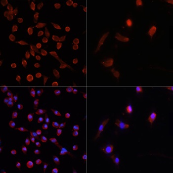
Immunofluorescence analysis of NIH/3T3 cells using LC3B antibody (orb1274430) at dilution of 1:100. NIH/3T3 cells were treated by Chloroquine (50 μM) for 20 hours (left). Blue: DAPI for nuclear staining.
Quick Database Links
Gene Symbol
MAP1LC3B
UniProt
UniProt Details
− No UniProt data available
Documents Download
Datasheet
Product Information
Request a Document
Protocol Information
WB
Western Blot (IB, immunoblot)
IHC
Immunohistochemistry
IF
Immunofluorescence
MAP1LC3B Antibody (orb1274430)

- 0.0
Based on 0 reviews
Participating in our Biorbyt product reviews program enables you to support fellow scientists by sharing your firsthand experience with our products.
Login to Submit a ReviewAvailable Sizes
Select a size below
Choose Conjugation or Carrier Free Version
Free Secondary Antibody (20 ul)0/0
Please add an antibody product to your cart first.