
You have no items in your shopping cart.
Red Fluorescent Protein Research
FusionRed Fluorescent Protein: Properties, Imaging and Detection
Explore the monomeric structure, low cytotoxicity and fusion-protein performance of FusionRed, compare FusionRed with mCherry and mKate2, and validate FusionRed-tagged proteins with Biorbyt FusionRed Antibody orb3188439.
1. What Is FusionRed Fluorescent Protein?
FusionRed is an engineered monomeric red fluorescent protein developed from the mKate2 protein scaffold. It was designed to address common problems associated with red fluorescent protein fusion constructs, including aggregation, abnormal localization and protein-associated cellular toxicity.
FusionRed is particularly valued for its low cytotoxicity and reliable performance when fused to cellular proteins. The original characterization evaluated FusionRed in numerous fusion constructs involving cytoskeletal proteins, membrane proteins, organelle markers and nuclear proteins.
The protein produces red fluorescence with excitation and emission maxima near 580 nm and 608 nm. These spectral properties support multicolour experiments alongside green and cyan fluorescent proteins, provided that channel overlap and bleed-through are properly controlled.
Key point: FusionRed was optimized primarily for monomeric behaviour, low cytotoxicity and reliable fusion-protein localization. It should not automatically be selected when maximum fluorescence brightness is the principal experimental requirement.
FusionRed Spectral Overview
580 nm
Excitation Maximum
Excitation is shifted toward the orange-red region
compared with many green fluorescent proteins.
608 nm
Emission Maximum
Red fluorescence can be detected using an
appropriately configured red-emission channel.
Red
Fluorescence Channel
Filter compatibility should be confirmed using the
available microscope or flow cytometer configuration.
FusionRed at a Glance
| Property | FusionRed |
|---|---|
| Protein class | Engineered monomeric red fluorescent protein |
| Protein origin | Developed from the mKate2 fluorescent protein scaffold |
| Approximate molecular weight | Approximately 26 kDa |
| Polypeptide length | Approximately 232 amino acids |
| Excitation / emission | Approximately 580 / 608 nm |
| Oligomeric state | Monomeric |
| Main advantages | Low cytotoxicity and reliable performance in fusion-protein constructs |
| Common applications | Live-cell imaging, stable cell lines, subcellular localization and fusion-protein labelling |
| Detection methods | Direct fluorescence and FusionRed antibody-based detection |
2. FusionRed vs mCherry and mKate2
FusionRed, mCherry and mKate2 are all red-emitting fluorescent proteins, but they were developed for different priorities. FusionRed emphasizes low cytotoxicity and fusion-protein performance, mCherry remains a widely used general-purpose red reporter, and mKate2 provides more red-shifted fluorescence.
| Feature | FusionRed | mCherry | mKate2 |
|---|---|---|---|
| Main design priority | Low cytotoxicity and reliable fusion performance | General-purpose monomeric red fluorescence | Bright, red-shifted fluorescence and imaging performance |
| Monomeric behaviour | Developed for strong monomeric behaviour | Generally regarded as monomeric | Can display concentration-dependent association under some conditions |
| Fusion-protein localization | Developed specifically for reliable fusion-protein labelling | Widely used, but performance can remain construct-dependent | Suitable for many fusion constructs |
| Brightness consideration | Useful signal, but brightness is not its main design advantage | Moderate brightness and broad experimental support | Strong brightness with more red-shifted emission |
| Best selected when | Low toxicity and correct fusion localization are priorities | A familiar and broadly supported red reporter is required | Red-shifted emission and stronger brightness are priorities |
Selection guidance: Select FusionRed when fusion-protein behaviour and low cellular toxicity are more important than maximum molecular brightness. Performance should still be validated using the exact fusion partner, linker and cell model.
3. FusionRed Antibody for Fusion-Protein Detection
Biorbyt offers a directly relevant FusionRed Antibody for Western blot detection of FusionRed and FusionRed-tagged fusion constructs.
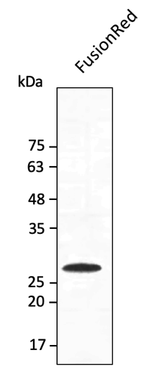
Product validation image for FusionRed Antibody orb3188439 .
Direct FusionRed Detection Reagent
View orb3188439
FusionRed Antibody — orb3188439
orb3188439 is a dedicated FusionRed antibody intended for Western blot detection. It can support confirmation of FusionRed reporter expression and assessment of the molecular weight of FusionRed-tagged fusion proteins.
Antibody-based detection complements direct red fluorescence by providing an independent method for evaluating protein expression, fusion integrity and potential cleavage or degradation products.
Product:
FusionRed Antibody
SKU:
orb3188439
Tested application:
WB
Format:
Unconjugated
Available size:
100 µg
Protocol:
Western blot protocol available
Document:
Product datasheet available
Custom option:
Custom production available
Recommended role: Use orb3188439 to confirm FusionRed-tagged protein expression and expected molecular weight by Western blot, especially when direct fluorescence alone does not provide enough information about fusion integrity.
Related Red Fluorescent Protein Antibodies
The following antibodies support broader RFP, DsRed, mCherry and tdTomato workflows. They are included as related reagents and should not replace the dedicated FusionRed antibody without application-specific validation.
| SKU | Product | Applications | Available Size |
|---|---|---|---|
| orb345392 | DsRed Antibody Rabbit polyclonal · Broad RFP detection | ELISA, FC, IF, IHC, WB | 25 µl |
| orb11618 | mCherry Antibody Goat polyclonal · mCherry detection | FC, ICC, IEM, IF, IHC-Fr, IHC-P, WB | 100 µg |
| orb182397 | tdTomato Antibody Goat polyclonal · tdTomato and mCherry detection | ELISA, FACS, IF, IHC-Fr, IHC-P, WB | 100 µg |
4. Key Applications of FusionRed
Fusion-Protein Localization
FusionRed can be attached to proteins of interest to monitor localization, trafficking and redistribution while reducing the risk of artefacts associated with fluorescent-protein oligomerization.
Long-Term Live-Cell Imaging
Its low reported cytotoxicity makes FusionRed useful for extended observation of living cells and experiments in which reporter expression must be maintained over longer periods.
Stable Cell-Line Generation
FusionRed can support stable expression workflows where continuous reporter production and cellular tolerance are important experimental requirements.
Multicolour Imaging
Red emission allows FusionRed to be combined with selected green, cyan or blue reporters. Spectral overlap, excitation bleed-through and detector settings should be validated for each combination.
5. Detecting and Validating FusionRed Constructs
FusionRed constructs can be analysed using direct red fluorescence and antibody-based Western blot detection. Biorbyt FusionRed Antibody orb3188439 provides a direct reagent for confirming reporter expression and evaluating FusionRed-tagged fusion proteins.
Direct FusionRed Fluorescence
Direct fluorescence is the primary approach for live-cell localization, trafficking studies and dynamic imaging.
- Suitable for living cells and time-course imaging
- No primary or secondary antibody required
- Preserves spatial and temporal information
- Supports multicolour reporter experiments
- Does not confirm fusion-protein molecular weight
FusionRed Antibody Detection
Western blot detection with orb3188439 provides an independent method for confirming FusionRed expression and evaluating fusion-protein integrity.
- Confirms the expected molecular weight by WB
- Helps identify truncated or degraded products
- Supports detection independently of chromophore maturation
- Can confirm weakly fluorescent FusionRed constructs
- Complements direct live-cell fluorescence analysis
Essential Experimental Controls
- Untransfected or untagged sample: measures cellular autofluorescence and non-specific antibody background.
- Known FusionRed-positive sample: confirms that the imaging and antibody-detection systems are functioning correctly.
- Fusion-partner or tag control: helps determine whether the complete fusion protein remains intact.
- Expected molecular-weight control: distinguishes full-length expression from cleavage or degradation products.
Interpretation: Red fluorescence confirms maturation of the FusionRed domain, but it does not independently prove that the attached protein is intact, correctly localized or biologically active. Western blotting with orb3188439 provides complementary expression and molecular-weight information.
6. FusionRed Experimental Design Considerations
Imaging Configuration
Configure excitation and emission detection around the approximate 580 nm and 608 nm maxima. Confirm performance using the available laser lines, filter sets and detector sensitivity.
Fusion Orientation
Test both N-terminal and C-terminal configurations when possible. Linker length and fusion position can affect localization, function and reporter maturation.
Expression Level
Avoid relying only on strongly overexpressing cells. Excessive expression can alter localization and cellular behaviour even when the fluorescent protein itself is monomeric.
Brightness consideration: FusionRed is valued for low toxicity and fusion performance rather than exceptionally high brightness. Exposure time, illumination intensity and expression level may require optimization, particularly for low-abundance proteins.
7. Companion Reagents and Related Resources
Red Fluorescent Protein
mCherry Research Resource
Review mCherry fluorescence, fusion applications and
Biorbyt mCherry detection reagents.
Red Fluorescent Protein
tdTomato Research Resource
Explore tdTomato imaging properties, experimental
applications and antibody-based detection.
RFP Antibodies
Browse Red Fluorescent Protein Products
Explore Biorbyt antibodies and reagents associated
with red fluorescent protein detection workflows.
Western Blot Detection
ECL Plus Substrate — orb527071
Chemiluminescent HRP substrate for sensitive Western
blot detection using compatible antibodies.
8. Why Choose Biorbyt for FusionRed Detection?
Dedicated FusionRed Antibody
orb3188439 is directly listed as a FusionRed Antibody rather than a general RFP detection reagent.
Western Blot Detection
The product supports WB confirmation of FusionRed expression and fusion-protein molecular weight.
Protocol and Datasheet Support
Product documentation and a Western blot protocol are available to support experimental setup.
Related RFP Portfolio
Additional RFP, DsRed, mCherry and tdTomato antibodies support broader fluorescent-protein workflows.
9. Frequently Asked Questions
What is FusionRed?
FusionRed is an engineered monomeric red fluorescent protein developed from the mKate2 scaffold. It was optimized for low cytotoxicity and reliable performance in fusion-protein constructs.
What are the excitation and emission maxima of FusionRed?
FusionRed has excitation and emission maxima near 580 nm and 608 nm. Actual imaging performance depends on the available laser, filter and detector configuration.
Is FusionRed brighter than mCherry?
FusionRed was not designed primarily to maximize brightness. Its principal advantages are low cytotoxicity, monomeric behaviour and fusion-protein performance.
Can FusionRed be detected by Western blot?
Yes. Biorbyt FusionRed Antibody orb3188439 is listed for Western blot detection and can support confirmation of FusionRed-tagged protein expression.
Can orb3188439 be used for immunofluorescence?
The current product information lists Western blot as the tested application. IF, ICC, IHC, IP and flow cytometry should not be presented as validated applications unless additional product data become available.
Does FusionRed fluorescence confirm that the complete fusion protein is intact?
No. Fluorescence confirms maturation of the FusionRed domain. Western blotting or another independent detection method is needed to confirm molecular weight and full-length fusion integrity.
10. Scientific References
- Shemiakina II, Ermakova GV, Cranfill PJ, et al. A monomeric red fluorescent protein with low cytotoxicity. Nature Communications. 2012;3:1204. doi:10.1038/ncomms2208
- Mukherjee S, Hung ST, Douglas N, et al. Engineering of a Brighter Variant of the FusionRed Fluorescent Protein Using Lifetime Flow Cytometry and Structure-Guided Mutations. Biochemistry. 2020;59:3669–3682. doi:10.1021/acs.biochem.0c00484
- Cranfill PJ, Sell BR, Baird MA, et al. Quantitative assessment of fluorescent proteins. Nature Methods. 2016;13:557–562. doi:10.1038/nmeth.3891
- Shaner NC, Campbell RE, Steinbach PA, et al. Improved monomeric red, orange and yellow fluorescent proteins derived from Discosoma sp. red fluorescent protein. Nature Biotechnology. 2004;22:1567–1572. doi:10.1038/nbt1037
Scientifically reviewed by:
Biorbyt Scientific Support Team
Last reviewed:
July 2026