You have no items in your shopping cart.
Featured
Description
Research Area
Cell Biology
Images & Validation
−Item 1 of 6
| Tested Applications | FC, ICC, IF, IHC-Fr, IHC-P, WB |
|---|---|
| Dilution Range | WB=1:500-2000, IHC-P=1:100-500, IHC-F=1:100-500, ICC/IF=1:100-500, IF=1:100-500, Flow-Cyt=1μg/Test |
| Reactivity | Human, Mouse |
| Predicted Reactivity | Rat |
Related Conjugates & Formulations
−Key Properties
−| Antibody Type | Primary Antibody |
|---|---|
| Host | Rabbit |
| Clonality | Polyclonal |
| Isotype | IgG |
| Immunogen | KLH conjugated synthetic peptide derived from human FGFR2 (21-120/821aa) |
| Target | FGFR2 |
| Molecular Weight | 142 kDa |
| Purification | Affinity purified by Protein A |
| Conjugation | Unconjugated |
Storage & Handling
−| Storage | Maintain refrigerated at 2-8°C for up to 2 weeks. For long term storage store at -20°C in small aliquots to prevent freeze-thaw cycles. |
|---|---|
| Form/Appearance | Liquid |
| Buffer/Preservatives | 0.01M TBS (pH7.4) with 1% rAlbumin, 0.02% Proclin300 and 50% Glycerol. |
| Concentration | 1mg/ml |
| Expiration Date | 12 months from date of receipt. |
| Disclaimer | For research use only |
Alternative Names
−BBDS; BEK; BFR-1; CD332; CEK3; CFD1; ECT1; JWS; K-SAM; KGFR; TK14; TK25; Fgfr-2; Fgfr-7; Fgfr2b; Fgfr7; KGFRTr; svs; FGFR2_HUMAN; FGFR2; K-sam (KGFR); Keratinocyte growth factor receptor; 2.7.10.1; KSAM; FGFR2_MOUSE; Keratinocyte growth factor receptor (KGFR); fibroblast growth factor receptor 2; bacteria-expressed kinase; craniofacial dysostosis 1; Jackson-Weiss syndrome; Crouzon syndrome; Pfeiffer syndrome
Similar Products
−- Item 1 of 7
FGFR2 Antibody (N-term) [orb1929046]
FC, IF, IHC-P, WB
Human, Mouse
Rabbit
Polyclonal
Unconjugated
50 μl, 100 μl - Item 1 of 7
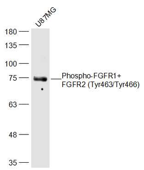
Phospho-FGFR1+FGFR2 (Tyr463/Tyr466) Rabbit Polyclonal Antibody [orb5241]
ELISA, IF, IHC-Fr, IHC-P, WB
Bovine, Equine, Gallus, Porcine, Rabbit
Human, Mouse, Rat
Rabbit
Polyclonal
Unconjugated
50 μl, 100 μl, 200 μl - Item 1 of 5
- Item 1 of 4
FGFR2 Antibody (N-term R22) [orb1929044]
FC, IF, WB
Mouse
Human
Rabbit
Polyclonal
Unconjugated
50 μl, 100 μl - Item 1 of 4
FGFR2 Rabbit Polyclonal Antibody [orb1152347]
ELISA, FC, IHC, WB
Human
Rabbit
Polyclonal
Unconjugated
100 μg
Quality Guarantee
Explore bioreagents carefree to elevate your research. All our products are rigorously tested for performance. If a product does not perform as described on its datasheet, our scientific support team will provide expert troubleshooting, a prompt replacement, or a refund. For full details, please see our Terms & Conditions and Buying Guide. Contact us at [email protected].
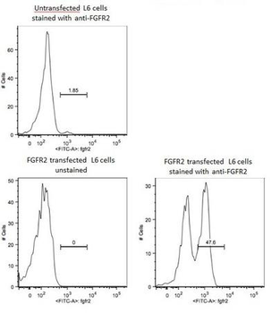
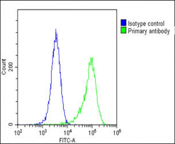
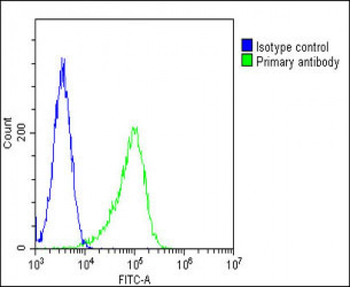
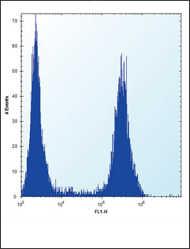
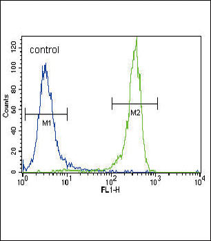
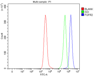
L6 cells were transfected with FGFR2, and stained with RABBIT ANTI-FGFR2 POLYCLONAL ANTIBODY, conjugated at 1:100 dilution.
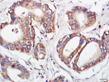
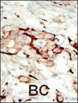
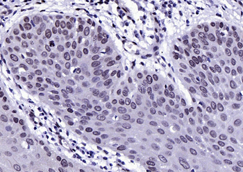
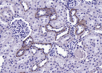
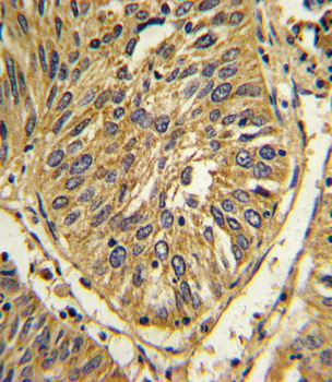
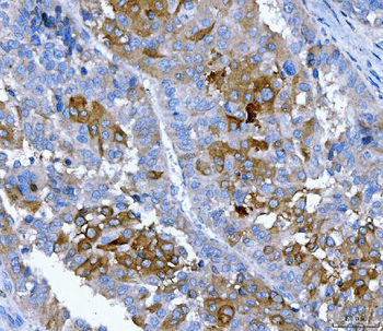
Paraformaldehyde-fixed, paraffin embedded (human stomach cancer), Antigen retrieval by boiling in sodium citrate buffer (pH6.0) for 15 min, Block endogenous peroxidase by 3% hydrogen peroxide for 20 minutes, Blocking buffer (normal goat serum) at 37°C for 30 min, Antibody incubation with (FGFR2) Polyclonal Antibody, Unconjugated (orb10656) at 1:400 overnight at 4°C, followed by a conjugated secondary for 20 minutes and DAB staining.
Paraformaldehyde-fixed, paraffin embedded (human stomach cancer), Antigen retrieval by boiling in sodium citrate buffer (pH6.0) for 15 min, Block endogenous peroxidase by 3% hydrogen peroxide for 20 minutes, Blocking buffer (normal goat serum) at 37°C for 30 min, Antibody incubation with (FGFR2) Polyclonal Antibody, Unconjugated (orb10656) at 1:400 overnight at 4°C, followed by a conjugated secondary for 20 minutes and DAB staining.
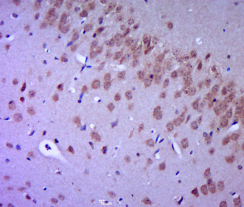
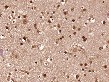
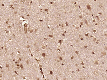
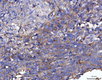
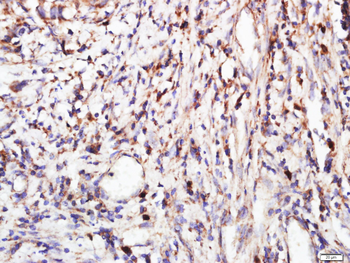
Paraformaldehyde-fixed, paraffin embedded (mouse brain tissue), Antigen retrieval by boiling in sodium citrate buffer (pH6.0) for 15 min, Block endogenous peroxidase by 3% hydrogen peroxide for 20 minutes, Blocking buffer (normal goat serum) at 37°C for 30 min, Antibody incubation with (FGFR2) Polyclonal Antibody, Unconjugated (orb10656) at 1:400 overnight at 4°C, followed by a conjugated secondary for 20 minutes and DAB staining.
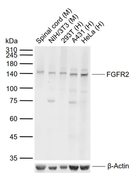
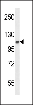
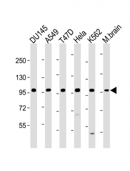
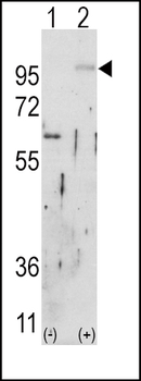
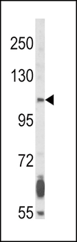
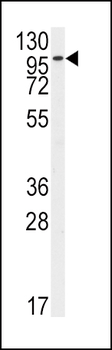
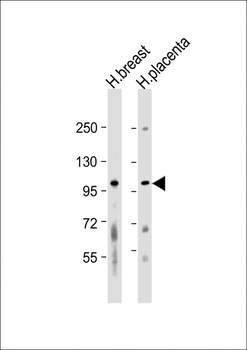
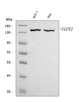
Sample: Lane 1: Mouse Spinal cord tissue lysates, Lane 2: Mouse NIH/3T3 cell lysates, Lane 3: Human 293T cell lysates, Lane 4: Human A431 cell lysates, Lane 5: Human HeLa cell lysates, Primary: Anti-FGFR2 (orb10656) at 1/1000 dilution, Secondary: IRDye800CW Goat Anti-Rabbit IgG at 1/20000 dilution, Predicted band size: 89 kDa, Observed band size: 142 kDa.
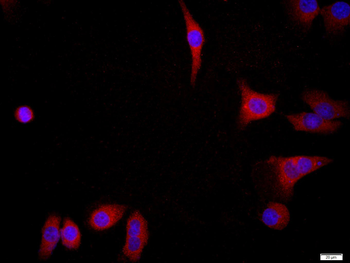
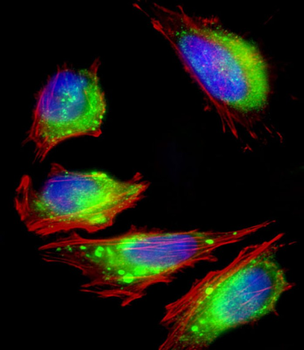
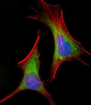
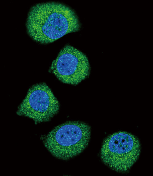
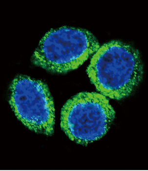
Tissue/Cell: MCF7, 4% Paraformaldehyde-fixed, Triton X-100 at room temperature for 20 min, Blocking buffer (normal goat serum) at 37°C for 20 min, Antibody incubation with (FGFR2) Polyclonal Antibody, Unconjugated (orb10656) 1:200, 90 minutes at 37°C, followed by a conjugated Goat Anti-Rabbit IgG antibody (orb868805) at 37°C for 90 minutes, DAPI (blue) was used to stain the cell nuclei.
Quick Database Links
Gene Symbol
FGFR2
UniProt
UniProt Details
− No UniProt data available
Documents Download
Datasheet
Product Information
Request a Document
Protocol Information
WB
Western Blot (IB, immunoblot)
IHC-P
Immunohistochemistry Paraffin
IHC-Fr
Immunohistochemistry Frozen
FC
Flow Cytometry
IF
Immunofluorescence
ICC
Immunocytochemistry
FGFR2 Rabbit Polyclonal Antibody (orb10656)

- 0.0
Based on 0 reviews
Participating in our Biorbyt product reviews program enables you to support fellow scientists by sharing your firsthand experience with our products.
Login to Submit a ReviewAvailable Sizes
Select a size below
Free Secondary Antibody (20 ul)0/0
Please add an antibody product to your cart first.