You have no items in your shopping cart.
Featured
Description
Research Area
Cancer Biology, Cardiovascular Research, Immunology & Inflammation, Neuroscience, Signal Transduction, Stem Cell & Developmental Biology
Images & Validation
−Item 1 of 5
| Tested Applications | FC, IF, IHC-P, WB |
|---|---|
| Dilution Range | IF - 1:10-50, WB - 1:1000, IHC-P - 1:10-50, FC - 1:10-50 |
| Reactivity | Human |
Key Properties
−| Host | Rabbit |
|---|---|
| Clonality | Polyclonal |
| Isotype | Rabbit IgG |
| Immunogen | Recombinant Protein |
| Target | FGFR2 |
| Molecular Weight | 92025 |
| Conjugation | Unconjugated |
Storage & Handling
−| Storage | Maintain refrigerated at 2-8°C for up to 2 weeks. For long term storage store at -20°C in small aliquots to prevent freeze-thaw cycles |
|---|---|
| Form/Appearance | Purified polyclonal antibody supplied in PBS with 0.09% (W/V) sodium azide. This antibody is prepared by Saturated Ammonium Sulfate (SAS) precipitation followed by dialysis against PBS. |
| Expiration Date | 12 months from date of receipt. |
| Disclaimer | For research use only |
Alternative Names
−Fibroblast growth factor receptor 2, FGFR-2, K-sam, KGFR, Keratinocyte growth factor receptor, CD332, FGFR2, BEK, KGFR, KSAM
Similar Products
−- Item 1 of 7
FGFR2 Antibody (N-term) [orb1929046]
FC, IF, IHC-P, WB
Human, Mouse
Rabbit
Polyclonal
Unconjugated
50 μl, 100 μl - Item 1 of 7
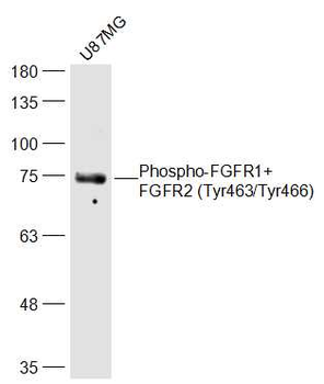
Phospho-FGFR1+FGFR2 (Tyr463/Tyr466) Rabbit Polyclonal Antibody [orb5241]
ELISA, IF, IHC-Fr, IHC-P, WB
Bovine, Equine, Gallus, Porcine, Rabbit
Human, Mouse, Rat
Rabbit
Polyclonal
Unconjugated
50 μl, 100 μl, 200 μl - Item 1 of 6
- Item 1 of 6
FGFR2 Rabbit Polyclonal Antibody [orb10656]
FC, ICC, IF, IHC-Fr, IHC-P, WB
Rat
Human, Mouse
Rabbit
Polyclonal
Unconjugated
50 μl, 100 μl, 200 μl - Item 1 of 4
FGFR2 Antibody (N-term R22) [orb1929044]
FC, IF, WB
Mouse
Human
Rabbit
Polyclonal
Unconjugated
50 μl, 100 μl
Quality Guarantee
Explore bioreagents carefree to elevate your research. All our products are rigorously tested for performance. If a product does not perform as described on its datasheet, our scientific support team will provide expert troubleshooting, a prompt replacement, or a refund. For full details, please see our Terms & Conditions and Buying Guide. Contact us at [email protected].
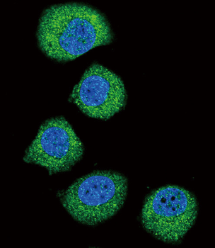
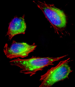
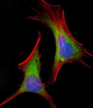
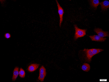
Confocal immunofluorescent analysis of FGFR2 Antibody with U251 cell followed by Alexa Fluor 488-conjugated goat anti-rabbit lgG (green). DAPI was used to stain the cell nuclear (blue).
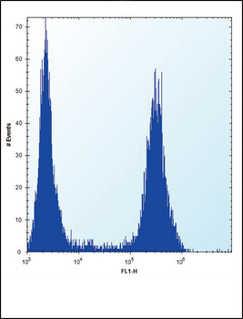
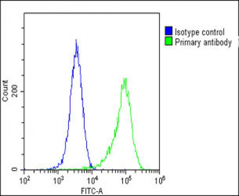
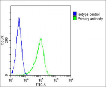
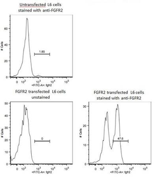
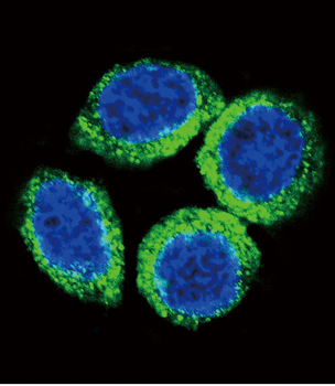
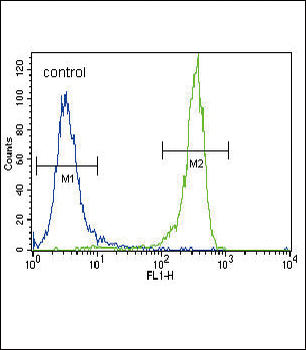
FGFR2-Antibody flow cytometric analysis of U251 cells (right histogram) compared to a negative control cell (left histogram). FITC-conjugated goat-anti-rabbit secondary antibodies were used for the analysis.
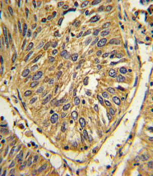
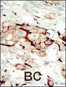
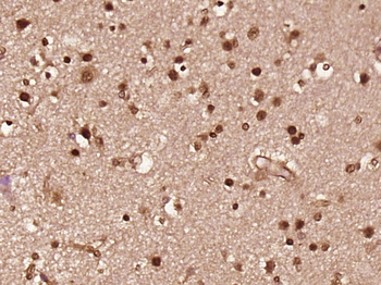
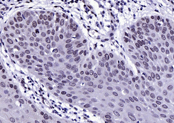
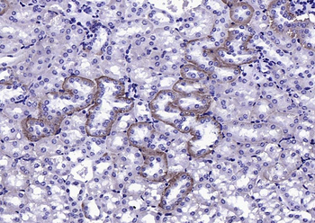
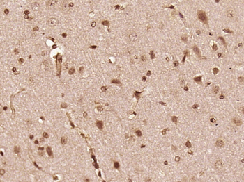
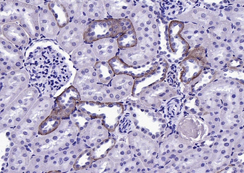
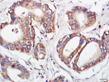
Formalin-fixed and paraffin-embedded human lung carcinoma with FGFR2 Antibody, which was peroxidase-conjugated to the secondary antibody, followed by DAB staining. This data demonstrates the use of this antibody for immunohistochemistry; clinical relevance has not been evaluated.
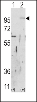
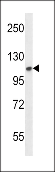
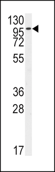
Western blot analysis of FGFR2 (arrow) using rabbit polyclonal FGFR2 Antibody. 293 cell lysates (2 ug/lane) either nontransfected (Lane 1) or transiently transfected with the FGFR2 gene (Lane 2).
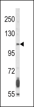
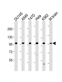
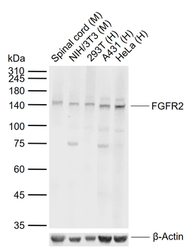
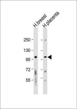
Western blot analysis of FGFR2 Antibody in NCI-H460 cell line lysates (35 ug/lane). SFGFR2 (arrow) was detected using the purified Pab.
Quick Database Links
Gene Symbol
FGFR2
UniProt
RefSeq (Protein):NP_001138389.1, NP_075259.4, NP_075418.1, NP_001138387.1, NP_001138386.1, NP_000132.3, NP_001138391.1, NP_001138388.1, NP_001138390.1, NP_001138385.1
UniProt Details
− No UniProt data available
NCBI Reference Sequences
−Associated Accession Numbers
Curated reference sequences for the gene transcript and protein productDocuments Download
Datasheet
Product Information
Request a Document
Protocol Information
WB
Western Blot (IB, immunoblot)
IHC-P
Immunohistochemistry Paraffin
FC
Flow Cytometry
IF
Immunofluorescence
FGFR2 Antibody (orb1428650)

- 0.0
Based on 0 reviews
Participating in our Biorbyt product reviews program enables you to support fellow scientists by sharing your firsthand experience with our products.
Login to Submit a ReviewAvailable Sizes
Select a size below
Choose Conjugation or Carrier Free Version
Free Secondary Antibody (20 ul)0/0
Please add an antibody product to your cart first.