You have no items in your shopping cart.
Description
Research Area
Cell Biology
Images & Validation
−Item 1 of 5
| Tested Applications | IF, IHC-P, WB |
|---|---|
| Dilution Range | IF - 1:200, WB - 1:500, IHC-P - 1:10-50 |
| Reactivity | Human |
Key Properties
−| Host | Rabbit |
|---|---|
| Clonality | Polyclonal |
| Isotype | Rabbit IgG |
| Immunogen | This ATG5 antibody is generated from rabbits immunized with a KLH conjugated synthetic peptide selected from the full length of human ATG5. |
| Target | ATG5 (HGNC:589) |
| Molecular Weight | 32447 Da |
| Conjugation | Unconjugated |
Storage & Handling
−| Storage | Maintain refrigerated at 2-8°C for up to 2 weeks. For long term storage store at -20°C in small aliquots to prevent freeze-thaw cycles |
|---|---|
| Form/Appearance | Purified monoclonal antibody supplied in PBS with 0.09% (W/V) sodium azide. This antibody is purified through a protein A column, followed by dialysis against PBS. |
| Expiration Date | 12 months from date of receipt. |
| Disclaimer | For research use only |
Alternative Names
−Autophagy protein 5, APG5-like, Apoptosis-specific protein, ATG5, APG5L, ASP
Similar Products
−- Item 1 of 6
ATG5/APG5L Rabbit Polyclonal Antibody [orb5704]
FC, IF, IHC-Fr, IHC-P, WB
Bovine, Canine, Equine, Gallus, Porcine, Rabbit
Human, Mouse, Rat
Rabbit
Polyclonal
Unconjugated
50 μl, 100 μl, 200 μl - Item 1 of 6
ATG5 Rabbit Polyclonal Antibody [orb330734]
IHC, WB
Bovine, Canine, Equine, Guinea pig, Mouse, Rabbit, Rat, Zebrafish
Human
Rabbit
Polyclonal
Unconjugated
100 μl - Item 1 of 5
ATG5 Antibody (C-term) [orb1933444]
IF, IHC-P, WB
Human, Mouse
Rabbit
Polyclonal
Unconjugated
50 μl, 100 μl - Item 1 of 5
ATG5/APG5L Mouse Monoclonal Antibody [orb500007]
ICC, IF, IHC-Fr, IHC-P, WB
Mouse
Human, Mouse, Rat
Mouse
Monoclonal
Unconjugated
50 μl, 100 μl, 200 μl, 200 μg - Item 1 of 1
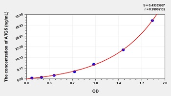
Human Autophagy Related Protein 5 (ATG5) ELISA Kit [orb781282]
Human
0.79-50 ng/mL
0.28 ng/mL
48 T, 96 T

Quality Guarantee
Explore bioreagents carefree to elevate your research. All our products are rigorously tested for performance. If a product does not perform as described on its datasheet, our scientific support team will provide expert troubleshooting, a prompt replacement, or a refund. For full details, please see our Terms & Conditions and Buying Guide. Contact us at [email protected].
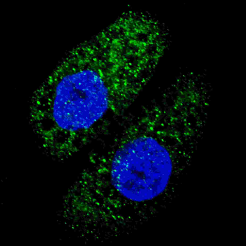
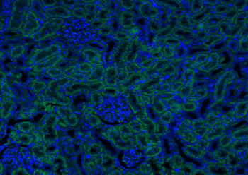
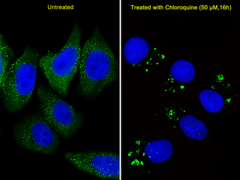
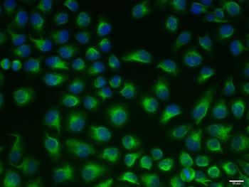
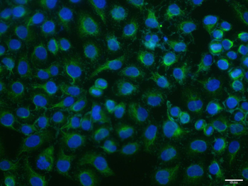
Fluorescent image of U251 cells stained with ATG5 antibody. U251 cells were treated with Chloroquine (50 μM, 16 h), then fixed with 4% PFA (20 min), permeabilized with Triton X-100 (0.2%, 30 min). Cells were then incubated with ATG5 primary antibody (1:200, 2 h at room temperature). For secondary antibody, Alexa Fluor 488 conjugated donkey anti-rabbit antibody (green) was used (1:1000, 1 h). Nuclei were counterstained with Hoechst 33342 (blue) (10 μg/ml, 5 min). ATG5 immunoreactivity is localized to autophagic vacuoles in the cytoplasm of U251 cells.
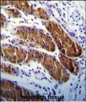
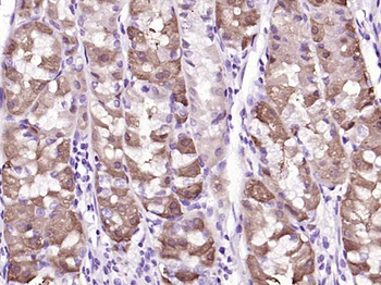
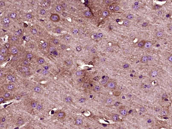
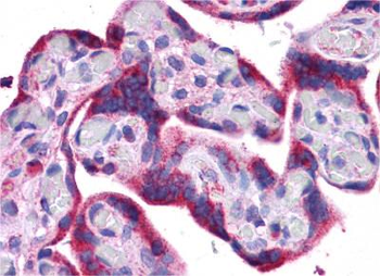
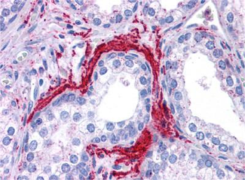
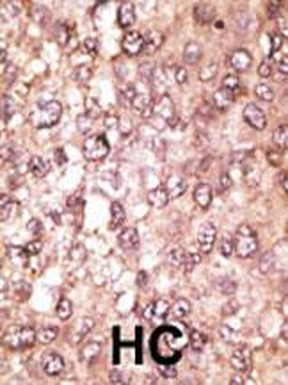
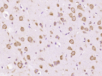
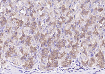
ATG5 Antibody immunohistochemistry analysis in formalin fixed and paraffin embedded human stomach tissue followed by peroxidase conjugation of the secondary antibody and DAB staining. This data demonstrates the use of ATG5 Antibody for immunohistochemistry. Clinical relevance has not been evaluated.
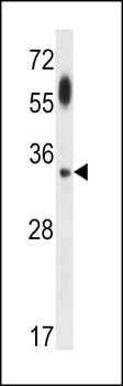
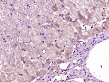
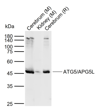
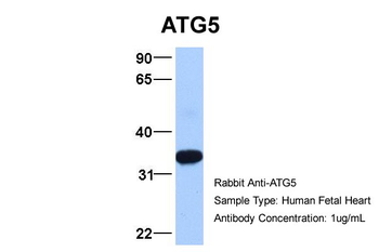
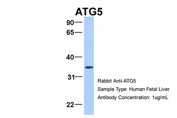
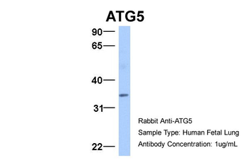
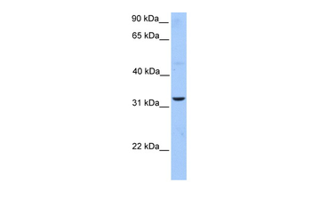
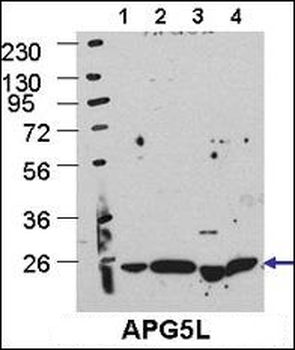
ATG5 Antibody western blot analysis in uterus tumor cell line lysates (35 ug/lane). This demonstrates the ATG5 antibody detected the ATG5 protein (arrow).
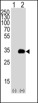
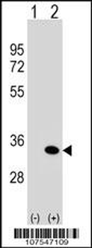
Western blot analysis of APG5 (arrow) using rabbit polyclonal APG5 Antibody. 293T cell lysates either nontransfected (Lane 1) or transiently transfected (Lane 2) with the APG5 gene.
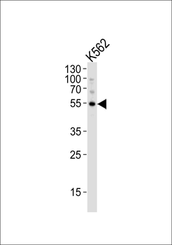
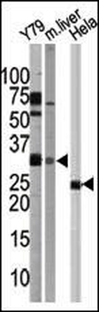
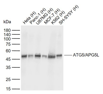
Western blot analysis of lysate from K562 cell line, using APG5 Antibody. Diluted at 1:1000. A goat anti-rabbit IgG H&L (HRP) at 1:10000 dilution was used as the secondary antibody. Lysate at 20 ug.
Quick Database Links
UniProt Details
− No UniProt data available
NCBI Reference Sequences
−Associated Accession Numbers
Curated reference sequences for the gene transcript and protein product| Protein | NP_004840.1 |
|---|
Documents Download
Datasheet
Product Information
Request a Document
Protocol Information
WB
Western Blot (IB, immunoblot)
IHC-P
Immunohistochemistry Paraffin
IF
Immunofluorescence
APG5 Antibody (orb1933443)

- 0.0
Based on 0 reviews
Participating in our Biorbyt product reviews program enables you to support fellow scientists by sharing your firsthand experience with our products.
Login to Submit a ReviewAvailable Sizes
Select a size below
Choose Conjugation or Carrier Free Version
Free Secondary Antibody (20 ul)0/0
Please add an antibody product to your cart first.