You have no items in your shopping cart.
Featured
Description
Research Area
Immunology & Inflammation
Images & Validation
−Item 1 of 8
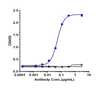
| Tested Applications | ELISA, IHC-P, WB |
|---|---|
| Reactivity | Human |
Key Properties
−| Antibody Type | Primary Antibody |
|---|---|
| Host | Rabbit |
| Clonality | Polyclonal |
| Isotype | IgG |
| Immunogen | Anti-TSLP antibody (orb1240270) was raised against a peptide corresponding to 17 amino acids near the carboxy terminus of human TSLP. The immunogen is located within the last 50 amino acids of TSLP. |
| Target | TSLP |
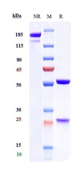
| Molecular Weight | Predicted: 18 kDObserved: 26 kD ( Post-modifications: 2 N-linked glycosylations). |
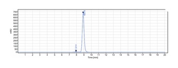
| Purification | TSLP Antibody is affinity chromatography purified via peptide column. |
| Conjugation | Unconjugated |
Storage & Handling
−| Storage | Maintain refrigerated at 2-8°C for up to 2 weeks. For long term storage store at -20°C in small aliquots to prevent freeze-thaw cycles. |
|---|---|
| Form/Appearance | Liquid |
| Buffer/Preservatives | TSLP Antibody is supplied in PBS containing 0.02% sodium azide. |
| Concentration | 1 mg/mL |
| Expiration Date | 12 months from date of receipt. |
| Disclaimer | For research use only |
Alternative Names
−TSLP Antibody: Thymic stromal lymphopoietin
Similar Products
−- Item 1 of 9
TSLP Antibody [orb1240256]
ELISA, IF, IHC-P, WB
Human, Mouse
Rabbit
Polyclonal
Unconjugated
0.02 mg, 0.1 mg - Item 1 of 4
TSLP Rabbit Polyclonal Antibody [orb11532]
IF, IHC-Fr, IHC-P
Human
Rabbit
Polyclonal
Unconjugated
50 μl, 100 μl, 200 μl - Item 1 of 6
- Item 1 of 1
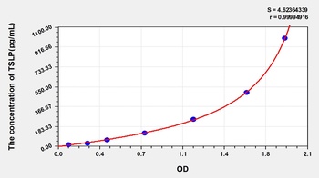
Mouse Thymic Stromal Lymphopoietin (TSLP) ELISA Kit [orb777789]
Mouse
15.63-1000 pg/mL
5.4 pg/mL
48 T, 96 T - Item 1 of 1
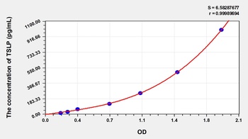
Human Thymic Stromal Lymphopoietin (TSLP) ELISA Kit [orb774948]
Human
15.63-1000 pg/mL
6.7 pg/mL
48 T, 96 T


Quality Guarantee
Explore bioreagents carefree to elevate your research. All our products are rigorously tested for performance. If a product does not perform as described on its datasheet, our scientific support team will provide expert troubleshooting, a prompt replacement, or a refund. For full details, please see our Terms & Conditions and Buying Guide. Contact us at [email protected].
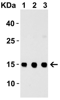
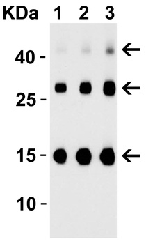
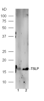
Western Blot Validation with Recombinant Protein. Loading: 30 ng of human TSLP recombinant protein per lane. Antibodies: TSLP orb1240270, 1h incubation at RT in 5% NFDM/TBST. Secondary: Goat anti-rabbit IgG HRP conjugate at 1:10000 dilution. Lane 1: 0.25 µg/mL, Lane 2: 0.5 µg/mL, Lane 3: 1 µg/mL.
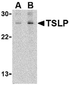
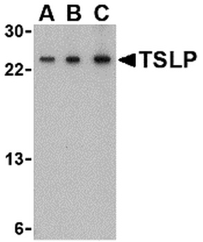
Western Blot Validation in Human Jurkat Cell Line. Loading: 15 µg of lysates per lane. Antibodies: TSLP orb1240270 (A: 1 µg/mL, B: 2 µg/mL), 1h incubation at RT in 5% NFDM/TBST. Secondary: Goat anti-rabbit IgG HRP conjugate at 1:10000 dilution.
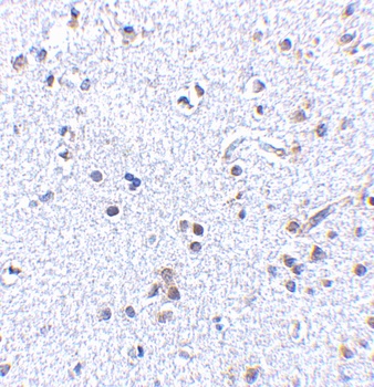
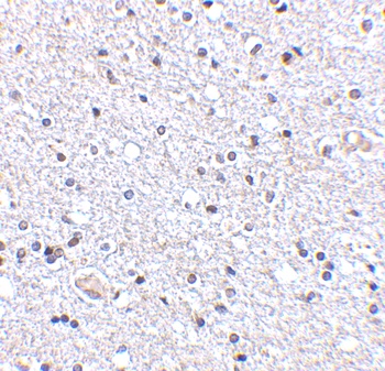
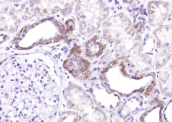
Immunohistochemistry Validation of TSLP in Human Brain Tissue. Immunohistochemical analysis of paraffin-embedded Human Brain Tissue using anti-TSLP antibody (orb1240270) at 2.5 µg/ml. Tissue was fixed with formaldehyde and blocked with 10% serum for 1 h at RT; antigen retrieval was by heat mediation with a citrate buffer (pH6). Samples were incubated with primary antibody overnight at 4°C. A goat anti-rabbit IgG H&L (HRP) at 1/250 was used as secondary. Counter stained with Hematoxylin.
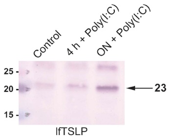
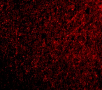
Induced Expression Validation in Human Oral Keratinocytes (Bjerkan et al., 2015). TSLP expression detected by anti-TSLP antibodies (orb1240270) was upregulated in cultured human oral keratinocytes after 24 h in response to poly (I: C).

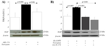
Regulated Expression Validation of TSLP in COPD Patients. (Anzalone et al., 2018). (A) shows NHBE cells, in the absence (Lane 2) or presence (Lane 3) of Tiotropium, were stimulated with rhIL-17A. (B) shows NHBE cells, with ISs from COPD patients untreated (Lane 2 and Lane 3) or treated (Lane 4) with anti-IL-17A antibody. (A) and (B) show TSLP expression decreases with the treatment of anti-cholinergic drugs.
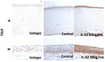
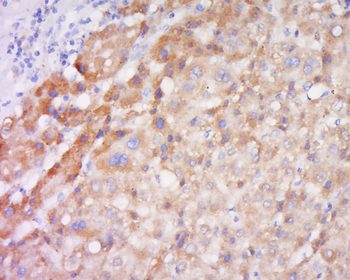
Induced Expression Validation of TSLP in Human Corneal Epithelium (Lin et al., 2018). Immunohistochemical images showing theTSLP protein detected by anti-TSLP antibodies in donor corneal tissues without (Control) or after exposure to IL-32 ex vivo; an isotype IgG antibody was used as a negative control. The production of TSLP was increased after IL-32 treatment.
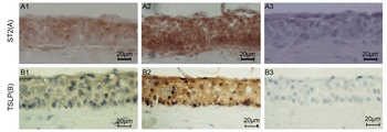
Induced Expression Validation of TSLP in Human Corneal Tissues (Lin et al., 2013). Immunohistochemical images showing TSLP protein detected by anti-TSLP antibodies in ex vivo donor human corneal tissues without (B1) or with exposure to IL-33 (B2). An isotype IgG antibody (B3) was used as a negative control. Magnificaiton 400X. The staining confirms the increased level of TSLP after IL-33 treatment.
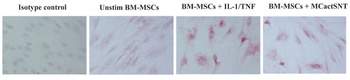
Induced Expression Validation of TSLP in Mesenchymal Stromal Cells (MSCs). (Allakhverdi et al., 2013). Immunocytochemical images showing TSLP protein detected by anti-TSLP antibodies in BM-MSCs unstimulated (control) or stimulated with IL-1/TNF or supernatants of activated MCs (MCactSNT). Isotype is used as a negative control. The production of TSLP increased with the stimulated conditions.
Documents Download
Datasheet
Product Information
Request a Document
Protocol Information
WB
Western Blot (IB, immunoblot)
IHC-P
Immunohistochemistry Paraffin
ELISA
Enzyme-linked Immunosorbent Assay (EIA)
TSLP Antibody (orb1240270)

- 0.0
Based on 0 reviews
Participating in our Biorbyt product reviews program enables you to support fellow scientists by sharing your firsthand experience with our products.
Login to Submit a ReviewAvailable Sizes
Select a size below
Free Secondary Antibody (20 ul)0/0
Please add an antibody product to your cart first.