You have no items in your shopping cart.
Streptavidin Peroxidase Conjugated
SKU: orb348767
Description
Research Area
Non-Animal
Images & Validation
−Item 1 of 10
| Application Notes |
|---|
Key Properties
−| Purity | Streptavidin-HRP was prepared from chromatographically purified streptavidin. Streptavidin Peroxidase conjugate was assayed by immunoelectrophoresis resulted in a single precipitin arc against anti-Peroxidase and anti-Streptavidin. |
|---|---|
| Conjugation | HRP |
Storage & Handling
−| Storage | Store vial at 4° C prior to restoration. For extended storage aliquot contents and freeze at -20° C or below. Avoid cycles of freezing and thawing. Centrifuge product if not completely clear after standing at room temperature. Streptavidin Peroxidase conjugated is stable for several weeks at 4° C as an undiluted liquid. Dilute only prior to immediate use. |
|---|---|
| Form/Appearance | Lyophilized |
| Buffer/Preservatives | Preservative: 0.01% (w/v) Gentamicin Sulfate. Do NOT add Sodium Azide!. Stabilizer: 10 mg/mL Bovine Serum Albumin (rAlbumin) - Immunoglobulin and Protease free; Buffer: 0.02 M Potassium Phosphate, 0.15 M Sodium Chloride, pH 7.2 |
| Concentration | 1.0 mg/mL |
| Expiration Date | 12 months from date of receipt. |
| Hazard Information | Non-Toxic |
| Disclaimer | For research use only |
Alternative Names
−HRP-SA, Horseradish Peroxidase conjugated S avidin, Streptavidin HRP, Streptavidin conjugated to horseradish peroxidase (HRP), HRP-linked Streptavidin
Similar Products
−
Mouse Human IgA1+IgA2 , conjugated with Horseradish peroxidase Antibody [orb22225]
Human
Mouse
Monoclonal
HRP
0.2 mg- Item 1 of 1
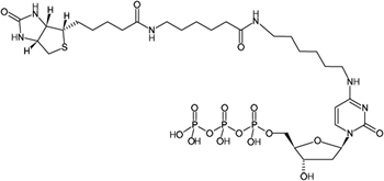
Biotin-14-dCTP [orb533284]
≥ 95% (HPLC)
Theoretical MW: 905.78 g/mol (free acid); Detected MW: 905.26 g/mol (free acid)
C31H54N7O16P3S
5 x 200 μl - Item 1 of 1
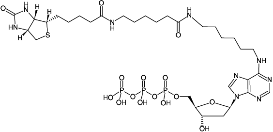
Biotin-14-dATP [orb533181]
≥ 95% (HPLC)
Theoretical MW: 929.81 g/mol (free acid); Detected MW: 929.27 g/mol (free acid)
C32H54N9O15P3S
5 x 200 μl - Item 1 of 1
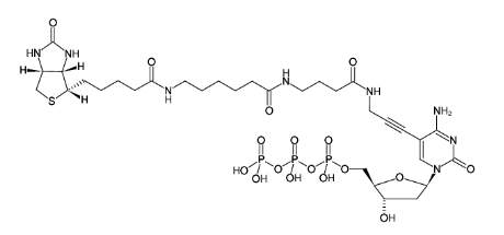
Biotin-16-dCTP [orb64049]
≥ 95% (HPLC)
Theoretical MW: 944.78 g/mol (free acid); Detected MW: 944.23 g/mol (free acid)
C32H51N8O17P3S (free acid)
5 x 200 μl - Item 1 of 1
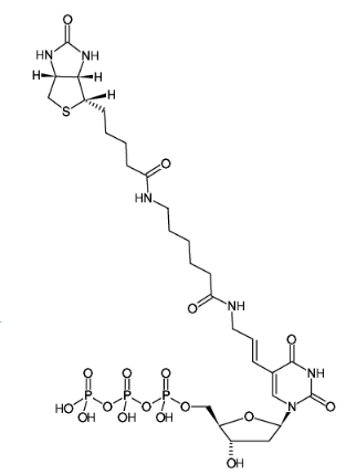
Biotin-11-dUTP [orb64042]
≥ 95% (HPLC)
Theoretical MW: 862.67 g/mol (free acid); Detected MW: 862.18 g/mol (free acid)
C28H45N6O17P3S
5 x 200 μl
Quality Guarantee
Explore bioreagents carefree to elevate your research. All our products are rigorously tested for performance. If a product does not perform as described on its datasheet, our scientific support team will provide expert troubleshooting, a prompt replacement, or a refund. For full details, please see our Terms & Conditions and Buying Guide. Contact us at [email protected].
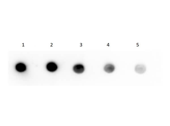
Dot Blot of Human IgG F(c) Fragment Biotin Conjugated using Streptavidin HRP. Human IgG F(c) Biotin Conjugate (1) 100 ng, (2) 33.33 ng, (3) 11.11 ng, (4) 3.70 ng, (5) 1.23 ng. Primary Antibody: none. Secondary Antibody: Streptavidin HRP (p/n orb348767) at 1:40000 for 30 mins at RT. Block: BlockOut buffer (p/n orb348644) at RT for 30 mins. Exposure: 1 sec.
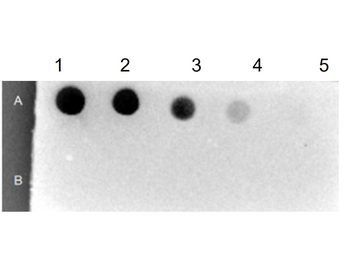
Dot Blot Results of Streptavidin Peroxidase Conjugate. Row A: BSA-Biotin Conjugated. Row B: BSA. Sample dilutions: 1- 100 ng, 2- 33.33 ng, 3- 11.11 ng, 4-3.7 ng, 5- 1.23 ng. Streptavidin Peroxidase Conjugated at 1.0 µg/ml for 30 mins at RT.
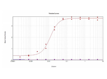
ELISA Results of Human IgG Whole Molecule Biotin Conjugated using Streptavidin-HRP. Each well was coated in duplicate with 1.0 µg of Human IgG Whole Molecule Biotin Conjugate. The working dilution is 82800. The starting dilution of antibody was 5 µg/ml and the X-axis represents the Log10 of a 3-fold dilution. This titration is a 4-parameter curve fit where the IC50 is defined as the titer of the antibody. Assay performed using Streptavidin-HRP (p/n orb348767) and TMB substrate (p/n orb348651).

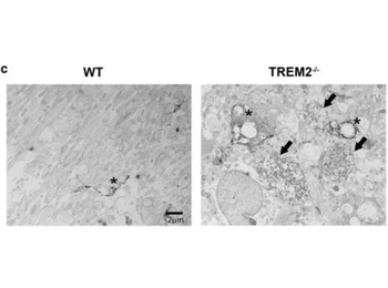
Immuno-electron microscopy using biotinylated anti-rabbit and streptavidin-HRP. Defect in myelin degradation in TREM2−/− microglia. (c) Immuno-EM images of TREM2−/− and WT microglia stained with Iba1 in the CC at 4 and 12 weeks on CPZ treatment. Images on the left in WT and TREM2−/−panels at week 4 and 12 (3000× magnification) depict Iba + microglial cells (asterisks). A higher magnification (15000×) for the boxed area is shown on the right of each image. Black arrows indicate phagosomes containing myelin debris. White arrows indicate pi granules.
Immuno-electron microscopy using biotinylated anti-rabbit and streptavidin-HRP. TREM2−/− mice show more severe axonal pathology after CPZ. (c) EM images of WT and TREM2−/− at 12 weeks of CPZ treatment. Black arrows indicate dystrophic autophagocytic axons and asterisks indicate Iba1 + immunolabeled microglia.
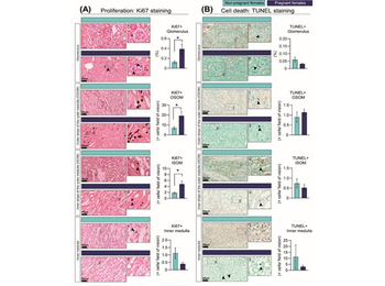
Percentage of cells showing cell-cycle activation as informed by Ki67 immunostaining (A) and cell death as identified by TUNEL assay (B) in the kidney in response to pregnancy in mice. Representative image of stained kidney from non-pregnant and pregnant female mice. Data are presented as mean ± SEM (n = 5/group). Asterisks represent significant differences between non-pregnant and pregnant mice as determined by Student's t-Test (* p < 0.05). Slides incubated with goat anti-rabbit secondary antibody (1:1000) and streptavidin-horseradish peroxidase (1:500, p/n orb348767). Images with the labels i and ii depict high magnification of the selected area. Arrow heads indicate positive DAB staining. Scale bar is 50 µm.
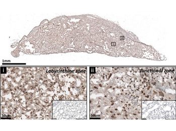
The expression of p110α protein by the mouse placenta on day 19 of pregnancy. Representative stained section shown with negative control shown in the figure inset. For localization of p110α, placental sections were washed with PBS to remove OCT and underwent antigen retrieval with citrate buffer before immunolabelling against p110α. Sections were treated with 0.5% Triton X-100 before immunolabelling. Bound antibody was detected using biotinylated goat anti-rabbit IgG followed by streptavidin-conjugated horseradish peroxidase (p/n orb348767) and 3, 3-diaminobenzidine (DAB). Sections were lightly counterstained with hematoxylin and mounted in DPX.
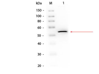
Western Blot of Goat anti-Glycerol Kinase Antibody Biotin Conjugated using Streptavidin HRP. Lane 1: Glycerol Kinase. Load: 50 ng per lane. Primary antibody: Glycerol Kinase Antibody Biotin Conjugated at 1:1000 overnight at 4°C. Secondary antibody: HRP Streptavidin (p/n orb348767) secondary antibody at 1:40000 for 30 min at RT. Block: orb348637 for 30 min at RT. Predicted/Observed size: 55 kDa, 55 kDa for Glycerol Kinase.
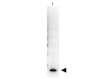
Western Blot of Peroxidase Conjugated Streptavidin. Lane 1: Human IL-7. Load: 50 ng per lane. Primary antibody: Human IL-7 Biotin Conjugated antibody at 1:1000 for overnight at 4°C. Secondary antibody: Peroxidase Conjugated Streptavidin at 1:40000 for 30 min at RT. Block: 5% BLOTTO 30 min at RT. Predicted/Observed size: 17 kDa, 17 kDa for Human IL-7. Other band(s): none.
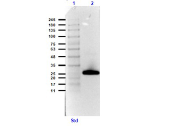
Western Blot Results using Streptavidin Peroxidase Conjugate and Goat Anti-GST Biotin Conjugate Antibodies. Lane 1: Opal prestained molecular weight ladder. Lane 2: GST (p/n orb345956) [0.05 µg]. Primary Antibody: Goat Anti-GST Biotin Conjugate (p/n orb345320) at 1.0 µg/ml overnight at 4°C. Secondary Antibody; Streptavidin Peroxidase Conjugate at 1:40000 for 30 mins at RT. Block: Blocking Buffer for Fluorescent Western Blotting (p/n orb348637) for 30 mins at RT. Exp: 5 sec.
Quick Database Links
UniProt
RefSeq:CAA00084.1
UniProt Details
− No UniProt data available
NCBI Reference Sequences
−Associated Accession Numbers
Curated reference sequences for the gene transcript and protein product| RefSeq | CAA00084.1 |
|---|
Documents Download
Datasheet
Product Information
Request a Document
Protocol Information
WB
Western Blot (IB, immunoblot)
IHC
Immunohistochemistry
ELISA
Enzyme-linked Immunosorbent Assay (EIA)
DOT
Dot Blot
Streptavidin Peroxidase Conjugated (orb348767)

- 0.0
Based on 0 reviews
Participating in our Biorbyt product reviews program enables you to support fellow scientists by sharing your firsthand experience with our products.
Login to Submit a ReviewAvailable Sizes
Select a size below