You have no items in your shopping cart.
Description
Research Area
Stem Cell & Developmental Biology
Images & Validation
−Item 1 of 4
| Tested Applications | ELISA, FC, IF, IHC, WB |
|---|---|
| Reactivity | Human |
| Predicted Reactivity | Bovine, Canine |
| Application Notes |
Key Properties
−| Antibody Type | Primary Antibody |
|---|---|
| Host | Goat |
| Clonality | Polyclonal |
| Immunogen | The immunogen for this antibody is: C-GRQGNFFASPMLK |
| Target | RACGAP1 |
| Purification | Purified from goat serum by ammonium sulphate precipitation followed by antigen affinity chromatography using the immunizing peptide. |
| Conjugation | Unconjugated |
Storage & Handling
−| Storage | Maintain refrigerated at 2-8°C for up to 2 weeks. For long term storage store at -20°C in small aliquots to prevent freeze-thaw cycles. |
|---|---|
| Form/Appearance | Liquid |
| Buffer/Preservatives | Supplied at 0.5 mg/ml in Tris saline, 0.02% sodium azide, pH 7.3 with 0.5% bovine serum albumin. Aliquot and store at -20°C. Minimize freezing and thawing. |
| Concentration | 500 ug/mL |
| Expiration Date | 12 months from date of receipt. |
| Disclaimer | For research use only |
Alternative Names
−RACGAP1, MgcRacGAP, Rac GTPase activating protein 1, ID-GAP, MGCRACGAP, GTPase activating protein, HsCYK-4, HsCYK-4
Similar Products
−- Item 1 of 6
RACGAP1/MgcRacGAP Antibody [orb18360]
ELISA, FC, IF, IHC, WB
Bovine, Canine
Human
Polyclonal
Unconjugated
100 μg - Item 1 of 4
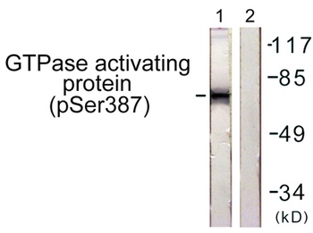
Rac GAP1 (phospho Ser387) rabbit pAb Antibody [orb768551]
ELISA, IF, IHC, WB
Human, Monkey, Mouse, Rat
Polyclonal
Unconjugated
50 μl, 100 μl - Item 1 of 4
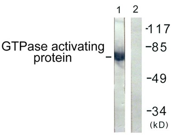
Rac GAP1 rabbit pAb Antibody [orb766174]
ELISA, IF, IHC, WB
Human, Monkey, Mouse, Rat
Polyclonal
Unconjugated
50 μl, 100 μl - Item 1 of 1
Human Rac-GTPase Activating Protein 1 (RACGAP1) ELISA Kit [orb777529]
Human
0.16-10 ng/mL
0.056 ng/mL
48 T, 96 T - Item 1 of 4
RACGAP1 Antibody (N-term) [orb1938178]
FC, IF, IHC-P, WB
Human
Rabbit
Polyclonal
Unconjugated
50 μl, 100 μl


Quality Guarantee
Explore bioreagents carefree to elevate your research. All our products are rigorously tested for performance. If a product does not perform as described on its datasheet, our scientific support team will provide expert troubleshooting, a prompt replacement, or a refund. For full details, please see our Terms & Conditions and Buying Guide. Contact us at [email protected].
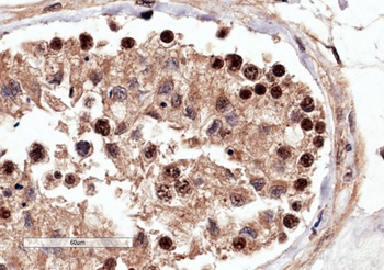
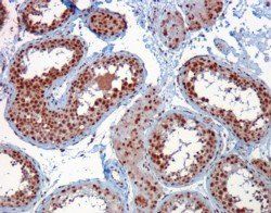
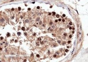
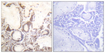
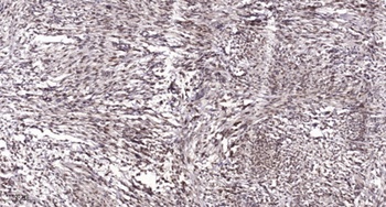
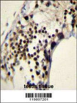
orb1247045 (4 ug/ml) staining of paraffin embedded Human Testis. Microwaved antigen retrieval with Tris/EDTA buffer pH9, HRP-staining.
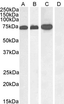
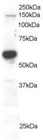
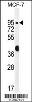
orb1247045 (1 ug/ml) staining of A431 nuclear (A), Jurkat (B), Jurkat nuclear (C) and negative control Human Pancreas (D) lysate. (35 ug protein in RIPA buffer) Detected by chemiluminescence.
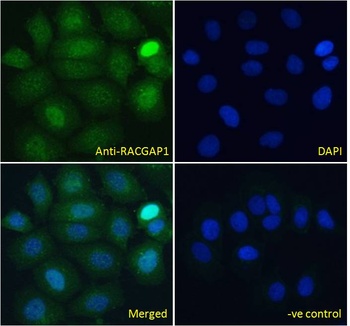
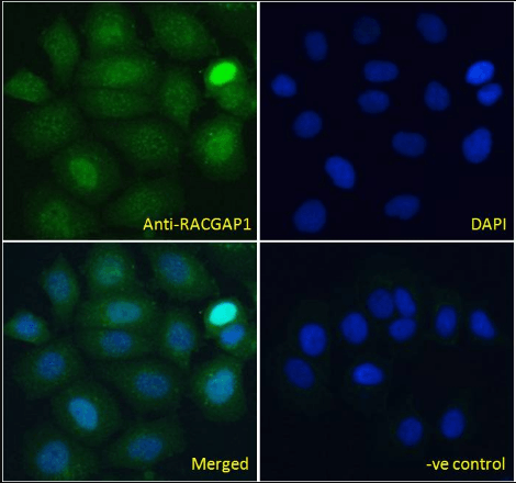
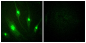
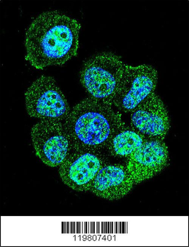
orb1247045 Immunofluorescence analysis of paraformaldehyde fixed MCF7 cells, permeabilized with 0.15% Triton. Primary incubation 1hr (10 ug/ml) followed by Alexa Fluor 488 secondary antibody (4 ug/ml), showing nuclear staining. The nuclear stain is DAPI (blue).
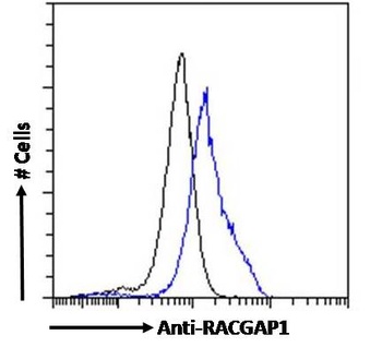
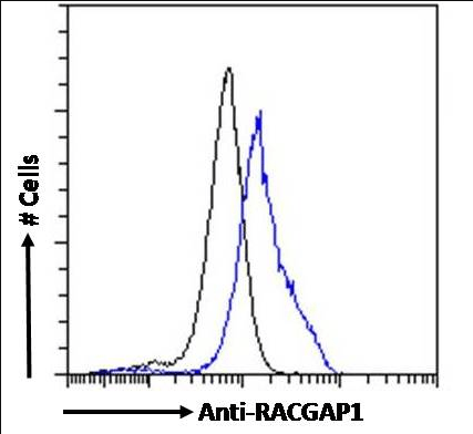
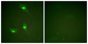
orb1247045 Flow cytometric analysis of paraformaldehyde fixed MCF7 cells (blue line), permeabilized with 0.5% Triton. Primary incubation 1hr (10 ug/ml) followed by Alexa Fluor 488 secondary antibody (4 ug/ml). IgG control: Unimmunized goat IgG (black line).
Quick Database Links
Gene Symbol
RACGAP1
UniProt
RefSeq (Protein):NP_001306936.1, NP_001306935.1, NP_001306934.1, NP_037409.2
UniProt Details
− No UniProt data available
NCBI Reference Sequences
−Associated Accession Numbers
Curated reference sequences for the gene transcript and protein productDocuments Download
Datasheet
Product Information
Request a Document
Protocol Information
WB
Western Blot (IB, immunoblot)
IHC
Immunohistochemistry
FC
Flow Cytometry
IF
Immunofluorescence
ELISA
Enzyme-linked Immunosorbent Assay (EIA)
RACGAP1 Antibody (orb1247045)

- 0.0
Based on 0 reviews
Participating in our Biorbyt product reviews program enables you to support fellow scientists by sharing your firsthand experience with our products.
Login to Submit a ReviewAvailable Sizes
Select a size below
Free Secondary Antibody (20 ul)0/0
Please add an antibody product to your cart first.