You have no items in your shopping cart.
Description
Research Area
Immunology & Inflammation
Images & Validation
−Item 1 of 6
| Tested Applications | ELISA, FC, IF, IHC, WB |
|---|---|
| Reactivity | Human |
| Application Notes |
Key Properties
−| Antibody Type | Primary Antibody |
|---|---|
| Host | Goat |
| Clonality | Polyclonal |
| Immunogen | The immunogen for this antibody is: CKKQSDTHLEET |
| Target | CD274 |
| Purification | Purified from goat serum by ammonium sulphate precipitation followed by antigen affinity chromatography using the immunizing peptide. |
| Conjugation | Unconjugated |
Storage & Handling
−| Storage | Maintain refrigerated at 2-8°C for up to 2 weeks. For long term storage store at -20°C in small aliquots to prevent freeze-thaw cycles. |
|---|---|
| Form/Appearance | Liquid |
| Buffer/Preservatives | Supplied at 0.5 mg/ml in Tris saline, 0.02% sodium azide, pH 7.3 with 0.5% bovine serum albumin. Aliquot and store at -20°C. Minimize freezing and thawing. |
| Concentration | 500 ug/mL |
| Expiration Date | 12 months from date of receipt. |
| Disclaimer | For research use only |
Alternative Names
−CD274, CD274 antigen, PD-L1, PDCD1LG1, B7-H, B7H1, PDL1, PDCD1L1, programmed cell death 1 ligand 1, PDL1, HGNC:17635, CD274 molecule, MGC142294, MGC142296
Similar Products
−- Item 1 of 14
CD274 Rabbit Polyclonal Antibody [orb10162]
ELISA, IHC-P, WB
Human, Mouse, Rat
Rabbit
Polyclonal
Unconjugated
200 μg, 100 μg - Item 1 of 14
CD274 Antibody [orb1239835]
ELISA, FC, IF, IHC-P, WB
Human, Mouse, Rat
Rabbit
Polyclonal
Unconjugated
0.02 mg, 0.1 mg - Item 1 of 10
- Item 1 of 9
- Item 1 of 10
PD-L1 Antibody / B7-H1 / CD274 [orb2641418]
ELISA, FACS, IF, IHC-P, WB
Human, Mouse
Mouse
Monoclonal
Unconjugated
100 μg


Quality Guarantee
Explore bioreagents carefree to elevate your research. All our products are rigorously tested for performance. If a product does not perform as described on its datasheet, our scientific support team will provide expert troubleshooting, a prompt replacement, or a refund. For full details, please see our Terms & Conditions and Buying Guide. Contact us at [email protected].

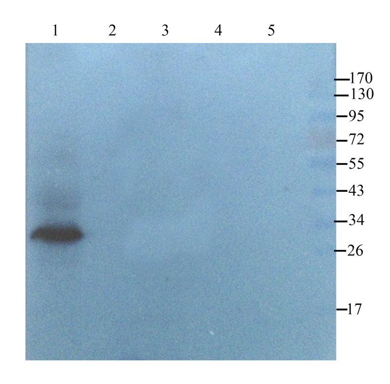
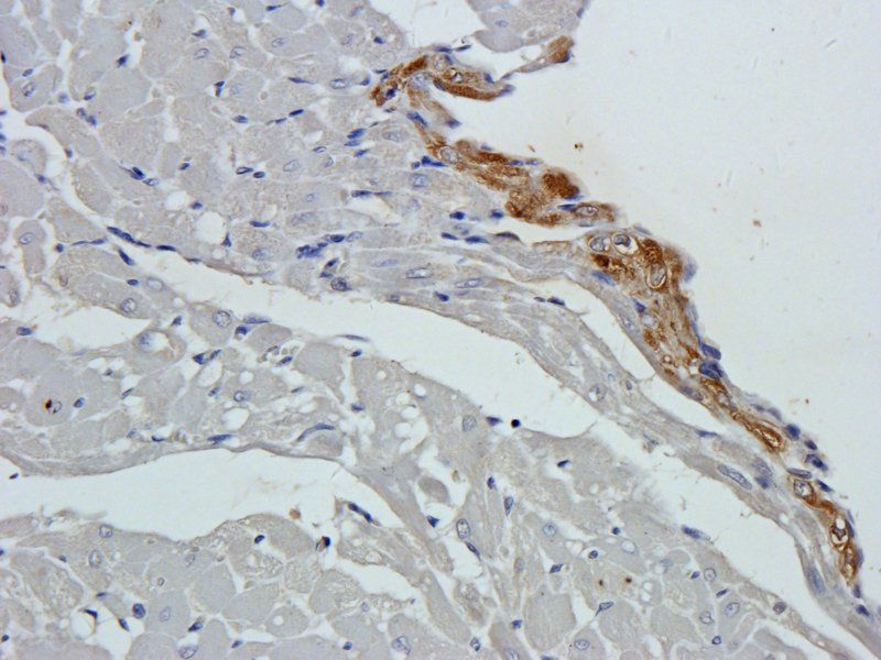
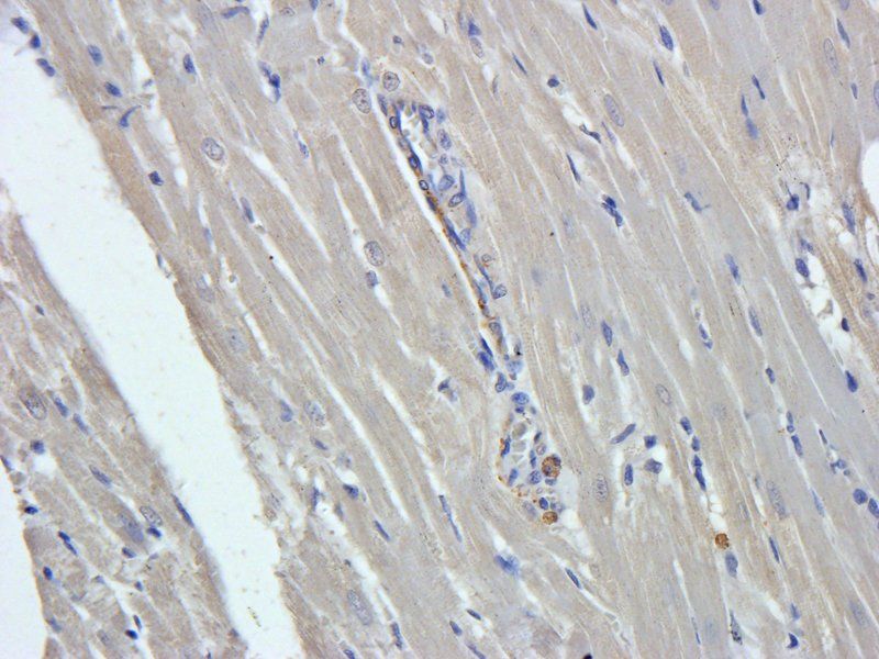
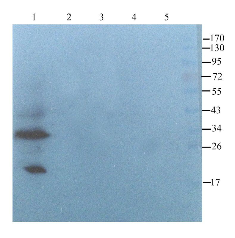
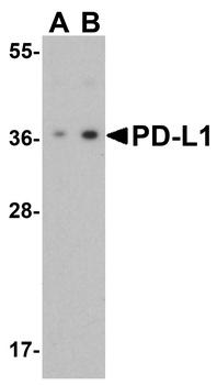
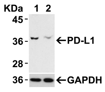
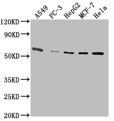
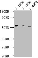
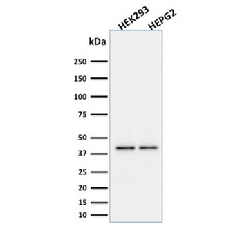
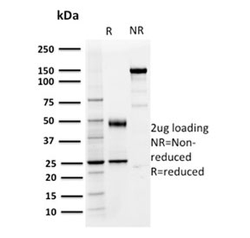
orb1247125 (0.3 ug/ml) staining of Human Heart (A) lysate + Blocking peptide (B) (35 ug protein in RIPA buffer). Detected by chemiluminescence.
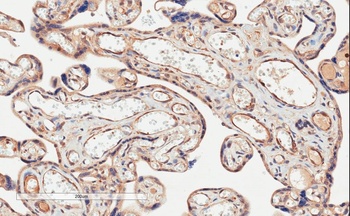
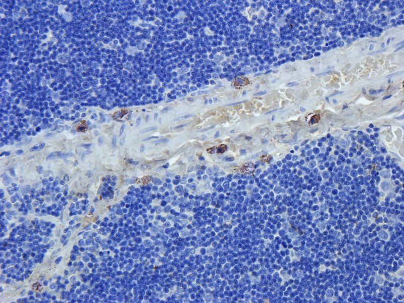
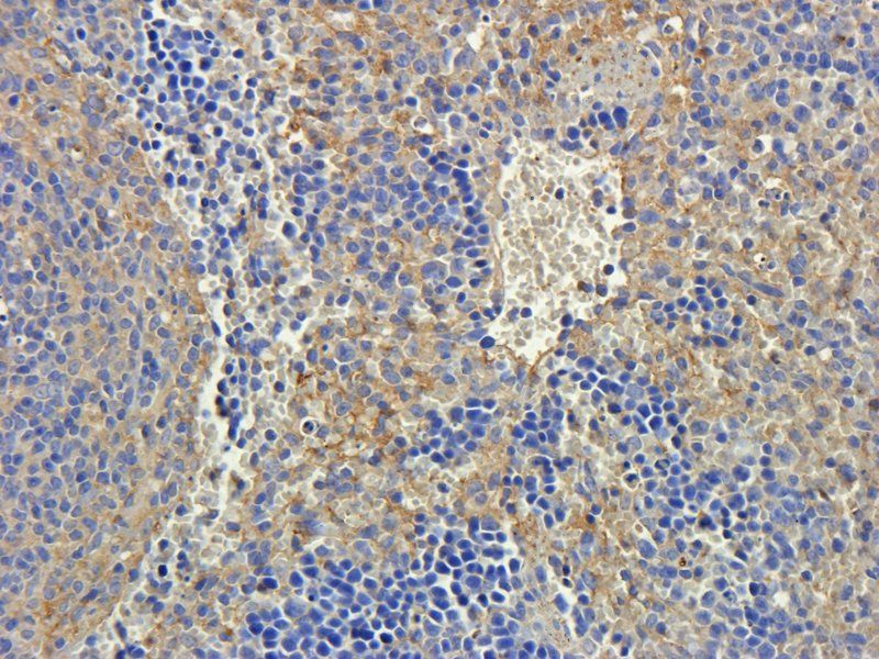
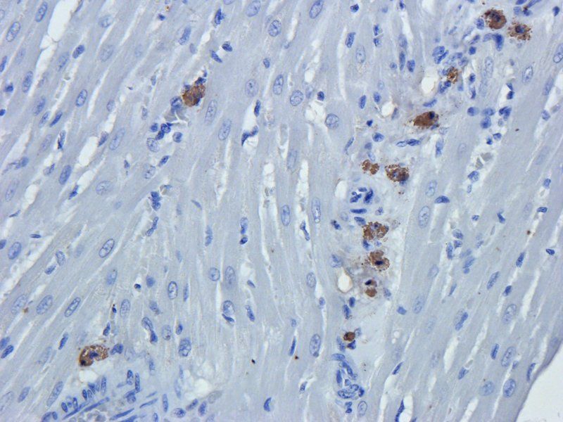
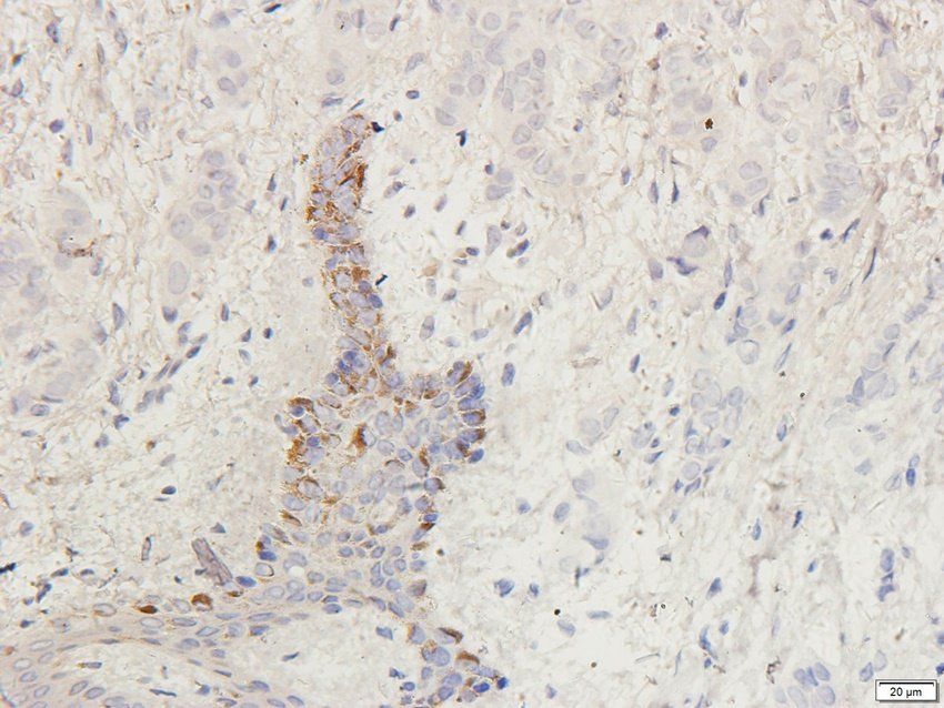
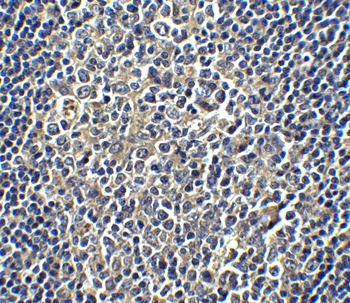
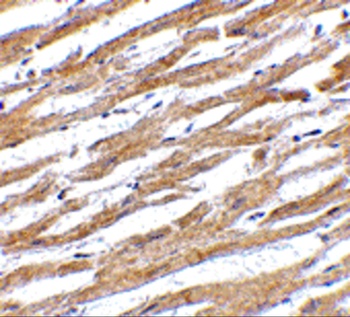
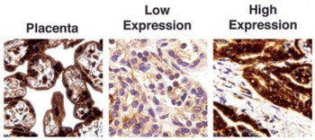
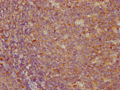
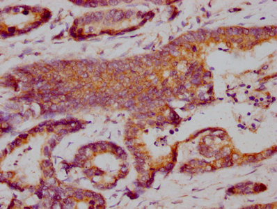
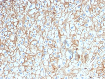
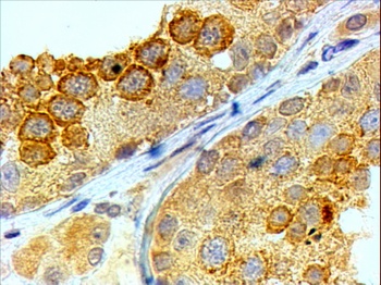
orb1247125 (2 ug/ml) staining of paraffin embedded Human Placenta. Microwaved antigen retrieval with citrate buffer pH 6, HRP-staining.
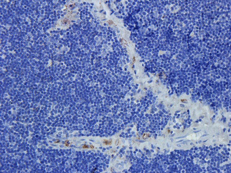
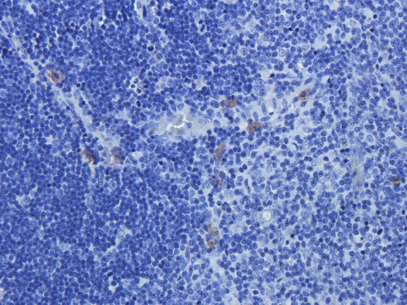
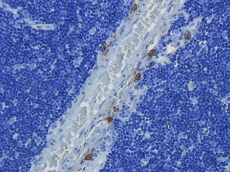
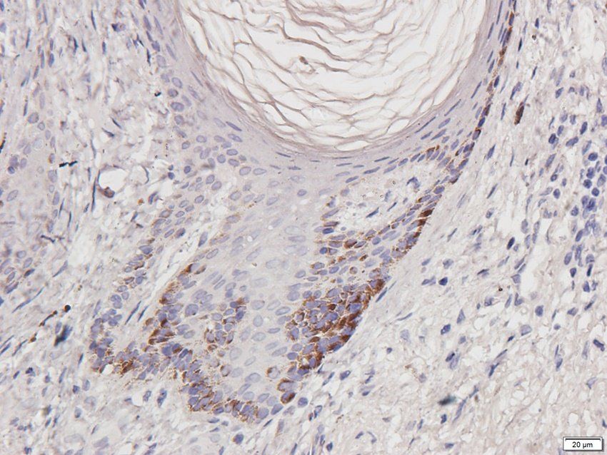
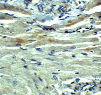
orb1247125 Negative Control showing staining of paraffin embedded Human Placenta, with no primary antibody.
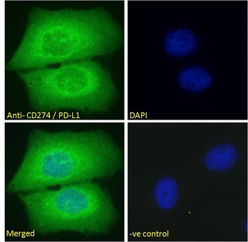
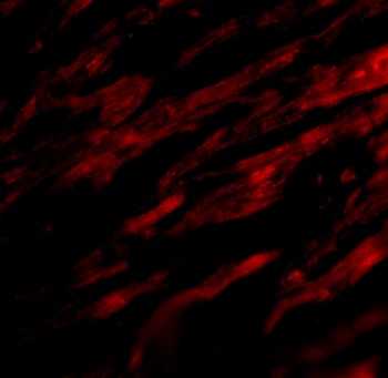
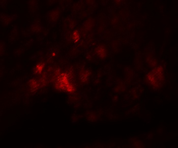
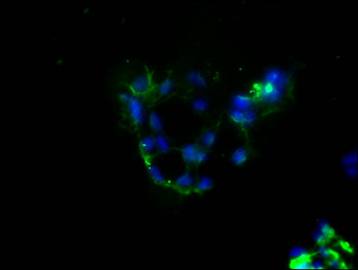
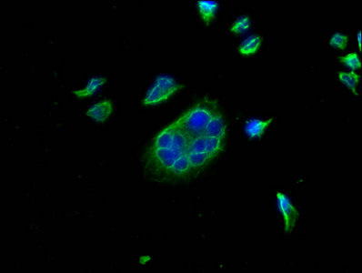
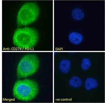
orb1247125 Immunofluorescence analysis of paraformaldehyde fixed A431 cells, permeabilized with 0.15% Triton. Primary incubation 1hr (10 ug/ml) followed by Alexa Fluor 488 secondary antibody (2 ug/ml), showing cytoplasmic staining. The nuclear stain is DAPI.
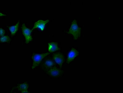
orb1247125 Immunofluorescence analysis of paraformaldehyde fixed U2OS cells, permeabilized with 0.15% Triton. Primary incubation 1hr (10 ug/ml) followed by Alexa Fluor 488 secondary antibody (2 ug/ml), showing cytoplasmic staining. The nuclear stain is DAPI.
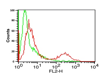
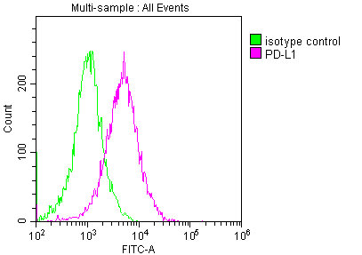
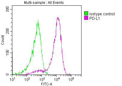
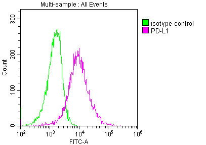
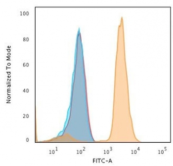
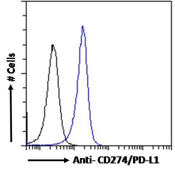
orb1247125 Flow cytometric analysis of paraformaldehyde fixed Jurkat cells (blue line), permeabilized with 0.5% Triton. Primary incubation 1hr (10 ug/ml) followed by Alexa Fluor 488 secondary antibody (1 ug/ml). IgG control: Unimmunized goat IgG (black line).
Quick Database Links
UniProt Details
− No UniProt data available
NCBI Reference Sequences
−Associated Accession Numbers
Curated reference sequences for the gene transcript and protein product| Protein | NP_001254635.1, NP_054862.1 |
|---|
Documents Download
Datasheet
Product Information
Request a Document
Protocol Information
WB
Western Blot (IB, immunoblot)
IHC
Immunohistochemistry
FC
Flow Cytometry
IF
Immunofluorescence
ELISA
Enzyme-linked Immunosorbent Assay (EIA)
CD274 Antibody (orb1247125)

- 0.0
Based on 0 reviews
Participating in our Biorbyt product reviews program enables you to support fellow scientists by sharing your firsthand experience with our products.
Login to Submit a ReviewAvailable Sizes
Select a size below
Free Secondary Antibody (20 ul)0/0
Please add an antibody product to your cart first.