You have no items in your shopping cart.
Featured
KO/KD
Validated
Validated
Description
Research Area
Immunology & Inflammation
Images & Validation
−Item 1 of 14
| Tested Applications | ELISA, FC, IF, IHC-P, KO/KD Validated, WB |
|---|---|
| Reactivity | Human, Mouse, Rat |
Key Properties
−| Antibody Type | Primary Antibody |
|---|---|
| Host | Rabbit |
| Clonality | Polyclonal |
| Isotype | IgG |
| Immunogen | Anti-PD-L1 antibody (orb1239835) was raised against a peptide corresponding to 17 amino acids near the center of human PD-L1 isoform 1. The immunogen is located within amino acids 60 - 110 of PD-L1. |
| Target | CD274 |
| Molecular Weight | Predicted: 33 kDa Observed: 37 kDa |
| Purification | PD-L1 Antibody is affinity chromatography purified via peptide column. |
| Conjugation | Unconjugated |
Storage & Handling
−| Storage | Maintain refrigerated at 2-8°C for up to 2 weeks. For long term storage store at -20°C in small aliquots to prevent freeze-thaw cycles. |
|---|---|
| Form/Appearance | Liquid |
| Buffer/Preservatives | PD-L1 Antibody is supplied in PBS containing 0.02% sodium azide. |
| Concentration | 1 mg/mL |
| Expiration Date | 12 months from date of receipt. |
| Disclaimer | For research use only |
Alternative Names
−PD-L1 Antibody: B7-H, B7H1, PDL1, PD-L1, PDCD1L1, PDCD1LG1, Programmed cell death 1 ligand 1, B7 homolog 1
Similar Products
−- Item 1 of 14
CD274 Rabbit Polyclonal Antibody [orb10162]
ELISA, IHC-P, WB
Human, Mouse, Rat
Rabbit
Polyclonal
Unconjugated
200 μg, 100 μg - Item 1 of 10
- Item 1 of 9
- Item 1 of 9
- Item 1 of 10
PD-L1 Antibody / B7-H1 / CD274 [orb606675]
ELISA, FACS, IF, IHC-P, WB
Human, Mouse
Mouse
Monoclonal
Unconjugated
100 μg, 20 μg


Quality Guarantee
Explore bioreagents carefree to elevate your research. All our products are rigorously tested for performance. If a product does not perform as described on its datasheet, our scientific support team will provide expert troubleshooting, a prompt replacement, or a refund. For full details, please see our Terms & Conditions and Buying Guide. Contact us at [email protected].
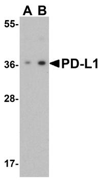
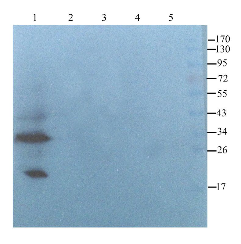
Western Blot Validation of PD-L1 in HeLa Cells. Loading: 15 µg of lysates per lane. Antibodies: orb1239835 (A: 1 µg/mL, B: 2 µg/mL), 1 h incubation at RT in 5% NFDM/TBST. Secondary: Goat anti-rabbit IgG HRP conjugate at 1:10000 dilution.
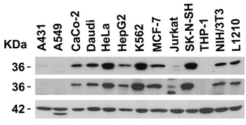
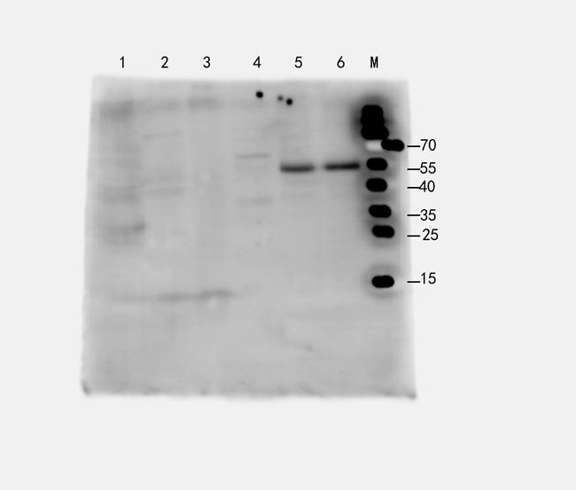
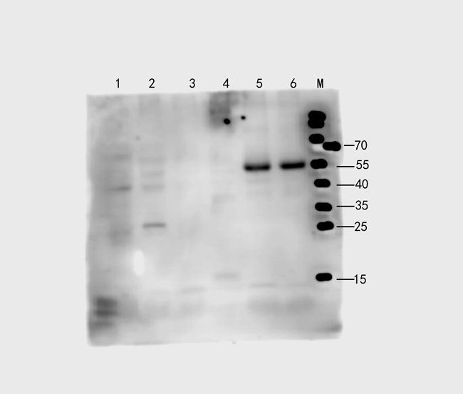
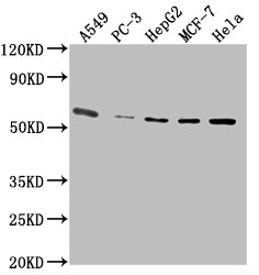
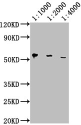
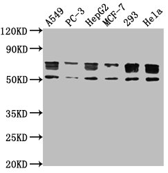
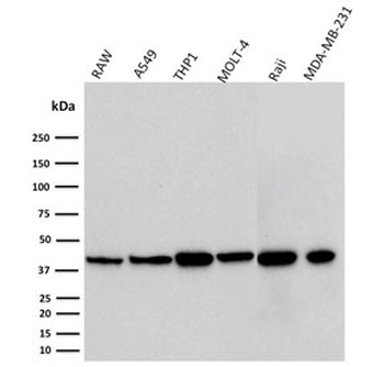
Independent Antibody Validation (IAV) via Protein Expression Profile in Human and Mouse cell lines. Loading: 15 µg of lysates per lane. Antibodies: orb1239835 (2 µg/mL), orb1239770 (2 µg/mL), and beta-actin (1 µg/mL), 1 h incubation at RT in 5% NFDM/TBST. Secondary: Goat anti-rabbit and or anti-mouse IgG HRP conjugate at 1:10000 and 1:5000 dilution, respectively.
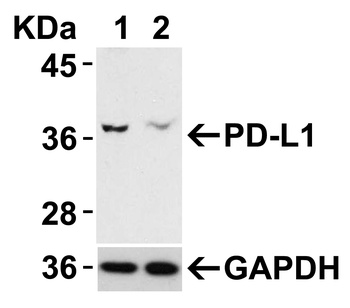
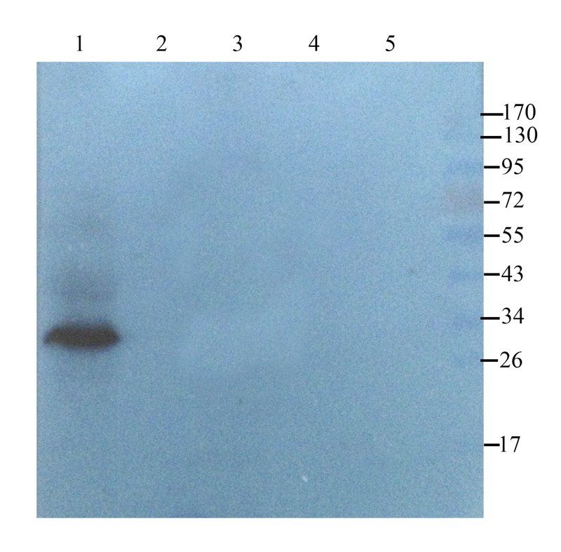
Validation with PD-L1 siRNA Knockdown in HeLa Cells. HeLa cells were transfected with control siRNAs (lane 1) or PD-L1 siRNAs (lane 2) Loading: 10 µg of HeLa whole cell lysates per lane. Antibodies: orb1239835 (2 µg/mL) and GAPDH (orb1239765, 0.02 µg/mL), 1 h incubation at RT in 5% NFDM/TBST. Secondary: Goat anti-mouse IgG HRP conjugate at 1:5000 dilution.
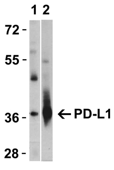
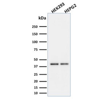
Validation with PD-L1 overexpression in 293 cells. Loading: Lysates/proteins at 15 µg per lane. Lane 1: non-transfected 293 cells, Lane 2: PD-L1 overexpressed 293 cells. Antibodies: orb1239835 (1 µg/mL). 1 h incubation at RT in 5% NFDM/TBST. Secondary: Goat anti-rabbit IgG HRP conjugate at 1:10000 dilution.
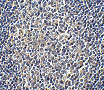
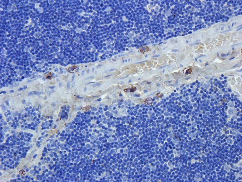
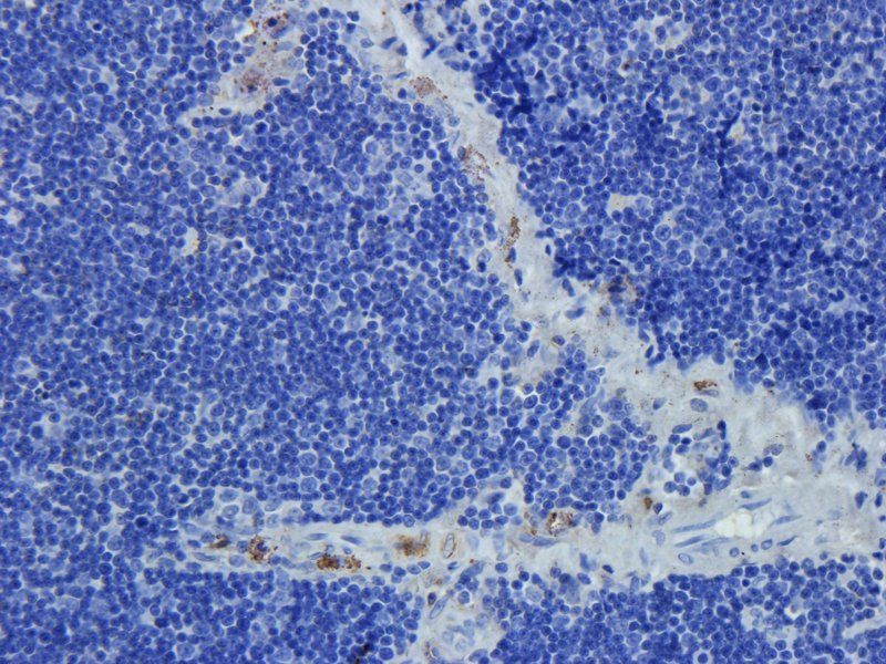
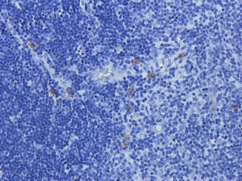
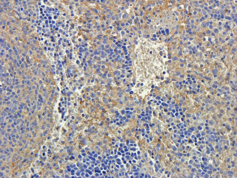
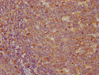
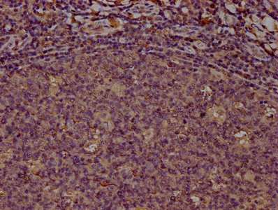
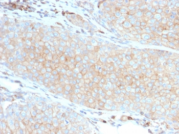
Immunohistochemistry Validation of PD-L1 in Human Tonsil Cells. Immunohistochemical analysis of paraffin-embedded human tonsil tissue using anti-PD-L1 antibody (orb1239835) at 5 µg/ml. Tissue was fixed with formaldehyde and blocked with 10% serum for 1 h at RT; antigen retrieval was by heat mediation with a citrate buffer (pH6). Samples were incubated with primary antibody overnight at 4°C. A goat anti-rabbit IgG H&L (HRP) at 1/250 was used as secondary. Counter stained with Hematoxylin.
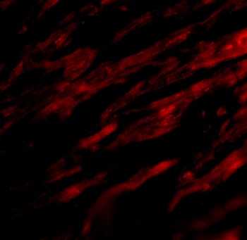
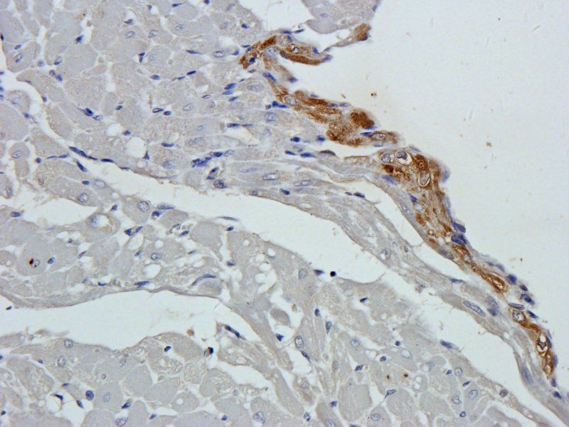
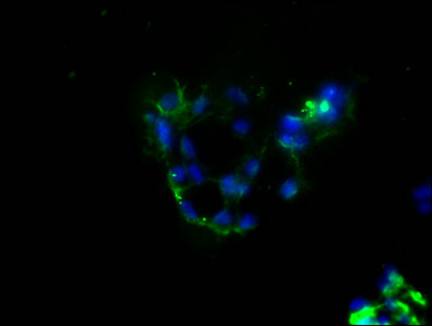
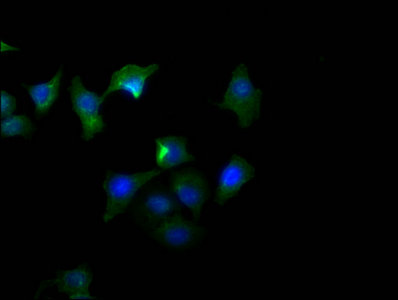
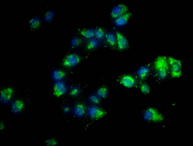
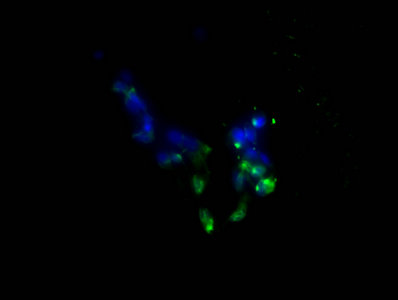
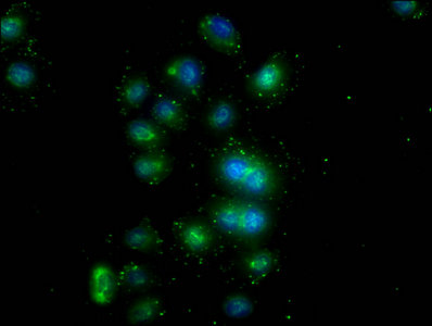
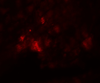
Immunofluorescence Validation of PD-L1 in Human Heart. Immunofluorescent analysis of 4% paraformaldehyde-fixed human heart tissue labeling PD-L1 with orb1239835 at 20 µg/mL, followed by goat anti-rabbit IgG secondary antibody at 1/500 dilution (red). Image showing both membrane and cytoplasmic staining on human heart tissue.
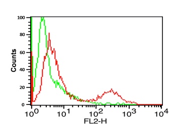
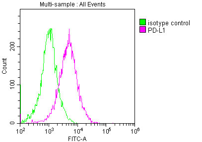
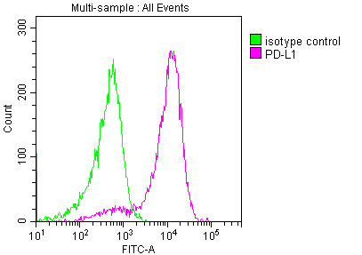
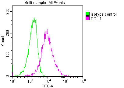
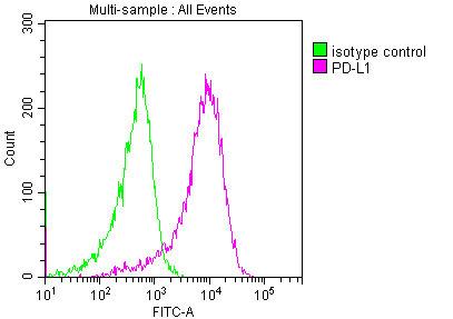
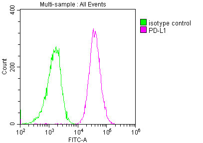
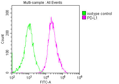
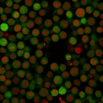
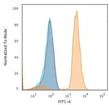
Flow Cytometry Validation of PD-L1. Overlay histogram showing A-20 cells stained with orb1239835 (red line, 1µg/1x10 6 cells). 1 h incubation at 4°C in 2% FBS/PBS. Followed by secondary antibody 488 goat anti-rabbit IgG (H+L) at 1/500 dilution for 1 h 4°C. Isotype control antibody (Green line) was mouse IgG1 (1µg/1x10 6 cells) used under the same conditions. Acquisition of > 10000 events was performed.
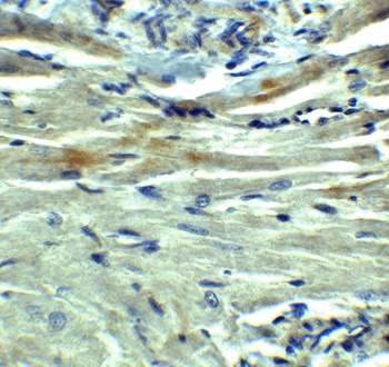
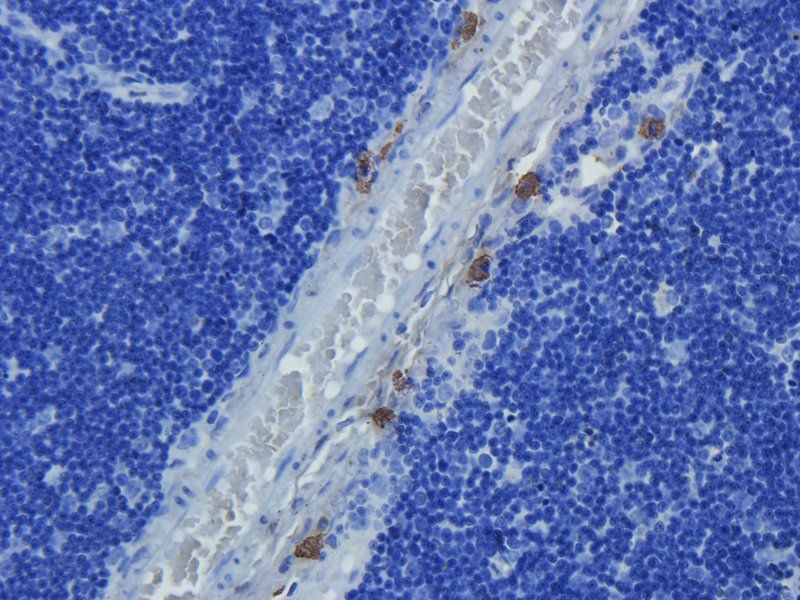
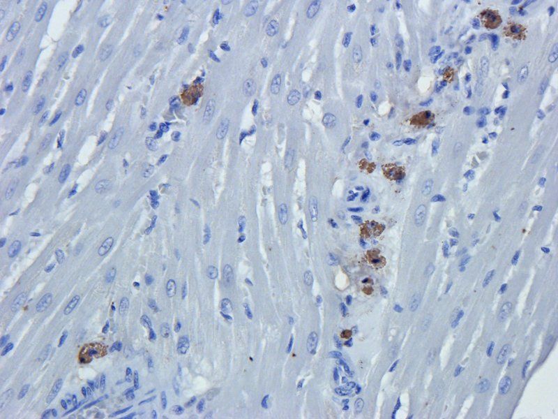
Immunohistochemistry Validation of PD-L1 in Rat Heart. Immunohistochemical analysis of paraffin-embedded rat heart tissue using anti-PD-L1 antibody (orb1239835) at 5 µg/ml. Tissue was fixed with formaldehyde and blocked with 10% serum for 1 h at RT; antigen retrieval was by heat mediation with a citrate buffer (pH6). Samples were incubated with primary antibody overnight at 4°C. A goat anti-rabbit IgG H&L (HRP) at 1/250 was used as secondary. Counter stained with Hematoxylin.
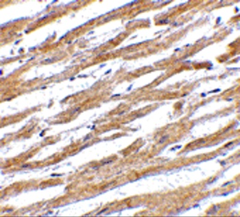
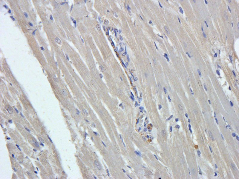
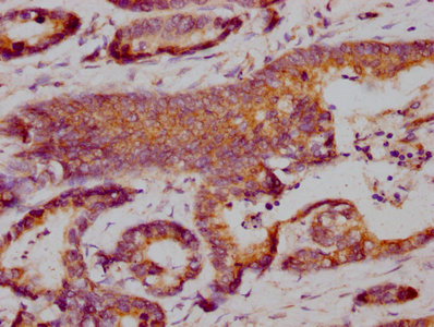
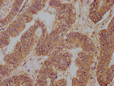
Immunohistochemistry Validation of PD-L1 in Human Heart. Immunohistochemical analysis of paraffin-embedded human heart tissue using anti-PD-L1 antibody (orb1239835). Tissue was fixed with formaldehyde and blocked with 10% serum for 1 h at RT; antigen retrieval was by heat mediation with a citrate buffer (pH6). Samples were incubated with primary antibody overnight at 4°C. A goat anti-rabbit IgG H&L (HRP) at 1/250 was used as secondary. Counter stained with Hematoxylin.
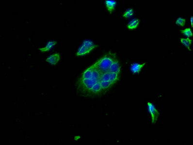
Immunofluorescence Validation of PD-L1 in Rat Heart. Immunofluorescence analysis of 4% paraformaldehyde-fixed rat heart tissue labeling PD-L1 with orb1239835 at 20 µg/ml, followed by goat anti-rabbit IgG secondary antibody at 1/250 dilution (red).
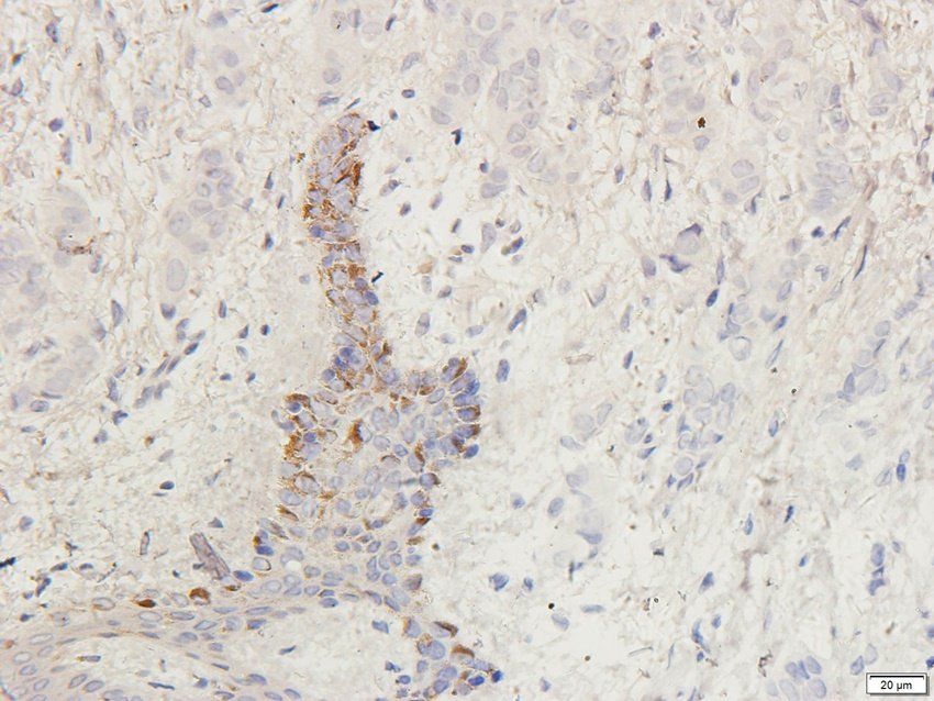
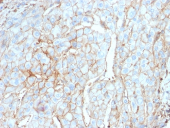
Immunohistochemistry Validation of PD-L1 in Human Tumors (Gadiot et al., 2011). Immunohistochemical analysis of patient tumors labeling PD-L1 with anti-PD-L1 antibodies (orb1239835). Several anti-PD-L1 antibodies were tested for staining.
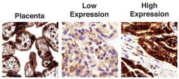
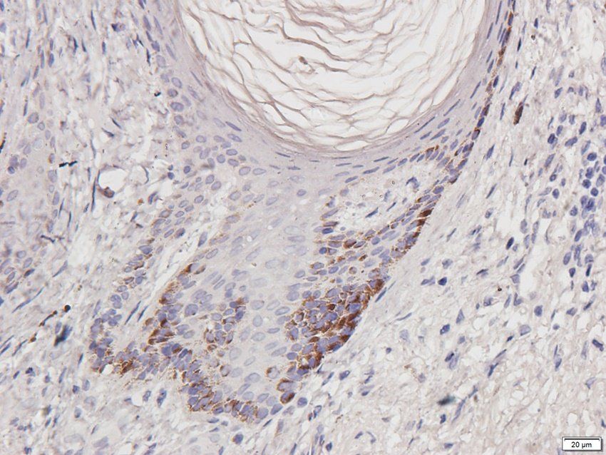
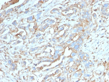
Immunohistochemistry Validation of PD-L1 in Human thyroid cancer (Angell et al., 2014). Immunohistochemical analysis of PD-L1 expression in human thyroid cancer with anti-PD-L1 antibodies (orb1239835). Placenta was used a positive control.
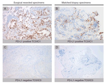
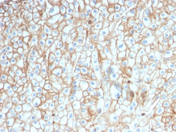
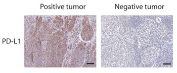
Immunohistochemistry Validation of PD-L1 in Human Lung Cancer (Ilie et al., 2015). Surgical specimens (left panel) and matched biopsy specimens (right panel). PD-L1-positive (A, B) and PD-L1-negative (C, D) tumors.
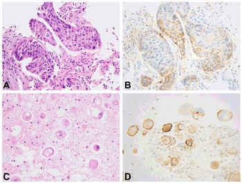
Immunohistochemistry Validation of PD-L1 in Human Lung adenocarcinoma (Heymann et al., 2017). HE staining (left panel) and PD-L1 expression (right panel) in the tumor and pleural fluid for a patient with lung adenocarcinoma. PD-L1 expression detected by anti-PD-L1 antibodies (orb1239835) demonstrated membranous staining in approximately 80% of tumor cells (C) and in approximately 75% of tumor cells (D), respectively.
Documents Download
Datasheet
Product Information
Request a Document
Protocol Information
WB
Western Blot (IB, immunoblot)
IHC-P
Immunohistochemistry Paraffin
FC
Flow Cytometry
IF
Immunofluorescence
ELISA
Enzyme-linked Immunosorbent Assay (EIA)
CD274 Antibody (orb1239835)

- 0.0
Based on 0 reviews
Participating in our Biorbyt product reviews program enables you to support fellow scientists by sharing your firsthand experience with our products.
Login to Submit a ReviewAvailable Sizes
Select a size below
Free Secondary Antibody (20 ul)0/0
Please add an antibody product to your cart first.