You have no items in your shopping cart.
Featured
Description
Research Area
Signal Transduction
Images & Validation
−Item 1 of 11
| Tested Applications | FC, ICC |
|---|---|
| Dilution Range | ICC/IF=1:50-200, Flow-Cyt=1μg/Test |
| Reactivity | Human, Mouse |
| Predicted Reactivity | Canine, Rabbit, Rat, Sheep |
Related Conjugates & Formulations
−Key Properties
−| Antibody Type | Primary Antibody |
|---|---|
| Host | Rabbit |
| Clonality | Polyclonal |
| Isotype | IgG |
| Immunogen | KLH conjugated synthetic peptide derived from human P38MAPK (141-240/360aa) |
| Target | MAPK14 |
| Molecular Weight | 41 kDa |
| Purification | Affinity purified by Protein A |
| Conjugation | Unconjugated |
Storage & Handling
−| Storage | Maintain refrigerated at 2-8°C for up to 2 weeks. For long term storage store at -20°C in small aliquots to prevent freeze-thaw cycles. |
|---|---|
| Form/Appearance | Liquid |
| Buffer/Preservatives | 0.01M TBS (pH7.4) with 1% rAlbumin, 0.02% Proclin300 and 50% Glycerol. |
| Concentration | 1mg/ml |
| Expiration Date | 12 months from date of receipt. |
| Disclaimer | For research use only |
Alternative Names
−CSBP; CSBP1; CSBP2; CSPB1; EXIP; Mxi2; PRKM14; PRKM15; RK; SAPK2A; p38; p38ALPHA; Crk1; p38-alpha; p38MAPK; p38a; Hog; p38Hog; MK14_CANLF; MAPK14; MAP kinase 14; MAPK 14; Mitogen-activated protein kinase p38 alpha (MAP kinase p38 alpha); 2.7.11.24; MK14_HUMAN; Cytokine suppressive anti-inflammatory drug-binding protein (CSAID-binding protein | CSBP); MAP kinase MXI2; MAX-interacting protein 2; Stress-activated protein kinase 2a (SAPK2a); MK14_MOUSE; MK14_RAT; mitogen-activated protein kinase 14; p38 MAP kinase
Similar Products
−- Item 1 of 16
Phospho-ERK1/2 (Thr202 + Tyr204) Rabbit Polyclonal Antibody [orb5178]
FC, ICC
Human
Human, Mouse
Rabbit
Polyclonal
Unconjugated
50 μl, 100 μl, 200 μl - Item 1 of 8
Phospho-p38 MAPK (Thr180 + Tyr182) Rabbit Polyclonal Antibody [orb6578]
ELISA, FC, ICC, IF, IHC-Fr, IHC-P, WB
Canine, Equine, Gallus, Human, Porcine, Rabbit
Mouse, Rat
Rabbit
Polyclonal
Unconjugated
50 μl, 100 μl, 200 μl - Item 1 of 10
Phospho-ERK1 (Thr202/Tyr204) + ERK2 (Thr183/Tyr185) Rabbit Polyclonal Antibody [orb783430]
FC, IF, IHC
Bovine, Canine, Equine, Gallus, Guinea pig, Porcine, Rabbit
Human, Mouse, Rat
Rabbit
Polyclonal
Unconjugated
50 μl, 100 μl, 200 μl - Item 1 of 8
Phospho-P38 MAPK (Thr180 + Tyr182) Rabbit Polyclonal Antibody [orb11208]
IF, IHC-Fr, IHC-P
Canine, Rabbit
Human, Mouse, Rat
Rabbit
Polyclonal
Unconjugated
50 μl, 100 μl, 200 μl - Item 1 of 4
ERK1 + ERK2 Rabbit Polyclonal Antibody [orb6018]
FC, ICC
Bovine, Canine, Equine, Gallus, Goat, Porcine, Rabbit, Sheep
Human, Mouse, Rat
Rabbit
Polyclonal
Unconjugated
50 μl, 100 μl, 200 μl


Quality Guarantee
Explore bioreagents carefree to elevate your research. All our products are rigorously tested for performance. If a product does not perform as described on its datasheet, our scientific support team will provide expert troubleshooting, a prompt replacement, or a refund. For full details, please see our Terms & Conditions and Buying Guide. Contact us at [email protected].
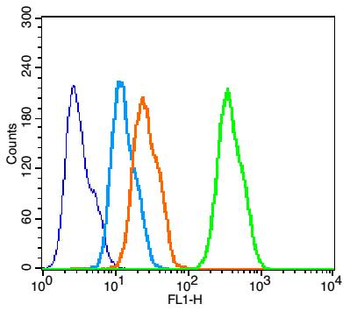
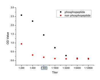
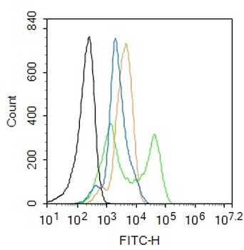
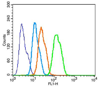
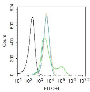
Blank control: HepG2 (blue). Primary Antibody: Rabbit Anti-P38 MAPK antibody (orb11207, Green), Dilution: 1 µg in 100 µl 1X PBS containing 0.5% BSA, Isotype Control Antibody: Rabbit IgG (orange), used under the same conditions, Secondary Antibody: Goat anti-rabbit IgG-FITC (white blue), Dilution: 1:200 in 1 X PBS containing 0.5% BSA. Protocol, The cells were fixed with 2% paraformaldehyde for 10 min at 37°C. Primary antibody (orb11207, 1 µg/1x10^6 cells) were incubated for 30 min at room temperature, followed by 1 X PBS containing 0.5% BSA + 1 0% goat serum (15 min) to block non-specific protein-protein interactions. Then the Goat Anti-rabbit IgG/FITC antibody was added into the blocking buffer mentioned above to react with the primary antibody at 1/200 dilution for 40 min at room temperature. Acquisition of 20000 events was performed.
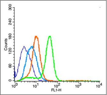
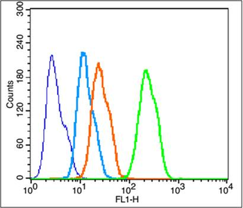
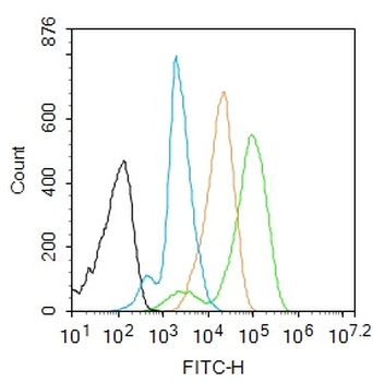
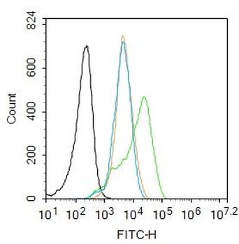
Blank control: MCF7. Primary Antibody (green line): Rabbit Anti-P38 MAPK antibody (orb11207), Dilution: 2 µg/10^6 cells, Isotype Control Antibody (orange line): Rabbit IgG. Secondary Antibody: Goat anti-rabbit IgG-FITC, Dilution: 1 µg/Test. Protocol, The cells were fixed with 4% PFA (10 min at room temperature) and then permeabilized with 90% ice-cold methanol for 20 min at -20°C. The cells were then incubated in 5% BSA to block non-specific protein-protein interactions for 30 min at room temperature. Cells stained with Primary Antibody for 30 min at room temperature. The secondary antibody used for 40 min at room temperature. Acquisition of 20000 events was performed.
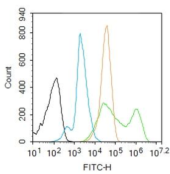
Blank control: Raw264.7. Primary Antibody (green line): Rabbit Anti-P38 MAPK antibody (orb11207), Dilution: 2 µg/10^6 cells, Isotype Control Antibody (orange line): Rabbit IgG. Secondary Antibody: Goat anti-rabbit IgG-AF488, Dilution: 1 µg/Test. Protocol, The cells were fixed with 4% PFA (10 min at room temperature) and then permeabilized with 90% ice-cold methanol for 20 min at -20°C. The cells were then incubated in 5% BSA to block non-specific protein-protein interactions for 30 min at room temperature. Cells stained with Primary Antibody for 30 min at room temperature. The secondary antibody used for 40 min at room temperature. Acquisition of 20000 events was performed.
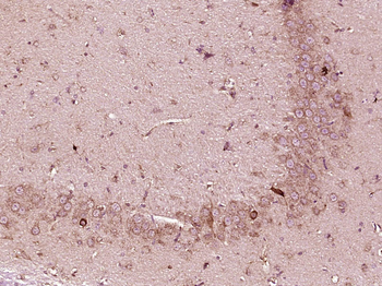
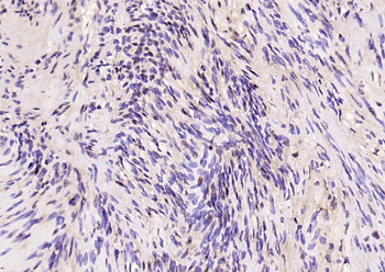
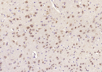
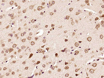
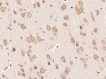
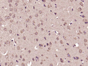
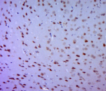
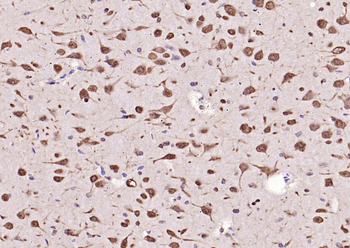
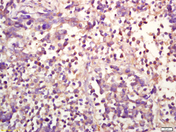
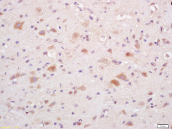
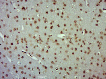
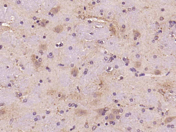
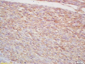
Paraformaldehyde-fixed, paraffin embedded (Rat brain), Antigen retrieval by boiling in sodium citrate buffer (pH6.0) for 15 min, Block endogenous peroxidase by 3% hydrogen peroxide for 20 minutes, Blocking buffer (normal goat serum) at 37°C for 30 min, Antibody incubation with (P38 MAPK) Polyclonal Antibody, Unconjugated (orb11207) at 1:400 overnight at 4°C, followed by operating according to SP Kit (Rabbit) instructionsand DAB staining.
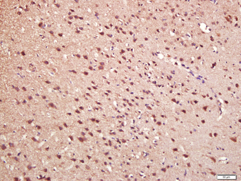
Paraformaldehyde-fixed, paraffin embedded (Rat brain), Antigen retrieval by boiling in sodium citrate buffer (pH6.0) for 15 min, Block endogenous peroxidase by 3% hydrogen peroxide for 20 minutes, Blocking buffer (normal goat serum) at 37°C for 30 min, Antibody incubation with (P38 MAPK) Polyclonal Antibody, Unconjugated (orb11207) at 1:500 overnight at 4°C, followed by a conjugated secondary for 20 minutes and DAB staining.
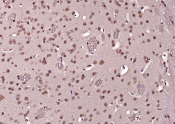
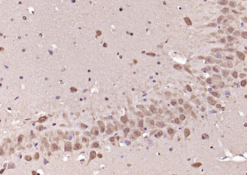
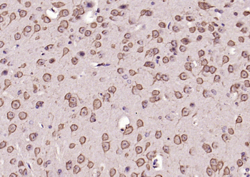
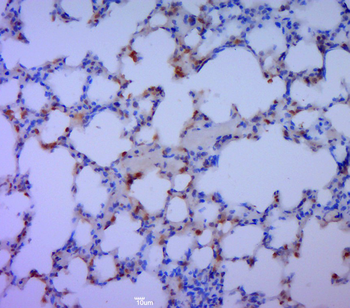
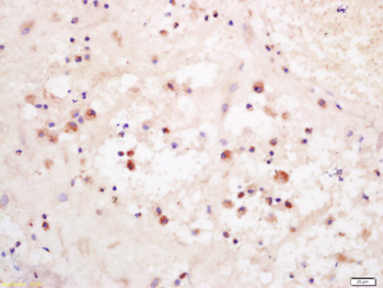
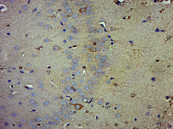
Paraformaldehyde-fixed, paraffin embedded (rat brain), Antigen retrieval by boiling in sodium citrate buffer (pH6.0) for 15 min, Block endogenous peroxidase by 3% hydrogen peroxide for 20 minutes, Blocking buffer (normal goat serum) at 37°C for 30 min, Antibody incubation with (Phospho-P38 MAPK (Thr180 + Tyr182)) Polyclonal Antibody, Unconjugated (orb11207) at 1:200 overnight at 4°C, followed by operating according to SP Kit (Rabbit) instructionsand DAB staining.
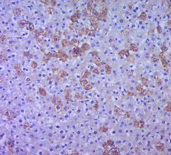
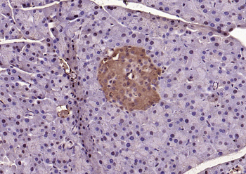
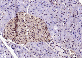
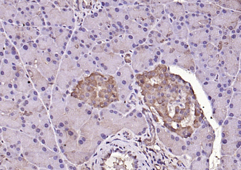
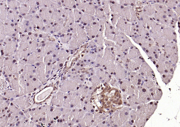
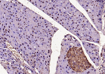
Paraformaldehyde-fixed, paraffin embedded (rat liver tissue), Antigen retrieval by boiling in sodium citrate buffer (pH6.0) for 15 min, Block endogenous peroxidase by 3% hydrogen peroxide for 20 minutes, Blocking buffer (normal goat serum) at 37°C for 30 min, Antibody incubation with (MAPK) Polyclonal Antibody, Unconjugated (orb11207) at 1:400 overnight at 4°C, followed by a conjugated secondary for 20 minutes and DAB staining.
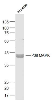
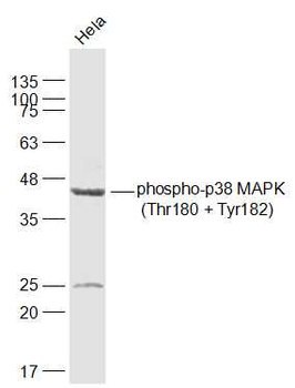
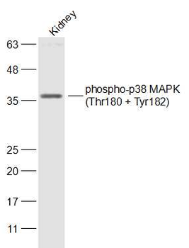
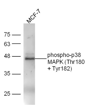
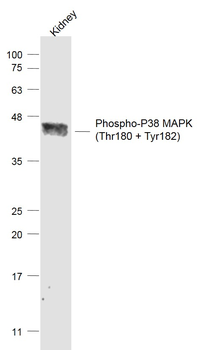
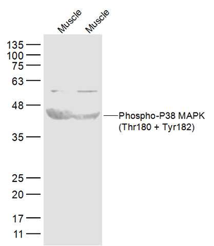
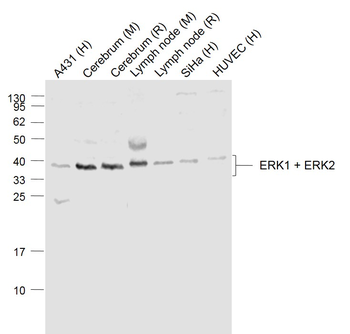
Sample: Muscle (Mouse) Lysate at 40 ug, Primary: Anti-P38 MAPK (orb11207) at 1/1000 dilution, Secondary: IRDye800CW Goat Anti-Rabbit IgG at 1/20000 dilution. Predicted band size: 41 kD, Observed band size: 41 kD.
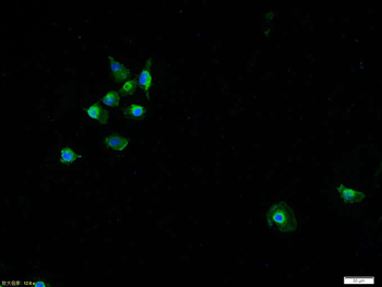
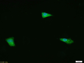
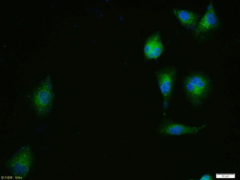
Tissue/Cell: HUVEC cell, 4% Paraformaldehyde-fixed, Triton X-100 at room temperature for 20 min, Blocking buffer (normal goat serum) at 37°C for 20 min, Antibody incubation with (P38 MAPK) polyclonal Antibody, Unconjugated (orb11207) 1:100, 90 minutes at 37°C, followed by a FITC conjugated Goat Anti-Rabbit IgG antibody (orb868805) at 37°C for 90 minutes, DAPI (blue) was used to stain the cell nuclei.
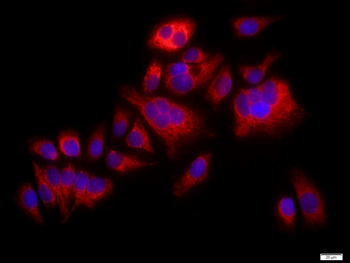
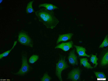
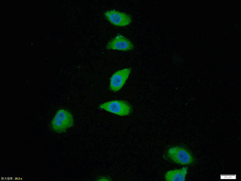
Tissue/Cell: MCF7 cell, 4% Paraformaldehyde-fixed, Triton X-100 at room temperature for 20 min, Blocking buffer (normal goat serum) at 37°C for 20 min, Antibody incubation with (P38 MAPK) polyclonal Antibody, Unconjugated (orb11207) 1:100, 90 minutes at 37°C, followed by a FITC conjugated Goat Anti-Rabbit IgG antibody at 37°C for 90 minutes, DAPI (blue) was used to stain the cell nuclei.
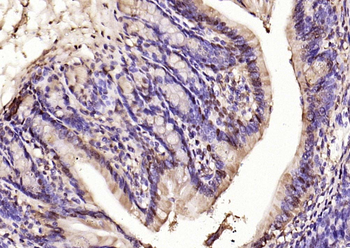
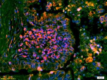
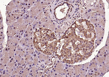
Tissue/Cell: mouse embryo tissue, 4% Paraformaldehyde-fixed and paraffin-embedded, Antigen retrieval: citrate buffer (0.01M, pH6.0), Boiling bathing for 15 min, Block endogenous peroxidase by 3% Hydrogen peroxide for 30 min, Blocking buffer (normal goat serum) at 37°C for 20 min, Incubation: Anti-p38MAPK/MAPK14/p38Alpha Polyclonal Antibody, Unconjugated (orb11207) 1:200, overnight at 4°C, followed by conjugation to the secondary antibody and DAB staining.
Quick Database Links
Gene Symbol
MAPK14
UniProt
UniProt Details
− No UniProt data available
Documents Download
Datasheet
Product Information
Request a Document
Protocol Information
FC
Flow Cytometry
ICC
Immunocytochemistry
Yu, Wu et al. BEX4 upregulation alters Sertoli cell growth properties and protein expression profiles: An explanation for cadmium-induced testicular Sertoli cell injury J Biochem Mol Toxicol, 31, (2017)
Li, Mengdi et al. Selenoprotein K Mediates the Proliferation, Migration, and Invasion of Human Choriocarcinoma Cells by Negatively Regulating Human Chorionic Gonadotropin Expression via ERK, p38 MAPK, and Akt Signaling Pathway Biol Trace Elem Res, 184, 47-59 (2018)
P38 MAPK Rabbit Polyclonal Antibody (orb11207)

- 0.0
Based on 0 reviews
Participating in our Biorbyt product reviews program enables you to support fellow scientists by sharing your firsthand experience with our products.
Login to Submit a ReviewAvailable Sizes
Select a size below
Free Secondary Antibody (20 ul)0/0
Please add an antibody product to your cart first.