You have no items in your shopping cart.
Featured
Description
Research Area
Stem Cell & Developmental Biology
Images & Validation
−Item 1 of 10
| Tested Applications | ELISA, IF, IHC-P, WB |
|---|---|
| Reactivity | Human, Mouse |
| Predicted Reactivity | Rat |
Key Properties
−| Antibody Type | Primary Antibody |
|---|---|
| Host | Rabbit |
| Clonality | Polyclonal |
| Isotype | IgG |
| Immunogen | Anti-Nephrin antibody (orb1239658) was raised against a peptide corresponding to 15 amino acids near the center of human Nephrin. The immunogen is located within amino acids 1100-1150 of Nephrin. |
| Target | NPHS1 |
| Molecular Weight | Predicted: 125, 137 kDa Observed: 125 kDa |
| Purification | Nephrin antibody is affinity chromatography purified via peptide column. |
| Conjugation | Unconjugated |
Storage & Handling
−| Storage | Maintain refrigerated at 2-8°C for up to 2 weeks. For long term storage store at -20°C in small aliquots to prevent freeze-thaw cycles. |
|---|---|
| Form/Appearance | Liquid |
| Buffer/Preservatives | Nephrin antibody is supplied in PBS containing 0.02% sodium azide. |
| Concentration | 1 mg/mL |
| Expiration Date | 12 months from date of receipt. |
| Disclaimer | For research use only |
Alternative Names
−NPHN, NPHS1, Renal glomerulus-specific cell adhesion receptor3
Similar Products
−- Item 1 of 14
NPHS1 Antibody [orb1239664]
ELISA, IF, IHC-P, KO/KD Validated, WB
Human, Mouse, Rat
Rabbit
Polyclonal
Unconjugated
0.1 mg, 0.02 mg - Item 1 of 9
Nephrin Rabbit Polyclonal Antibody [orb322977]
ELISA, IHC-P, WB
Human, Mouse, Rat
Rabbit
Polyclonal
Unconjugated
100 μg - Item 1 of 7
Nephrin Rabbit Polyclonal Antibody [orb185637]
FC, IF, IHC-Fr, IHC-P, WB
Rabbit
Canine, Human, Mouse, Rat
Rabbit
Polyclonal
Unconjugated
1 ml (Carrier free), 50 μl, 100 μl, 200 μl - Item 1 of 1
- Item 1 of 1


Quality Guarantee
Explore bioreagents carefree to elevate your research. All our products are rigorously tested for performance. If a product does not perform as described on its datasheet, our scientific support team will provide expert troubleshooting, a prompt replacement, or a refund. For full details, please see our Terms & Conditions and Buying Guide. Contact us at [email protected].
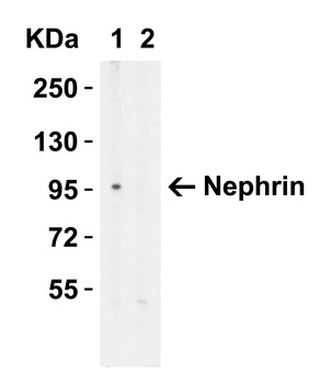
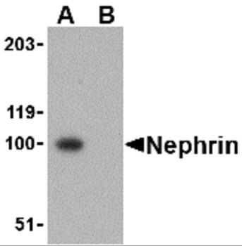
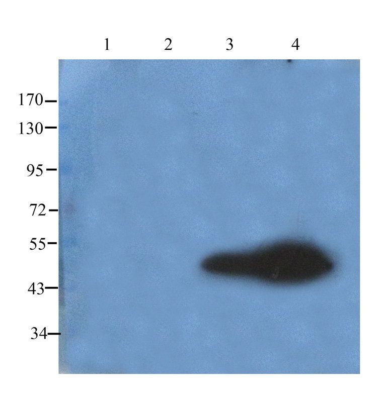
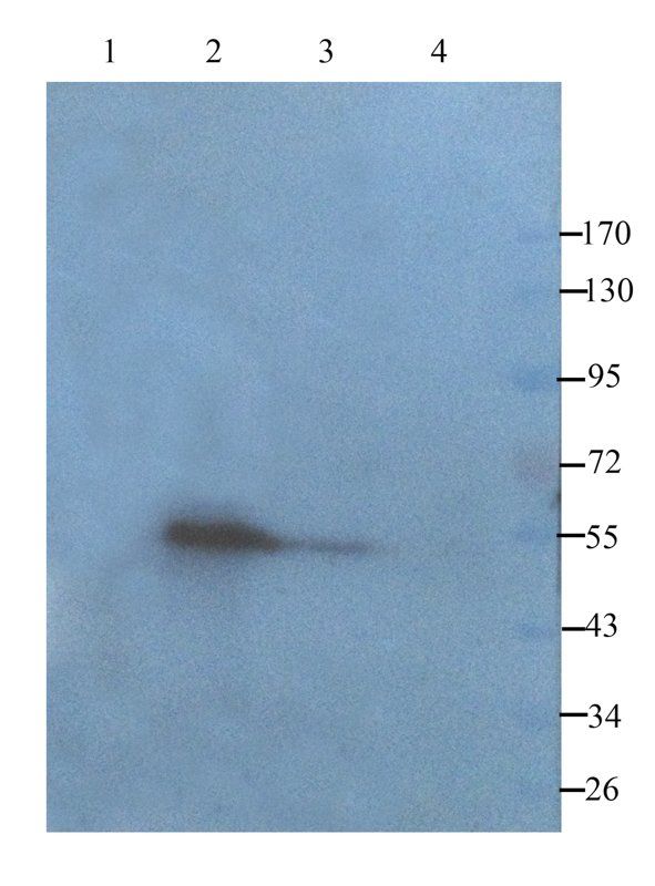
Western Blot Validation in HeLa Cell Lysate with (A) the Absence and (B) the Presence of Blocking Peptide. Loading: 15 µg of lysates per lane. Antibodies: Nephrin orb1239658 (1 µg/mL), 1h incubation at RT in 5% NFDM/TBST. Secondary: Goat anti-rabbit IgG HRP conjugate at 1:10000 dilution.
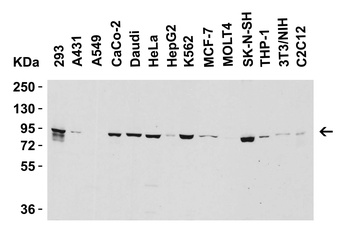
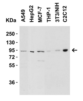
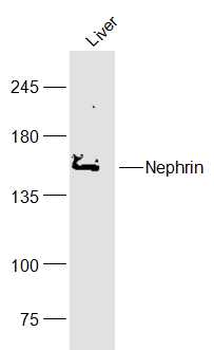
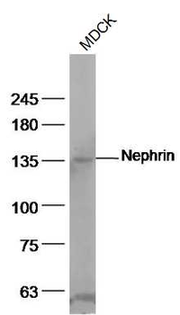
Western Blot Validation in Human and Mouse Cell Lines. Loading: 15 µg of lysates per lane. Antibodies: Nephrin orb1239658 (2 µg/mL), 1h incubation at RT in 5% NFDM/TBST. Secondary: Goat anti-rabbit IgG HRP conjugate at 1:10000 dilution.
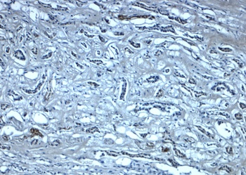
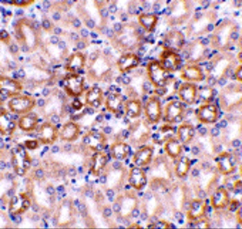
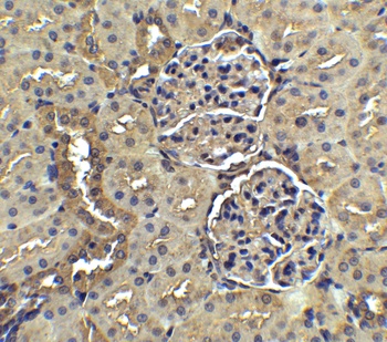
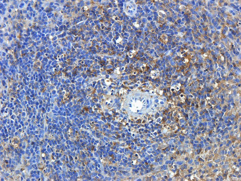
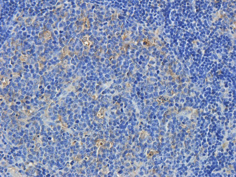
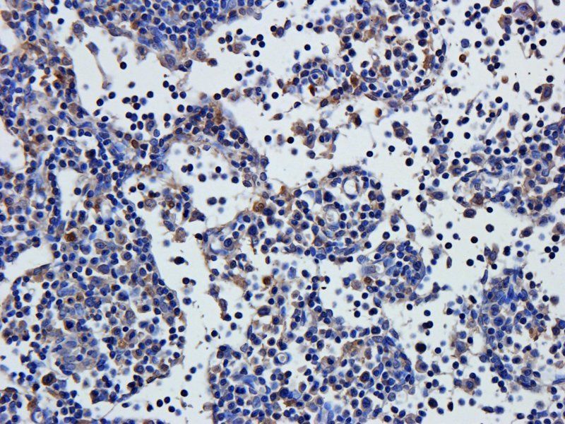
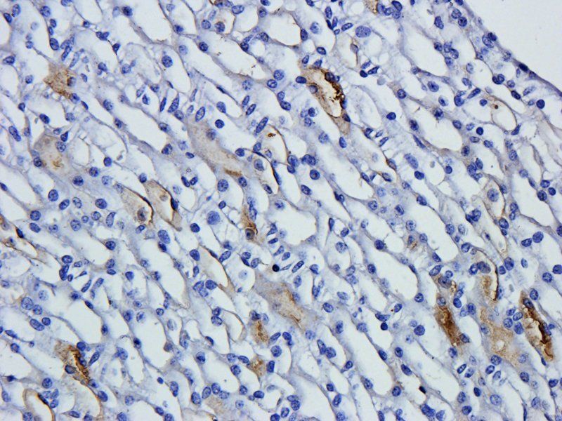
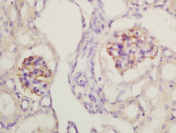
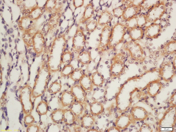
Immunohistochemistry Validation of Nephrin in Human Kidney Tissue. Immunohistochemical analysis of paraffin-embedded human kidney tissue using anti-Nephrin antibody (orb1239658) at 5 µg/ml. Tissue was fixed with formaldehyde and blocked with 10% serum for 1 h at RT; antigen retrieval was by heat mediation with a citrate buffer (pH6). Samples were incubated with primary antibody overnight at 4°C. A goat anti-rabbit IgG H&L (HRP) at 1/250 was used as secondary. Counter stained with Hematoxylin.
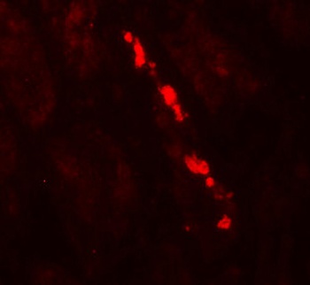
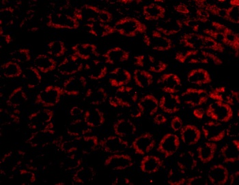
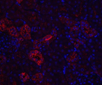
Immunofluorescence Validation of Nephrin in Human Kidney Tissue. Immunofluorescent analysis of 4% paraformaldehyde-fixed human kidney tissue labeling Nephrin with orb1239658 at 20 µg/mL, followed by goat anti-rabbit IgG secondary antibody at 1/500 dilution (red).
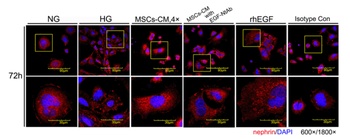
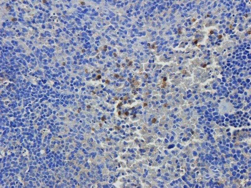
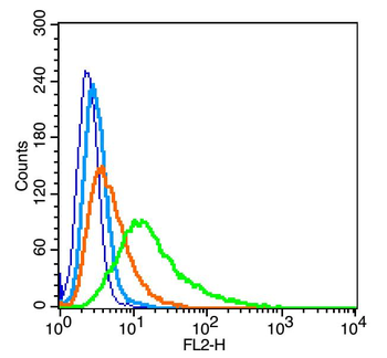
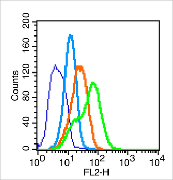
Immunofluorescence Validation of Nephrin in mouse podocyte clone 5 (MPC5) cells (Li et al, 2013). Double immunofluorescence analysis of podocytic membrane protein nephrin (red) and nuclei stained with DAPI (blue). The presence of high glucose (HG) and neutralizing antibody (NtAb) which blocked epithelial growth factor (EGF) decreased nephrin expression while mesenchymal stem cells-conditioned medium (MSCs-CM) and recombinant human EGF (rhEGF) prevented the effect.

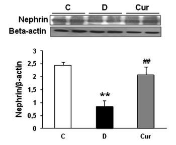
Induced Expression of Nephrin by curcumin treatment in the renal tissues of type 1 diabetic rats (Soetikno et al., 2013). Nephrin expression detected by anti-nephrin antibodies in type 1 diabetic rats. Nephrin was down-regulated in the vehicle-treated diabetic rats as compared to the control nondiabetic rats. However, this decreasein nephrin protein expression was markedly increased by curcumin treatment (P<.05) to near-normal levels. (n=5 in each group).

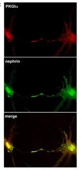
Immunofluorescence and Localization Validation of Nephrin in cultured rat podocytes (Piwkowska et al., 2012). Immunofluorescence staining showed Nephrin expression (green) detected by anti-nephrin antibodies and PKGIalpha (red). The co-localization of two antibodies (yellow) in rat podocytes was observed particularly at the tips of the cell processes.

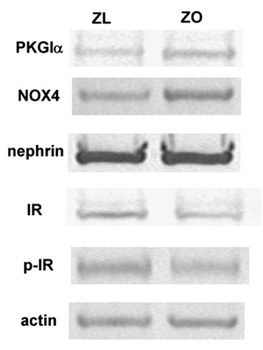
WB Validation of Nephrin in glomeruli of Zucker obese (ZO) and Zucker lean (ZL) rats (Piwkowska et al., 2013). The expression of nephrin detected by anti-nephrin antibodies did not change in ZO rats as compared to the control rats.
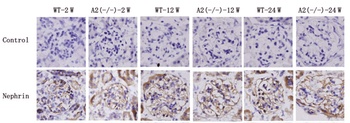
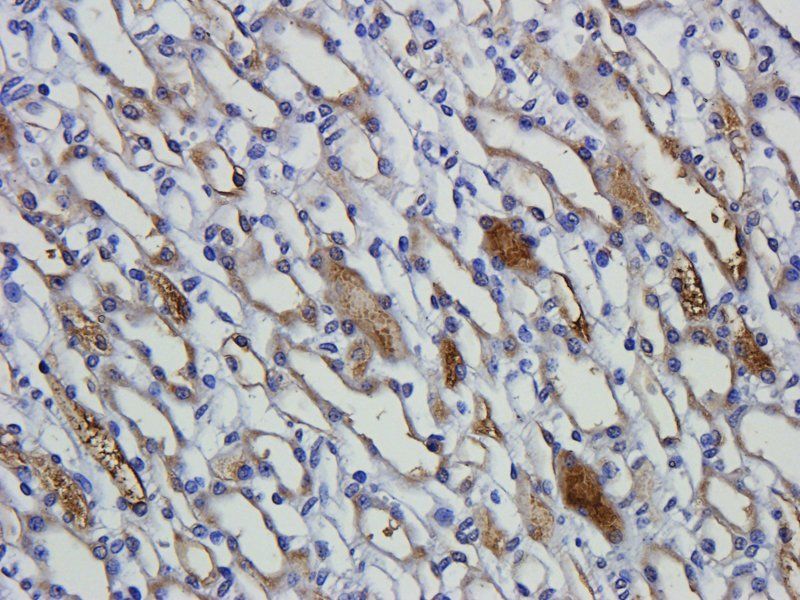
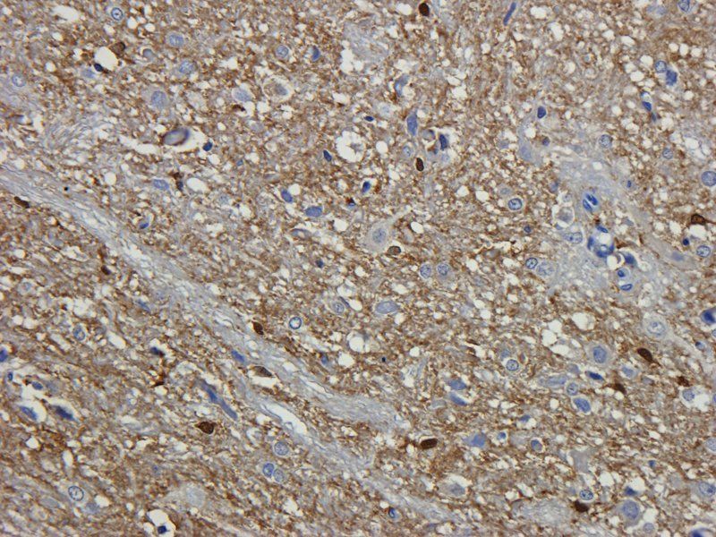
Immunohistochemistry Validation of Nephrin in mouse kidneys (Toyama et al., 2012). Protein analysis for nephrin by immunohistochemistry with anti-nephrin antibodies in kidneys of wild-type or AMPD2-deficient mice at 2, 12 or 24 weeks of age. No difference between wild-type andAMPD2-deficient mice at any age was observed.
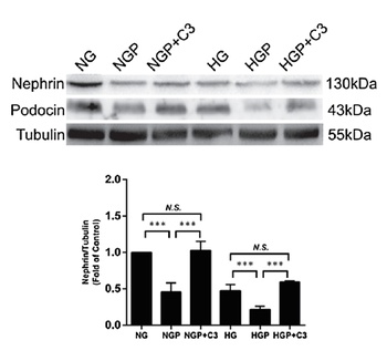
Regulated Expression Validation of Nephrin in mouse podocyte cells cultured in normal glucose (NG) medium or high glucose (HG) medium (Huang et al., 2019). Western Blot analysis was used to access the protein expression level of nephrin with anti-nephrin antibodies. Nephrin expression was down-regulated by PEGF treatment (NGP or HGP), which was reversed by the addition of C3 transferase.
Documents Download
Datasheet
Product Information
Request a Document
Protocol Information
WB
Western Blot (IB, immunoblot)
IHC-P
Immunohistochemistry Paraffin
IF
Immunofluorescence
ELISA
Enzyme-linked Immunosorbent Assay (EIA)
NPHS1 Antibody (orb1239658)

- 0.0
Based on 0 reviews
Participating in our Biorbyt product reviews program enables you to support fellow scientists by sharing your firsthand experience with our products.
Login to Submit a ReviewAvailable Sizes
Select a size below
Free Secondary Antibody (20 ul)0/0
Please add an antibody product to your cart first.