You have no items in your shopping cart.
KO/KD
Validated
Validated
Description
Research Area
Infectious Disease & Virology
Images & Validation
−Item 1 of 13
| Tested Applications | ELISA, IF, IHC-P, IP, KO/KD Validated, WB |
|---|---|
| Reactivity | Human, Mouse, Rat |
| Predicted Reactivity | Bovine, Gallus, Porcine, Sheep |
Key Properties
−| Antibody Type | Primary Antibody |
|---|---|
| Host | Rabbit |
| Clonality | Polyclonal |
| Isotype | IgG |
| Immunogen | Anti-MYD88 antibody (orb1239647) was raised against a peptide corresponding to 16 amino acids near the center of human MYD88 isoform 1. The immunogen is located within amino acids 220 - 270 of MYD88. |
| Target | MYD88 |
| Molecular Weight | Predicted: 35kD Observed: 35kD |
| Purification | MYD88 Antibody is affinity chromatography purified via peptide column. |
| Conjugation | Unconjugated |
Storage & Handling
−| Storage | Maintain refrigerated at 2-8°C for up to 2 weeks. For long term storage store at -20°C in small aliquots to prevent freeze-thaw cycles. |
|---|---|
| Form/Appearance | Liquid |
| Buffer/Preservatives | MYD88 Antibody is supplied in PBS containing 0.02% sodium azide. |
| Concentration | 1 mg/mL |
| Expiration Date | 12 months from date of receipt. |
| Disclaimer | For research use only |
Alternative Names
−MYD88 Antibody: Myeloid differentiation primary response 88, MYD88D
Similar Products
−- Item 1 of 12
MyD88 Rabbit Polyclonal Antibody [orb215902]
ELISA, FC, ICC, IF, IHC, IHC-Fr, WB
Human, Mouse, Rat
Rabbit
Polyclonal
Unconjugated
100 μg - Item 1 of 13
MYD88 Antibody [orb1239635]
ELISA, IF, IP, KO/KD Validated, WB
Bovine, Gallus, Porcine, Sheep
Human, Mouse, Rat
Rabbit
Polyclonal
Unconjugated
0.1 mg, 0.02 mg - Item 1 of 6
MyD88 Rabbit Polyclonal Antibody [orb11091]
FC, IF, IHC-Fr, IHC-P, WB
Human, Rat
Human, Mouse, Rat
Rabbit
Polyclonal
Unconjugated
50 μl, 100 μl, 200 μl - Item 1 of 6
MYD88 Antibody [orb18963]
ELISA, FC, IF, IHC, WB
Canine, Mouse, Rat
Human
Polyclonal
Unconjugated
100 μg - Item 1 of 6
MYD88 Rabbit Polyclonal Antibody [orb420074]
IHC-P, WB
Guinea pig, Human, Mouse, Rat
Rabbit
Polyclonal
Unconjugated
100 μg
Quality Guarantee
Explore bioreagents carefree to elevate your research. All our products are rigorously tested for performance. If a product does not perform as described on its datasheet, our scientific support team will provide expert troubleshooting, a prompt replacement, or a refund. For full details, please see our Terms & Conditions and Buying Guide. Contact us at [email protected].
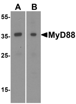
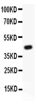
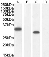
Western Blot Validation of MyD88 in HeLa (A) and Jurket (B) Cells. Loading: 15 µg of lysates per lane. Antibodies: orb1239647 (1 µg/mL) 1 h incubation at RT in 5% NFDM/TBST. Secondary: Goat anti-rabbit IgG HRP conjugate at 1:10000 dilution.
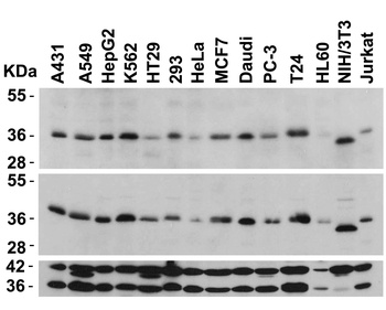
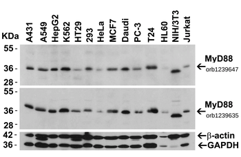
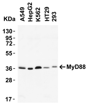
Independent Antibody Validation (IAV) via Protein Expression Profile in Cell Lines. Loading: 15 µg of lysates per lane. Antibodies: MyD88 orb1239647 (2 µg/mL), MyD88 orb1239635 (2 µg/mL), beta-actin (1 µg/mL), and GAPDH (0.02 µg/mL), 1 h incubation at RT in 5% NFDM/TBST. Secondary: Goat anti-rabbit IgG HRP conjugate at 1:10000 dilution.
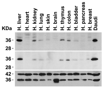
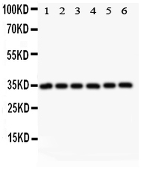
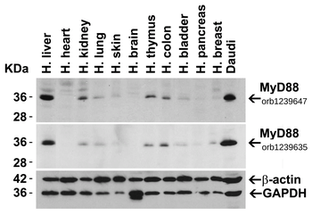
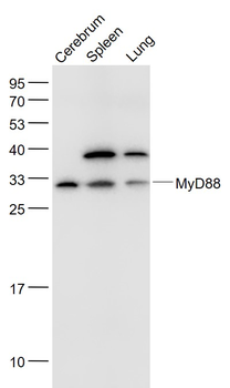
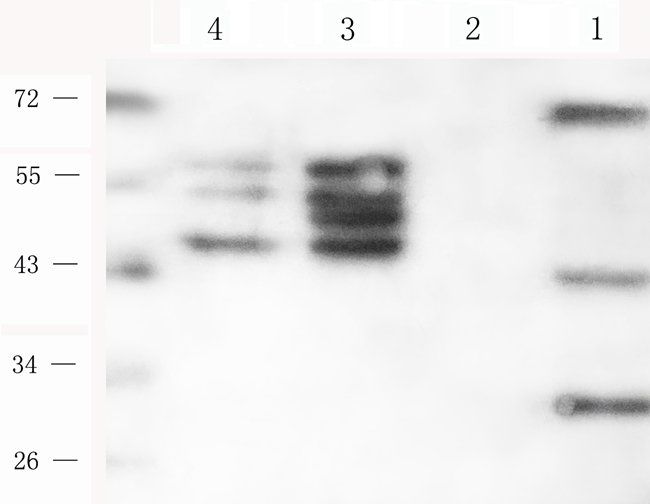
Independent Antibody Validation (IAV) via Protein Expression Profile in Human Tissues. Loading: 15 µg of lysates per lane. Antibodies: MyD88 orb1239647 (2 µg/mL), MyD88 orb1239635 (2 µg/mL), beta-actin (1 µg/mL), and GAPDH (0.02 µg/mL), 1 h incubation at RT in 5% NFDM/TBST. Secondary: Goat anti-rabbit IgG HRP conjugate at 1:10000 dilution.
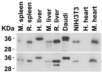
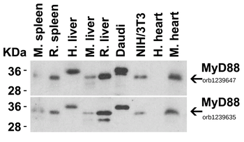
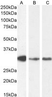
Animal Species Reactivity. Loading: Lysates/proteins at 15 µg per lane. Antibodies: orb1239647 (2 µg/mL) or orb1239635 (2 µg/mL). 1 h incubation at RT in 5% NFDM/TBST. Secondary: Goat anti-rabbit IgG HRP conjugate at 1:10000 dilution.
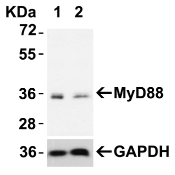
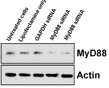
Validation with MyD88 siRNA Knockdown in HeLa Cells. HeLa cells were transfected with control siRNAs (lane 1) or MyD88 siRNAs (lane 2) Loading: 10 µg of HeLa whole cell lysates per lane. Antibodies: orb1239647 (2 µg/mL), 1 h incubation at RT in 5% NFDM/TBST. Secondary: Goat anti-rabbit IgG HRP conjugate at 1:10000 dilution.
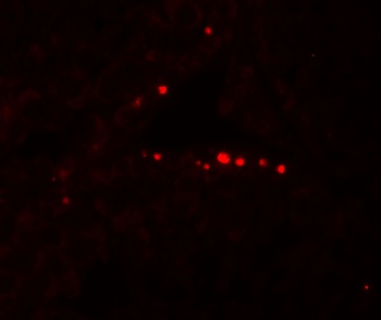
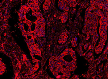
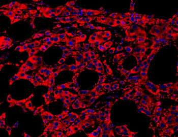
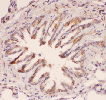
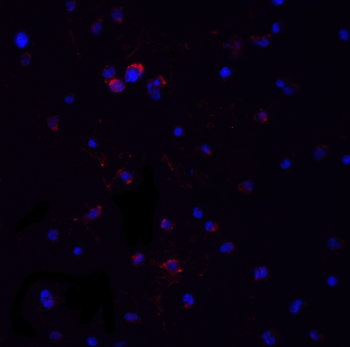
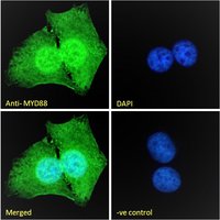
Immunofluorescence Validation of MyD88 n Human Testis. Immunofluorescent analysis of 4% paraformaldehyde-fixed human testis tissue labeling MyD88 with orb1239647 at 20 µg/mL, followed by goat anti-rabbit IgG secondary antibody at 1/500 dilution (red). Image showing nucleus staining on human testis cells.
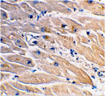
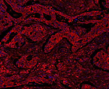
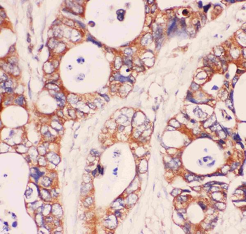
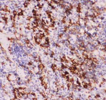
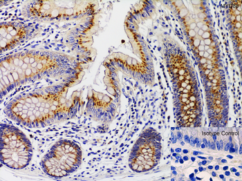
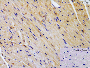
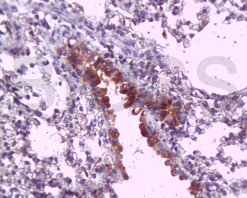
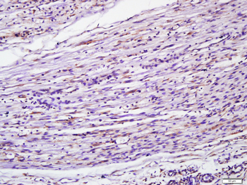
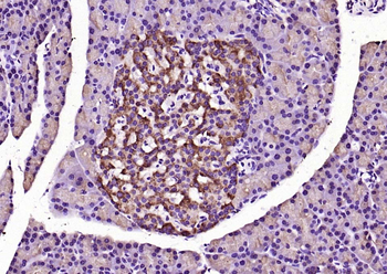
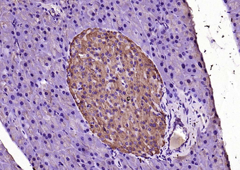
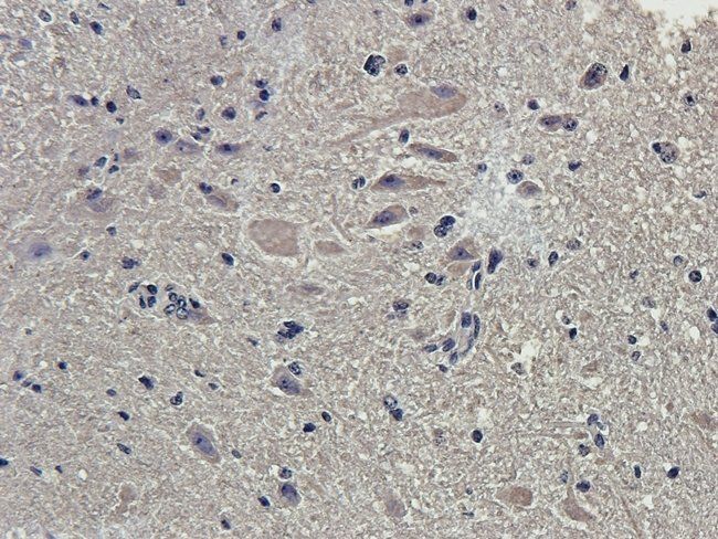
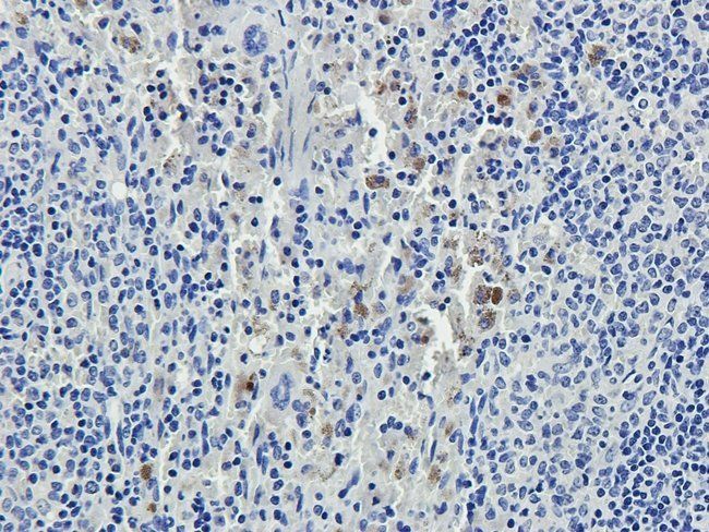
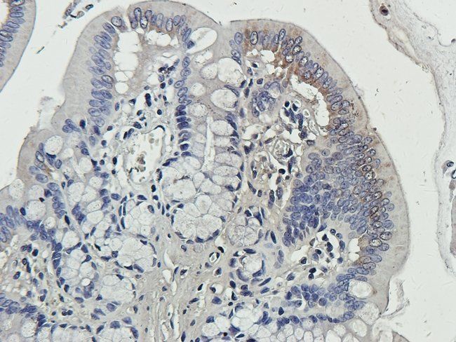
Immunohistochemistry Validation of MyD88 in Human Heart. Immunohistochemical analysis of paraffin-embedded human heart tissue using anti-MyD88 antibody (orb1239647) at 2 µg/ml. Tissue was fixed with formaldehyde and blocked with 10% serum for 1 h at RT; antigen retrieval was by heat mediation with a citrate buffer (pH6). Samples were incubated with primary antibody overnight at 4°C. A goat anti-rabbit IgG H&L (HRP) at 1/250 was used as secondary. Counter stained with Hematoxylin.
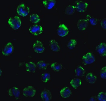
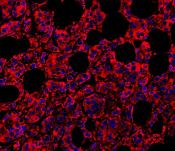
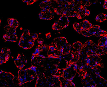
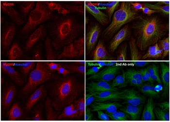
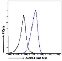
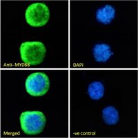
Immunofluorescence Validation of MyD88 in K562 Cells. Immunofluorescent analysis of 4% paraformaldehyde-fixed K562 cells labeling MyD88 with orb1239647 at 10 µg/mL, followed by Goat anti-rabbit IgG secondary antibody at 1/500 dilution (green) and DAPI staining (blue).
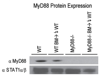
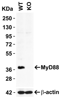
KO Validation in Mouse Macrophages (Miller et al., 2006). Bone marrow-derived macrophages from wild type (WT) mice and MyD88 knockout mice were assessed for MyD88 protein expression by anti-MyD88 antibodies. MyD88 expression was detected in WT mice, but not in MyD88 knockout mice.

KO Validation in MyD88-deficient MEF cell line (Burns et al., 2003). MyD88−/− deficient MEF cell line was reconstituted by retroviral infection with an empty vector, MyD88, or MyD88s expression vectors. The levels of MyD88 (isoform 1) or MyD88s (isoform 3) were confirmed with anti-MyD88 antibodies and MyD88 expression was not detected in the MyD88-deficient cells.
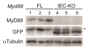
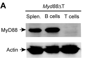
KO Validation in Mouse IECs (Vlantis et al., 2014). Western blot with anti-MyD88 antibodies on intestinal epithelial cells (IECs) showing efficient deletion of MyD88 and concomitant expression of GFP in MyD88IEC-KO mice, but not in MyD88 knockout mice.
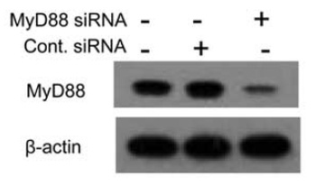
KD Validation in Chondrocytes (Ahmad et al., 2009). Chondrocytes were transfected with either MyD88 siRNA or control siRNA and analyzed for MyD88 expression by immunoblotting with anti-Myd88 antibodies that confirmed inhibition of the target proteins.
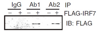
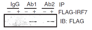
Immunoprecipitation Validation in HEK293 cells (Kawai et al., 2004). HEK293 cells were transiently transfected with FLAG-IRF7. Cell lysates were immunoprecipitated with control rabbit anti-mouse immunoglobulin serum (IgG) or anti-MyD88 (Ab1 and Ab2), followed by immunoblotting with anti-FLAG.
Quick Database Links
UniProt Details
− No UniProt data available
NCBI Reference Sequences
−Associated Accession Numbers
Curated reference sequences for the gene transcript and protein product| RefSeq | AAB49967.1 |
|---|
Documents Download
Datasheet
Product Information
Request a Document
Protocol Information
WB
Western Blot (IB, immunoblot)
IHC-P
Immunohistochemistry Paraffin
IF
Immunofluorescence
ELISA
Enzyme-linked Immunosorbent Assay (EIA)
IP
Immunoprecipitation
MYD88 Antibody (orb1239647)

- 0.0
Based on 0 reviews
Participating in our Biorbyt product reviews program enables you to support fellow scientists by sharing your firsthand experience with our products.
Login to Submit a ReviewAvailable Sizes
Select a size below
Free Secondary Antibody (20 ul)0/0
Please add an antibody product to your cart first.