You have no items in your shopping cart.
KO/KD
Validated
Validated
Description
Research Area
Infectious Disease & Virology
Images & Validation
−Item 1 of 13
| Tested Applications | ELISA, IF, IP, KO/KD Validated, WB |
|---|---|
| Reactivity | Human, Mouse, Rat |
| Predicted Reactivity | Bovine, Gallus, Porcine, Sheep |
Key Properties
−| Antibody Type | Primary Antibody |
|---|---|
| Host | Rabbit |
| Clonality | Polyclonal |
| Isotype | IgG |
| Immunogen | Anti-MYD88 antibody (orb1239635) was raised against a peptide corresponding to 17 amino acids near carboxy terminus of human MYD88 isoform 1. The immunogen is located within the last 50 amino acids of MYD88. |
| Target | MYD88 |
| Molecular Weight | Predicted: 35kD Observed: 35kD |
| Purification | MYD88 Antibody is affinity chromatography purified via peptide column. |
| Conjugation | Unconjugated |
Storage & Handling
−| Storage | Maintain refrigerated at 2-8°C for up to 2 weeks. For long term storage store at -20°C in small aliquots to prevent freeze-thaw cycles. |
|---|---|
| Form/Appearance | Liquid |
| Buffer/Preservatives | MYD88 Antibody is supplied in PBS containing 0.02% sodium azide. |
| Concentration | 1 mg/mL |
| Expiration Date | 12 months from date of receipt. |
| Disclaimer | For research use only |
Alternative Names
−MYD88 Antibody: MYD88D
Similar Products
−- Item 1 of 12
MyD88 Rabbit Polyclonal Antibody [orb215902]
ELISA, FC, ICC, IF, IHC, IHC-Fr, WB
Human, Mouse, Rat
Rabbit
Polyclonal
Unconjugated
100 μg - Item 1 of 13
MYD88 Antibody [orb1239647]
ELISA, IF, IHC-P, IP, KO/KD Validated, WB
Bovine, Gallus, Porcine, Sheep
Human, Mouse, Rat
Rabbit
Polyclonal
Unconjugated
0.1 mg, 0.02 mg - Item 1 of 6
MyD88 Rabbit Polyclonal Antibody [orb11091]
FC, IF, IHC-Fr, IHC-P, WB
Human, Rat
Human, Mouse, Rat
Rabbit
Polyclonal
Unconjugated
50 μl, 100 μl, 200 μl - Item 1 of 6
MYD88 Antibody [orb18963]
ELISA, FC, IF, IHC, WB
Canine, Mouse, Rat
Human
Polyclonal
Unconjugated
100 μg - Item 1 of 6
MYD88 Rabbit Polyclonal Antibody [orb420074]
IHC-P, WB
Guinea pig, Human, Mouse, Rat
Rabbit
Polyclonal
Unconjugated
100 μg

Quality Guarantee
Explore bioreagents carefree to elevate your research. All our products are rigorously tested for performance. If a product does not perform as described on its datasheet, our scientific support team will provide expert troubleshooting, a prompt replacement, or a refund. For full details, please see our Terms & Conditions and Buying Guide. Contact us at [email protected].
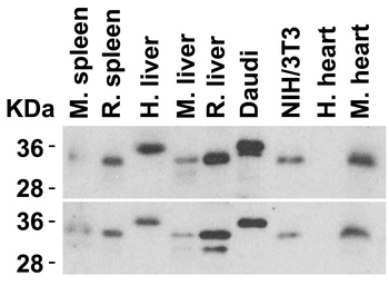
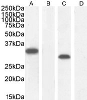
Animal Species Reactivity. Loading: Lysates/proteins at 15 µg per lane. Antibodies: orb1239647 (2 µg/mL) or orb1239635 (2 µg/mL). 1 h incubation at RT in 5% NFDM/TBST. Secondary: Goat anti-rabbit IgG HRP conjugate at 1:10000 dilution.
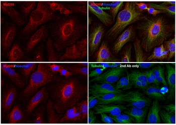
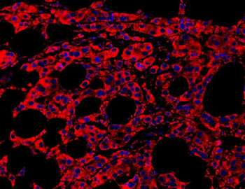
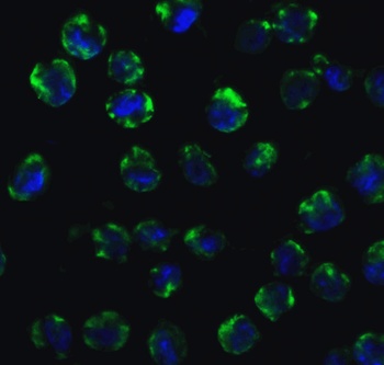
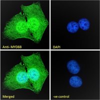
Immunofluorescence Validation of MyD88 In HeLa Cells. Immunofluorescent analysis of methanol-fixed HeLa cells labeling MyD88 with orb1239635 at 20 µg/mL, followed by goat anti-rabbit IgG secondary antibody at 1/1000 dilution (red) and Hoechst staining (blue). Alpha tubulin was stained with anti-alpha tubulin antibody following by goat anti-mouse IgG secondary antibody (green). Images were captured with confocal microscopy.
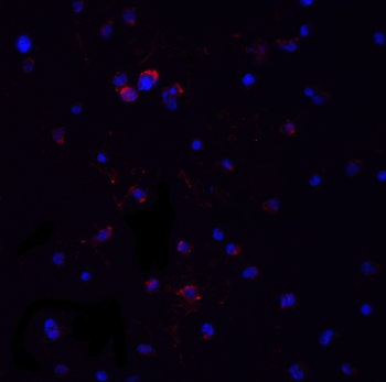
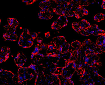
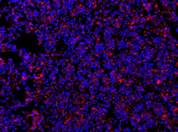
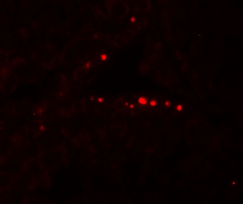
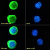
Immunofluorescence Validation of MyD88 in Jurkat Cells. Immunofluorescent analysis of 4% paraformaldehyde-fixed Jurkat cells labeling MyD88 with orb1239635 at 20 µg/mL, followed by goat anti-rabbit IgG secondary antibody at 1/500 dilution (red) and DAPI staining (blue).
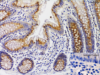
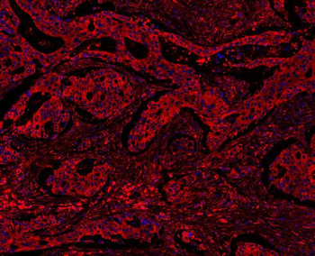
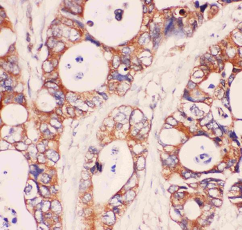
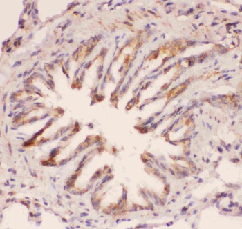
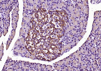
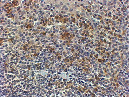
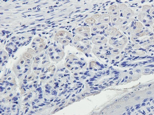
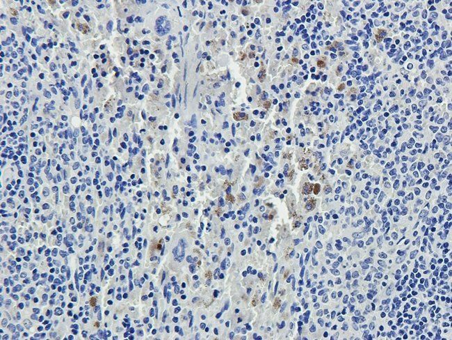
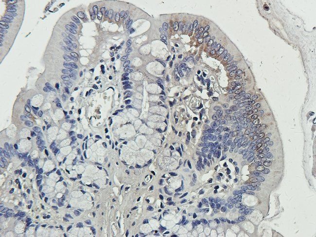
Immunohistochemistry Validation of MyD88 in Human Colon. Immunohistochemical analysis of paraffin-embedded human colon tissue using anti-MYD88 antibody (orb1239635) at 1 µg/mL. Tissue was fixed with formaldehyde and blocked with 10% serum for 1 h at RT; antigen retrieval was by heat mediation with a citrate buffer (pH6). Samples were incubated with primary antibody overnight at 4°C. A goat anti-rabbit IgG H&L (HRP) at 1/250 was used as secondary. Counter stained with Hematoxylin.
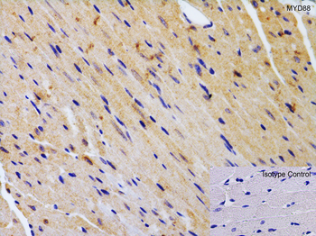
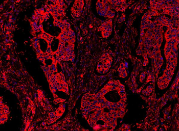
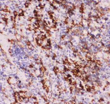
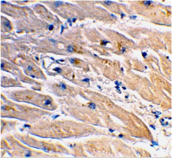
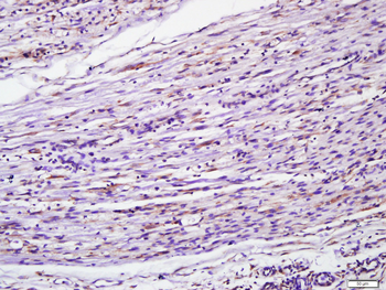
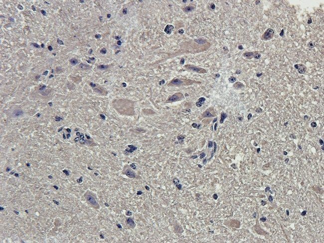
Immunohistochemistry Validation of MyD88 in Mouse Heart. Immunohistochemical analysis of paraffin-embedded mouse heart tissue using anti-MYD88 antibody (orb1239635) at 2 µg/mL. Tissue was fixed with formaldehyde and blocked with 10% serum for 1 h at RT; antigen retrieval was by heat mediation with a citrate buffer (pH6). Samples were incubated with primary antibody overnight at 4°C. A goat anti-rabbit IgG H&L (HRP) at 1/250 was used as secondary. Counter stained with Hematoxylin.
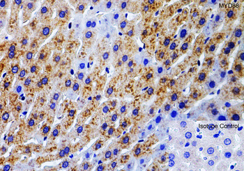
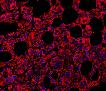
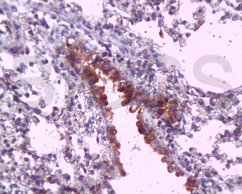
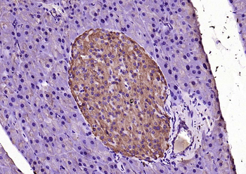
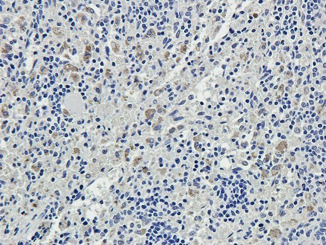
Immunohistochemistry Validation of MyD88 in Rat Liver. Immunohistochemical analysis of paraffin-embedded rat liver tissue using anti-MYD88 antibody (orb1239635) at 1 µg/mL. Tissue was fixed with formaldehyde and blocked with 10% serum for 1 h at RT; antigen retrieval was by heat mediation with a citrate buffer (pH6). Samples were incubated with primary antibody overnight at 4°C. A goat anti-rabbit IgG H&L (HRP) at 1/250 was used as secondary. Counter stained with Hematoxylin.
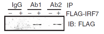
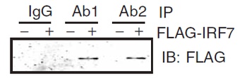
Immunoprecipitation Validation in HEK293 cells. HEK293 cells were transiently transfected with FLAG-IRF7. Cell lysates were immunoprecipitated with control rabbit anti-mouse immunoglobulin serum (IgG) or anti-MyD88 (Ab1 and Ab2), followed by immunoblotting with anti-FLAG.
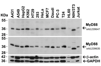
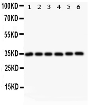
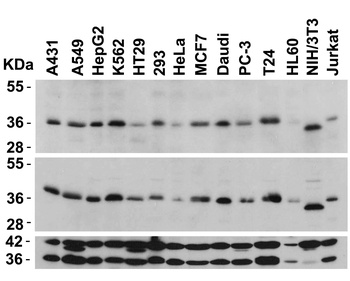
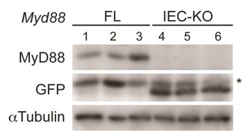
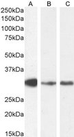
Independent Antibody Validation (IAV) via Protein Expression Profile in Cell Lines. Loading: 15 µg of lysates per lane. Antibodies: MyD88 orb1239647 (2 µg/mL), MyD88 orb1239635 (2 µg/mL), beta-actin (1 µg/mL), and GAPDH (0.02 µg/mL), 1 h incubation at RT in 5% NFDM/TBST. Secondary: Goat anti-rabbit IgG HRP conjugate at 1:10000 dilution.
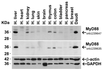
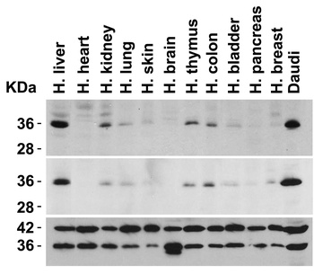
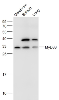
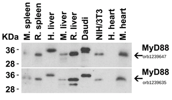
Independent Antibody Validation (IAV) via Protein Expression Profile in Human Tissues. Loading: 15 µg of lysates per lane. Antibodies: MyD88 orb1239647 (2 µg/mL), MyD88 orb1239635 (2 µg/mL), beta-actin (1 µg/mL), and GAPDH (0.02 µg/mL), 1 h incubation at RT in 5% NFDM/TBST. Secondary: Goat anti-rabbit IgG HRP conjugate at 1:10000 dilution.
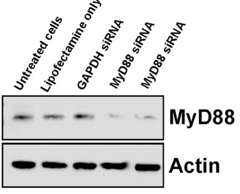
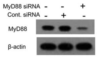
KD Validation in Raw 264.7 cells. The Transfection of RAW 264.7 cells with MyD88-specific siRNA resulted in attenuation of MyD88 protein by Western blot analysis with anti-Myd88 antibodies (orb1239635).
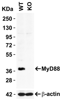
KO Validation in HeLa Cells. Loading: 10 µg of HeLa WT cell lysate or MyD88 KO cell lysate. Antibodies: MyD88 orb1239635 (2 µg/mL) and beta-actin orb1240312 (1 µg/mL), 1 h incubation at RT in 5% NFDM/TBST. Secondary: Goat Anti-Rabbit IgG HRP conjugate at 1:10000 dilution.
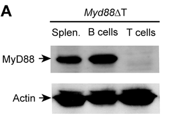
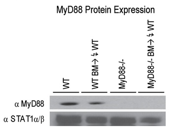
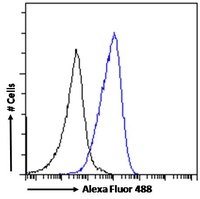
KO Validation of MyD88 in Mouse T cells. Splenocytes were isolated from a Myd88ΔT mouse in which MyD88 was specifically disrupted in T cells. T and B cells were FACS purified, and MyD88 expression was examined by Western blot with anti-MyD88 antibodies (orb1239635). MyD88 expression was detected in Splenocytes and B cells, but not in T cells.
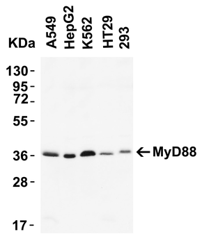
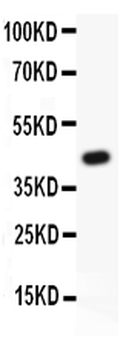
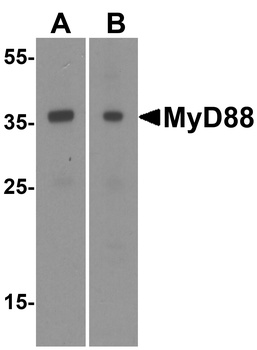
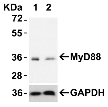
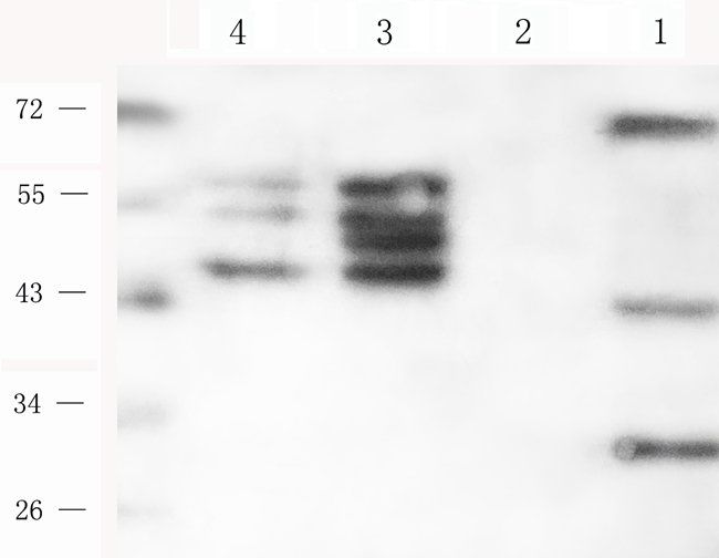
Western Blot Validation of MyD88 in human cell lines. Loading: 15 µg of lysates per lane. Antibodies: orb1239635 (2 µg/mL) 1 h incubation at RT in 5% NFDM/TBST. Secondary: Goat anti-rabbit IgG HRP conjugate at 1:10000 dilution. Predicted band size: 35 kDa.
Quick Database Links
UniProt Details
− No UniProt data available
NCBI Reference Sequences
−Associated Accession Numbers
Curated reference sequences for the gene transcript and protein product| RefSeq | AAB49967.1 |
|---|
Documents Download
Datasheet
Product Information
Request a Document
Protocol Information
WB
Western Blot (IB, immunoblot)
IF
Immunofluorescence
ELISA
Enzyme-linked Immunosorbent Assay (EIA)
IP
Immunoprecipitation
MYD88 Antibody (orb1239635)

- 0.0
Based on 0 reviews
Participating in our Biorbyt product reviews program enables you to support fellow scientists by sharing your firsthand experience with our products.
Login to Submit a ReviewAvailable Sizes
Select a size below
Free Secondary Antibody (20 ul)0/0
Please add an antibody product to your cart first.