You have no items in your shopping cart.
Description
Research Area
Immunology & Inflammation
Images & Validation
−Item 1 of 5
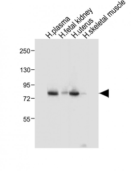
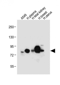
| Tested Applications | IF, IHC-P, WB |
|---|---|
| Dilution Range | IF: 1:10~50, IF: 1:10~50, IF: 1:25, WB: 1:1000, IHC-P: 1:50~100 |
| Reactivity | Human |
Key Properties
−| Antibody Type | Primary Antibody |
|---|---|
| Host | Mouse |
| Clonality | Monoclonal |
| Isotype | IgG1κ |
| Immunogen | This LTF Monoclonal antibody is generated from mouse immunized with LTF recombinant protein. Antigen Region: 249-712 aa. |
| Target | LTF (HGNC:6720) |
| Molecular Weight | 78182 Da |
| Conjugation | Unconjugated |
Storage & Handling
−| Storage | Maintain refrigerated at 2-8°C for up to 2 weeks. For long term storage store at -20°C in small aliquots to prevent freeze-thaw cycles |
|---|---|
| Form/Appearance | Purified monoclonal antibody supplied in PBS with 0.09% (W/V) sodium azide. This antibody is purified through a protein G column, followed by dialysis against PBS. |
| Expiration Date | 12 months from date of receipt. |
| Disclaimer | For research use only |
Alternative Names
−Lactotransferrin, Lactoferrin, 3421-, Growth-inhibiting protein 12, Talalactoferrin, Lactoferricin-H, Lfcin-H, Kaliocin-1, Lactoferroxin-A, Lactoferroxin-B, Lactoferroxin-C, LTF, GIG12, LF
Similar Products
−- Item 1 of 6
- Item 1 of 6
Lactoferrin/LTF Rabbit Polyclonal Antibody [orb413008]
ELISA, IF, IHC, WB
Human, Mouse, Rat
Rabbit
Polyclonal
Unconjugated
100 μg - Item 1 of 5
LTF Antibody [orb1410325]
FC, IF, IHC
Human
Mouse
Monoclonal
Unconjugated
20 μg, 100 μg, 100 μg (without BSA and Azide) - Item 1 of 5
- Item 1 of 3
Lactoferrin Rabbit Polyclonal Antibody [orb100096]
WB
Bovine, Canine, Equine, Mouse, Porcine, Rat, Sheep
Human
Rabbit
Polyclonal
Unconjugated
50 μl, 100 μl, 200 μl


Quality Guarantee
Explore bioreagents carefree to elevate your research. All our products are rigorously tested for performance. If a product does not perform as described on its datasheet, our scientific support team will provide expert troubleshooting, a prompt replacement, or a refund. For full details, please see our Terms & Conditions and Buying Guide. Contact us at [email protected].
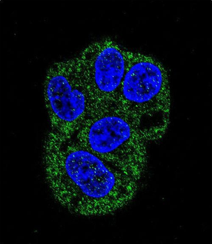
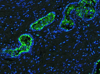
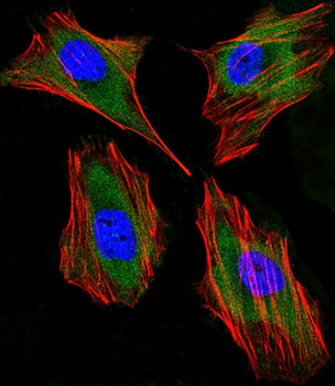
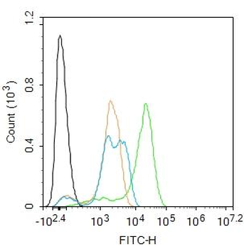
Confocal immunofluorescent analysis of LTF Antibody with HepG2 cell followed by Alexa Fluor 488-conjugated goat anti-mouse lgG (green). DAPI was used to stain the cell nuclear (blue).
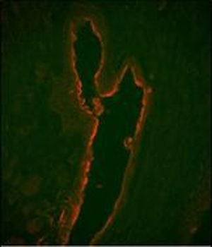
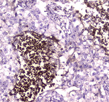
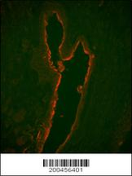
Immunofluorescence analysis of LTF Monoclonal Antibody with paraffin-embedded human prostate carcinoma tissue.0.05 mg/ml primary antibody was followed by PE-conjugated goat anti-mouse lgG (whole molecule). PE emits orange fluorescence.
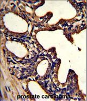
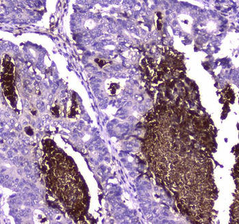
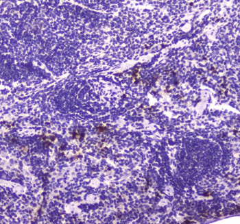
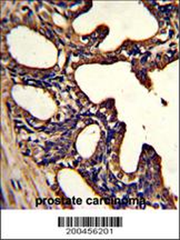
Formalin-fixed and paraffin-embedded human prostate carcinoma with LTF Monoclonal Antibody, which was peroxidase-conjugated to the secondary antibody, followed by DAB staining. This data demonstrates the use of this antibody for immunohistochemistry; clinical relevance has not been evaluated.
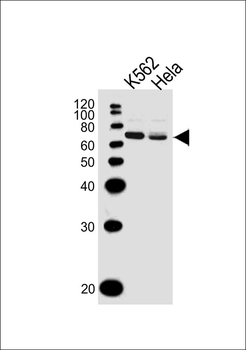
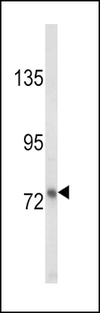
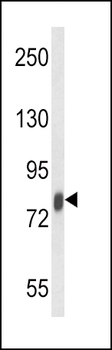
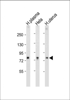
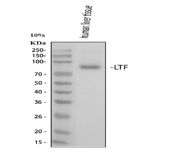
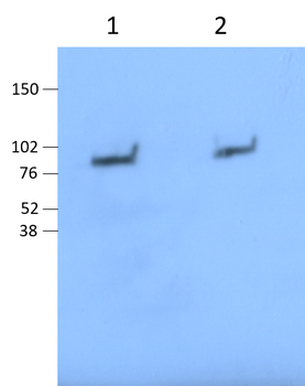
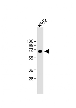
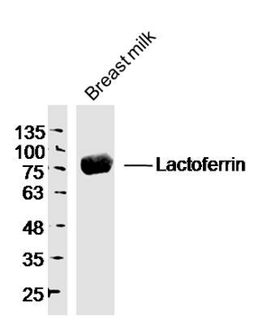
All lanes: Anti-LTF Antibody at 1:1000 dilution. Lane 1: K562 whole cell lysates. Lane 2: Hela whole cell lysates. Lysates/proteins at 20 µg per lane. Secondary Goat Anti-Mouse IgG, (H+L), Peroxidase conjugated at 1/10000 dilution. Predicted band size: 78 kDa. Blocking/Dilution buffer: 5% NFDM/TBST.

Immunofluorescent analysis of 4% paraformaldehyde-fixed, 0.1% Triton X-100 permeabilized Hela (Human Cervical epithelial adenocarcinoma cell line) cells labeling Pdx1 at 1/25 dilution, followed by Dylight 488-conjugated goat anti-mouse IgG secondary antibody at 1/200 dilution (green). Immunofluorescence image showing cytoplasm staining on Hela cell line. Cytoplasmic actin is detected with Dylight 554 Phalloidin at 1/100 dilution (red). The nuclear counter stain is DAPI (blue).
Quick Database Links
UniProt Details
− No UniProt data available
NCBI Reference Sequences
−Associated Accession Numbers
Curated reference sequences for the gene transcript and protein product| Protein | NP_001186078.1, NP_002334.2 |
|---|
Documents Download
Datasheet
Product Information
Request a Document
Protocol Information
WB
Western Blot (IB, immunoblot)
IHC-P
Immunohistochemistry Paraffin
IF
Immunofluorescence
LTF Antibody (orb1788113)

- 0.0
Based on 0 reviews
Participating in our Biorbyt product reviews program enables you to support fellow scientists by sharing your firsthand experience with our products.
Login to Submit a Review