You have no items in your shopping cart.
Description
Research Area
Immunology & Inflammation
Images & Validation
−Item 1 of 6
| Tested Applications | FC, WB |
|---|---|
| Dilution Range | WB - 1:4000, FC - 1:10-50 |
| Reactivity | Human, Mouse |
Key Properties
−| Host | Rabbit |
|---|---|
| Clonality | Polyclonal |
| Isotype | Rabbit IgG |
| Immunogen | This LTF antibody is generated from rabbits immunized with LTF recombinant protein. |
| Target | LTF (HGNC:6720) |
| Molecular Weight | 78182 Da |
| Conjugation | Unconjugated |
Storage & Handling
−| Storage | Maintain refrigerated at 2-8°C for up to 2 weeks. For long term storage store at -20°C in small aliquots to prevent freeze-thaw cycles |
|---|---|
| Form/Appearance | Purified polyclonal antibody supplied in PBS with 0.09% (W/V) sodium azide. This antibody is purified through a protein A column, followed by peptide affinity purification. |
| Expiration Date | 12 months from date of receipt. |
| Disclaimer | For research use only |
Alternative Names
−Lactotransferrin, Lactoferrin, 3421-, Growth-inhibiting protein 12, Talalactoferrin, Lactoferricin-H, Lfcin-H, Kaliocin-1, Lactoferroxin-A, Lactoferroxin-B, Lactoferroxin-C, LTF, GIG12, LF
Similar Products
−- Item 1 of 6
Lactoferrin/LTF Rabbit Polyclonal Antibody [orb413008]
ELISA, IF, IHC, WB
Human, Mouse, Rat
Rabbit
Polyclonal
Unconjugated
100 μg - Item 1 of 5
LTF Antibody [orb1410325]
FC, IF, IHC
Human
Mouse
Monoclonal
Unconjugated
20 μg, 100 μg, 100 μg (without BSA and Azide) - Item 1 of 5
- Item 1 of 3
Lactoferrin Rabbit Polyclonal Antibody [orb100096]
WB
Bovine, Canine, Equine, Mouse, Porcine, Rat, Sheep
Human
Rabbit
Polyclonal
Unconjugated
50 μl, 100 μl, 200 μl - Item 1 of 4
LTF Antibody [orb1410102]
FC, IF, IHC
Human
Mouse
Monoclonal
Unconjugated
20 μg, 100 μg, 100 μg (without BSA and Azide)


Quality Guarantee
Explore bioreagents carefree to elevate your research. All our products are rigorously tested for performance. If a product does not perform as described on its datasheet, our scientific support team will provide expert troubleshooting, a prompt replacement, or a refund. For full details, please see our Terms & Conditions and Buying Guide. Contact us at [email protected].
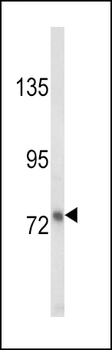
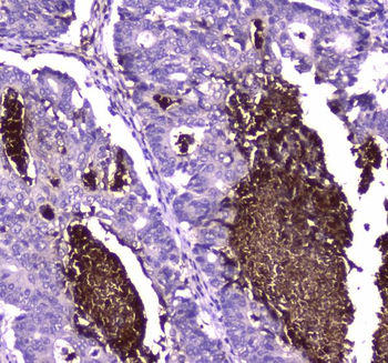
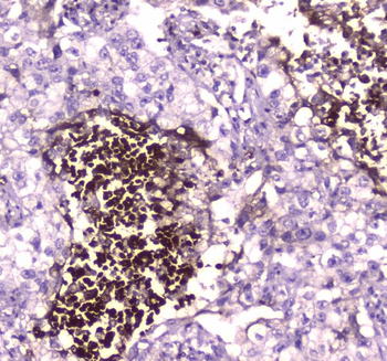
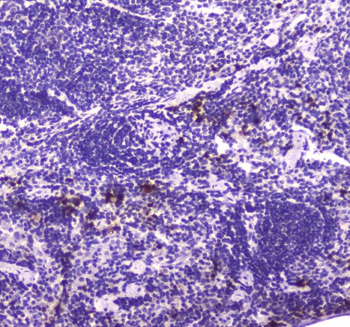
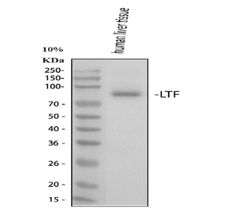
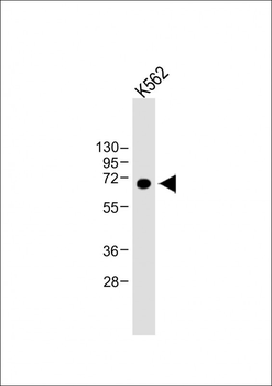
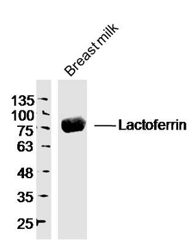
Western blot analysis of LTF Antibody in MDA-MB231 cell line lysates (35 ug/lane). LTF (arrow) was detected using the purified Pab.
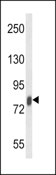
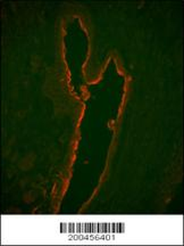
Western blot analysis of LTF Antibody in mouse spleen tissue lysates (35 ug/lane). LTF (arrow) was detected using the purified Pab.
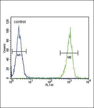
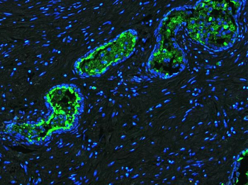
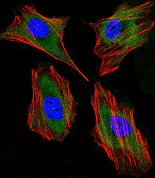
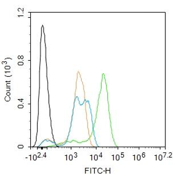
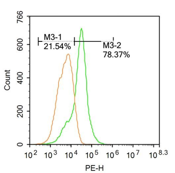
LTF Antibody flow cytometric analysis of MDA-MB231 cells (right histogram) compared to a negative control cell (left histogram). FITC-conjugated goat-anti-rabbit secondary antibodies were used for the analysis.
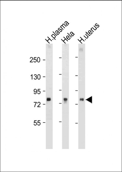
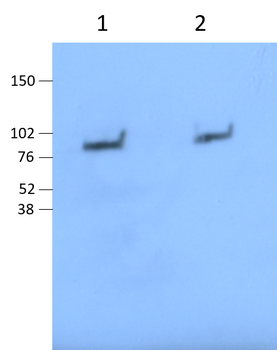
All lanes: Anti-LTF Antibody at 1:1000-2000 dilution. Lane 1: Human plasma tissue lysate. Lane 2: Hela whole cell lysate. Lane 3: Human uterus tissue lysate. Lysates/proteins at 20 µg per lane. Secondary Goat Anti-Rabbit IgG, (H+L), Peroxidase conjugated at 1/10000 dilution. Predicted band size: 78 kDa. Blocking/Dilution buffer: 5% NFDM/TBST.
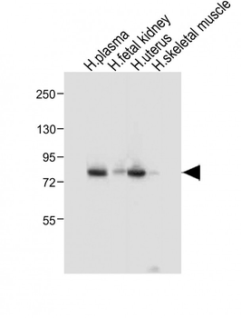
All lanes: Anti-LTF Antibody at 1:1000 dilution. Lane 1: Human plasma tissue lysate. Lane 2: Human fetal kidney tissue lysate. Lane 3: Human uterus tissue lysate. Lane 4: Human skeletal muscle tissue lysate. Lysates/proteins at 20 µg per lane. Secondary Goat Anti-Rabbit IgG, (H+L), Peroxidase conjugated at 1/10000 dilution. Predicted band size: 78 kDa. Blocking/Dilution buffer: 5% NFDM/TBST.
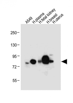
All lanes: Anti-LTF Antibody at 1:4000 dilution. Lane 1: A549 whole cell lysate. Lane 2: Human plasma tissue lysate. Lane 3: Human fetal kidney tissue lysate. Lane 4: Human breast tissue lysate. Lane 5: Human uterus tissue lysate. Lysates/proteins at 20 µg per lane. Secondary Goat Anti-Rabbit IgG, (H+L), Peroxidase conjugated at 1/10000 dilution. Predicted band size: 78 kDa. Blocking/Dilution buffer: 5% NFDM/TBST.
Quick Database Links
UniProt Details
− No UniProt data available
NCBI Reference Sequences
−Associated Accession Numbers
Curated reference sequences for the gene transcript and protein product| Protein | NP_001186078.1, NP_002334.2 |
|---|
Documents Download
Datasheet
Product Information
Request a Document
Protocol Information
WB
Western Blot (IB, immunoblot)
FC
Flow Cytometry
LTF Antibody (orb1928337)

- 0.0
Based on 0 reviews
Participating in our Biorbyt product reviews program enables you to support fellow scientists by sharing your firsthand experience with our products.
Login to Submit a ReviewAvailable Sizes
Select a size below
Free Secondary Antibody (20 ul)0/0
Please add an antibody product to your cart first.