You have no items in your shopping cart.
Description
Research Area
Cell Biology
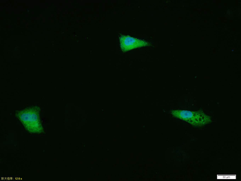
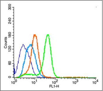
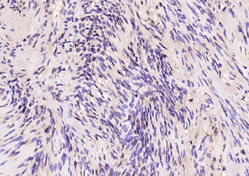
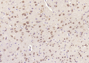
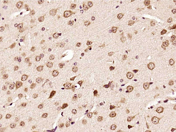
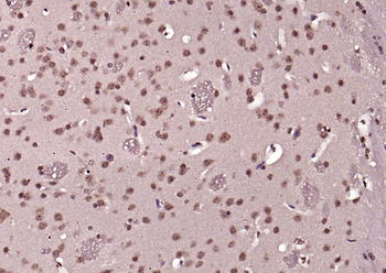
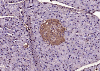
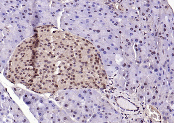
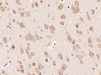
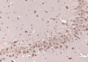
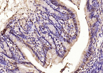
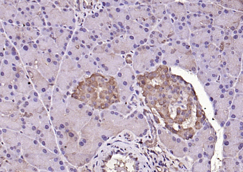
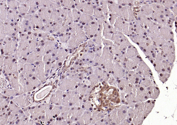
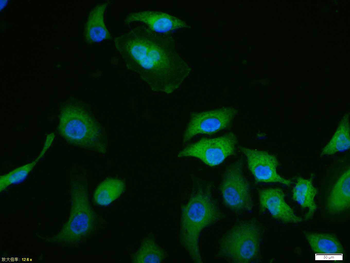
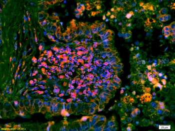
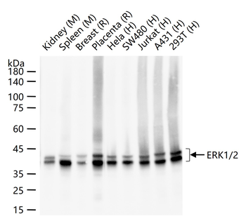
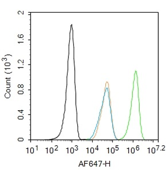
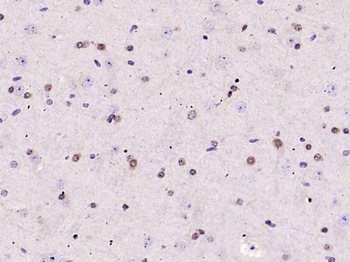
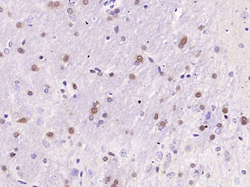
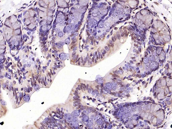
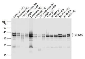
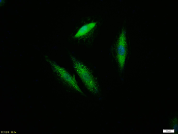
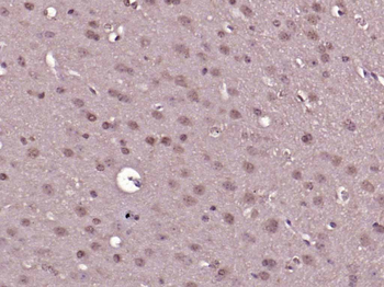
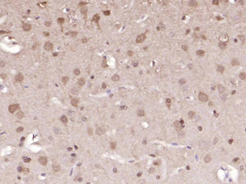
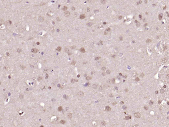
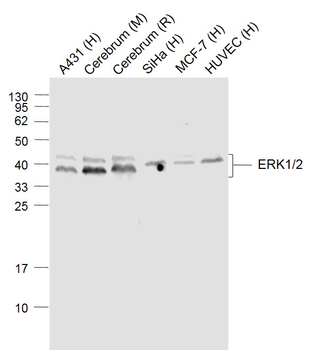
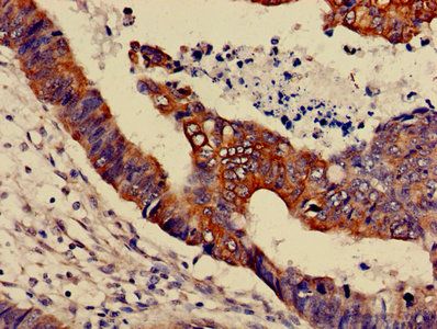
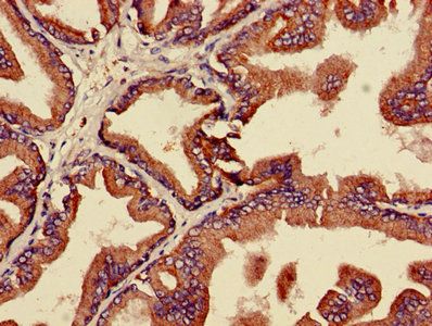
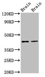
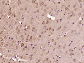
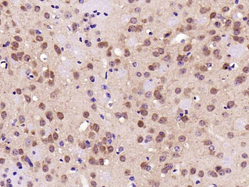
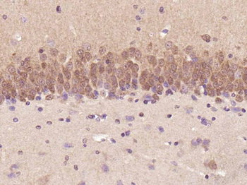
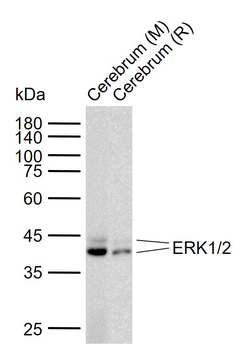
Images & Validation
−
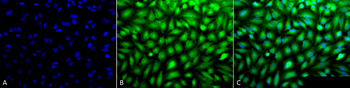
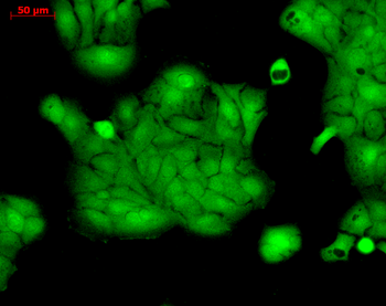
Item 1 of 6
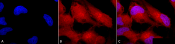
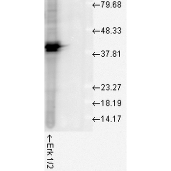
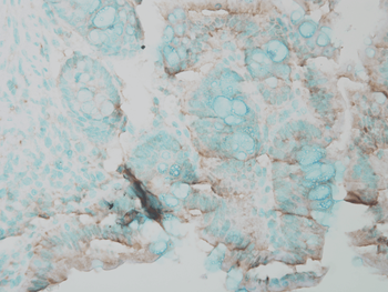
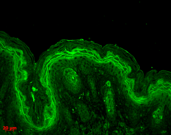
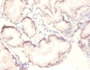
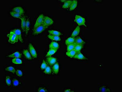
| Tested Applications | FC, ICC, IF, IHC, WB |
|---|---|
| Dilution Range | WB (1:1000), IHC (1:100), ICC/IF (1:100), FCM (1:100); optimal dilutions for assays should be determined by the user. |
| Reactivity | Bovine, Drosophila, Frog, Gallus, Human, Mouse, Rat, Sheep |
| Application Notes |
Key Properties
−| Host | Rabbit |
|---|---|
| Clonality | Polyclonal |
| Immunogen | A 35 residue synthetic peptide, corresponding to Rat Erk1 MAP kinase with the CGG spacer group added and the peptide coupled to KLH. |
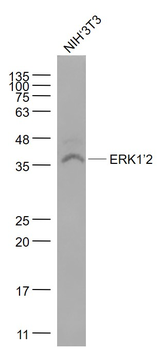
| Target | Erk1/2 |
| Molecular Weight | 42kDa (ERK2). |
| Purification | Peptide Affinity Purified |
| Conjugation | Unconjugated |
Storage & Handling
−| Storage | Maintain refrigerated at 2-8°C for up to 2 weeks. For long term storage store at -20°C in small aliquots to prevent freeze-thaw cycles. |
|---|---|
| Buffer/Preservatives | PBS pH 7.4, 50% glycerol, 0.09% sodium azide. Storage buffer changes when conjugated. |
| Concentration | 1 mg/ml |
| Expiration Date | 12 months from date of receipt. |
| Disclaimer | For research use only |
Alternative Names
−ERK1, ERK2, ERT1, ERT2, MAP kinase 1, MAP kinase 2, MAPK1, MAPK2, MAPK3, p38, p40, p41, p41mapk, p42 MAPK, p44 ERK1, p44 MAPK, PRKM1, PRKM2, PRKM3
Similar Products
−- Item 1 of 16
Phospho-ERK1/2 (Thr202 + Tyr204) Rabbit Polyclonal Antibody [orb5178]
FC, ICC
Human
Human, Mouse
Rabbit
Polyclonal
Unconjugated
50 μl, 100 μl, 200 μl - Item 1 of 7
ERK1/2 Recombinant Rabbit Monoclonal Antibody [orb704524]
FC, ICC, IF, IHC-Fr, IHC-P, WB
Zebrafish
Human, Mouse, Rat
Rabbit
Recombinant
Unconjugated
50 μl, 100 μl, 25 μl - Item 1 of 5
ERK1/2 Mouse Monoclonal Antibody [orb500888]
FC, KO/KD Validated, WB
Mouse, Rat
Human
Mouse
Monoclonal
Unconjugated
50 μl, 100 μl, 200 μl, 200 μg - Item 1 of 6
MAPK3 Antibody [orb238890]
ELISA, IF, IHC, WB
Human, Mouse, Rat
Rabbit
Polyclonal
Unconjugated
100 μg, 50 μg - Item 1 of 4
ERK1/2 Mouse Monoclonal Antibody [orb500875]
FC, IF, IHC-Fr, IHC-P, WB
Human
Human, Mouse, Rat
Mouse
Monoclonal
Unconjugated
50 μl, 100 μl, 200 μl, 200 μg

Quality Guarantee
Explore bioreagents carefree to elevate your research. All our products are rigorously tested for performance. If a product does not perform as described on its datasheet, our scientific support team will provide expert troubleshooting, a prompt replacement, or a refund. For full details, please see our Terms & Conditions and Buying Guide. Contact us at [email protected].
Quick Database Links
UniProt Details
− No UniProt data available
NCBI Gene Details
− No NCBI Gene data available
NCBI Reference Sequences
−Associated Accession Numbers
Curated reference sequences for the gene transcript and protein product| Protein | NP_059043.1 |
|---|
Protocol Information
WB
Western Blot (IB, immunoblot)
IHC
Immunohistochemistry
FC
Flow Cytometry
IF
Immunofluorescence
ICC
Immunocytochemistry
Filter by Applications
Filter by Species
Su-Ji Min, Hye-Won Hyun, Tae-Cheon Kang Leptomycin B attenuates neuronal death via PKA- and PP2B-mediated ERK1/2 activation in the rat hippocampus following status epilepticus Brain Research, 1670, 14 (2017)
Applications
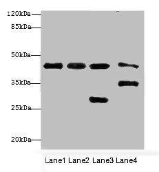
WB
Reactivity
Rat
Available Sizes
Select a size below
Choose Conjugation or Carrier Free Version
Free Secondary Antibody (20 ul)0/0
Please add an antibody product to your cart first.