You have no items in your shopping cart.
Featured
Description
Research Area
Cell Biology
Images & Validation
−Item 1 of 7
| Tested Applications | ELISA, FC, ICC, WB |
|---|---|
| Dilution Range | WB=1:500-2000, ICC/IF=1:100-500, Flow-Cyt=1μg/Test |
| Reactivity | Human |
| Predicted Reactivity | Canine, Mouse, Porcine |
Related Conjugates & Formulations
−Key Properties
−| Antibody Type | Primary Antibody |
|---|---|
| Host | Rabbit |
| Clonality | Polyclonal |
| Isotype | IgG |
| Immunogen | KLH conjugated synthetic peptide derived from human EGFR (951-1050/1210aa) |
| Target | EGFR |
| Molecular Weight | 170 kDa |
| Purification | Affinity purified by Protein A |
| Conjugation | Unconjugated |
Storage & Handling
−| Storage | Maintain refrigerated at 2-8°C for up to 2 weeks. For long term storage store at -20°C in small aliquots to prevent freeze-thaw cycles. |
|---|---|
| Form/Appearance | Liquid |
| Buffer/Preservatives | 0.01M TBS (pH7.4) with 1% rAlbumin, 0.02% Proclin300 and 50% Glycerol. |
| Concentration | 1mg/ml |
| Expiration Date | 12 months from date of receipt. |
| Disclaimer | For research use only |
Alternative Names
−ERBB; ERBB1; ERRP; HER1; NISBD2; NNCIS; PIG61; mENA; 9030024J15Rik; Errb1; Wa5; wa-2; wa2; ErbB-1; EGFR_HUMAN; EGFR; Proto-oncogene c-ErbB-1; Receptor tyrosine-protein kinase erbB-1; 2.7.10.1; EGFR_MOUSE; epidermal growth factor receptor; epidermal growth factor receptor (avian erythroblastic leukemia viral (v-erb-b) oncogene homolog); erythroblastic leukemia viral (v-erb-b) oncogene homolog (avian); erb-b2 receptor tyrosine kinase 1
Similar Products
−- Item 1 of 18
EGFR isoform a variant Rabbit Polyclonal Antibody [orb308736]
ELISA, ICC, IF, IHC-P, WB
Human, Mouse, Porcine, Rat
Rabbit
Polyclonal
Unconjugated
100 μg - Item 1 of 9
EGFRvIII Rabbit Polyclonal Antibody [orb191506]
IHC-P, WB
Human, Mouse
Rabbit
Polyclonal
Unconjugated
200 μg, 100 μg - Item 1 of 8
EGFR Rabbit Polyclonal Antibody [orb654422]
ELISA, FC, ICC, IF, IHC, WB
Human, Mouse, Rat
Rabbit
Polyclonal
Unconjugated
100 μg - Item 1 of 7
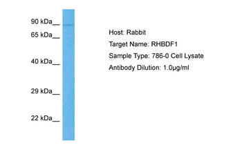
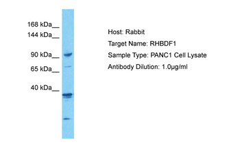
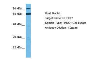
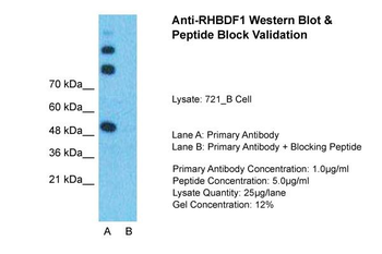
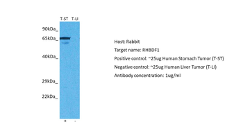
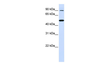
RHBDF1 Rabbit Polyclonal Antibody [orb580834]
WB
Bovine, Canine, Equine, Guinea pig, Mouse, Rabbit, Rat, Zebrafish
Human
Rabbit
Polyclonal
Unconjugated
100 μl - Item 1 of 5
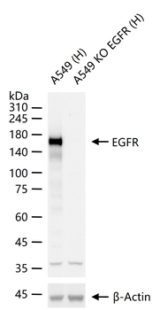
EGFR Rabbit Polyclonal Antibody [orb704502]
IF, IHC-Fr, IHC-P, KO/KD Validated, WB
Mouse, Rat
Human, Mouse, Rat
Rabbit
Polyclonal
Unconjugated
50 μl, 100 μl
Quality Guarantee
Explore bioreagents carefree to elevate your research. All our products are rigorously tested for performance. If a product does not perform as described on its datasheet, our scientific support team will provide expert troubleshooting, a prompt replacement, or a refund. For full details, please see our Terms & Conditions and Buying Guide. Contact us at [email protected].
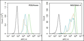
Black line: Positive blank control (HUVEC), Negative blank control (Molt-4), Green line: Primary Antibody (Rabbit Anti-EGFR antibody (orb10580)), Orange line: Isotype Control Antibody (Rabbit IgG). Blue line: Secondary Antibody (Goat anti-rabbit IgG-AF647), HUVEC (Positive) and Molt-4 (Negative control) cells (black) were fixed with 4% PFA for 10 min at room temperature, permeabilized with 90% ice-cold methanol for 20 min at -20°C, and incubated in 5% BSA blocking buffer for 30 min at room temperature. Cells were then stained with EGFR Antibody (orb10580) at 1:50 dilution in blocking buffer and incubated for 30 min at room temperature, washed twice with 2% BSA in PBS, followed by secondary antibody (blue) incubation for 40 min at room temperature. Acquisitions of 20000 events were performed. Cells stained with primary antibody (green), and isotype control (orange).
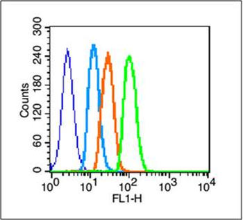
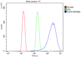
Blank control (blue line): A431 (blue). Primary Antibody (green line): Rabbit Anti-EGFRantibody (orb10580), Dilution: 3 µg/10^6 cells, Isotype Control Antibody (orange line): Rabbit IgG. Secondary Antibody (white blue line): Goat anti-rabbit IgG-FITC, Dilution: 1 µg/Test. Protocol, The cells were fixed with 2% paraformaldehyde (10 min), then permeabilized with 90% ice-cold methanol for 30 min on ice. Cells stained with Primary Antibody for 30 min at room temperature. The cells were then incubated in 1 X PBS/2% BSA/10% goat serum to block non-specific protein-protein interactions followed by the antibody for 15 min at room temperature. The secondary antibody used for 40 min at room temperature. Acquisition of 20000 events was performed.
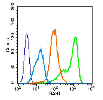
Blank control: HUVEC cells (blue). Primary Antibody: Rabbit Anti-EGFR antibody (orb10580), Dilution: 1 µg in 100 µl 1X PBS containing 0.5% BSA, Isotype Control Antibody: Rabbit IgG (orange), used under the same conditions, Secondary Antibody: Goat anti-rabbit IgG-PE (white blue), Dilution: 1:200 in 1 X PBS containing 0.5% BSA. Protocol, The cells were fixed with 2% paraformaldehyde (10 min), then permeabilized with 90% ice-cold methanol for 30 min on ice. Primary antibody (orb10580, 1 µg/1x10^6 cells) were incubated for 30 min on the ice, followed by 1 X PBS containing 0.5% BSA + 10% goat serum (15 min) to block non-specific protein-protein interactions. Then the Goat Anti-rabbit IgG/PE antibody was added into the blocking buffer mentioned above to react with the primary antibody at 1/200 dilution for 30 min on ice. Acquisition of 20000 events was performed.
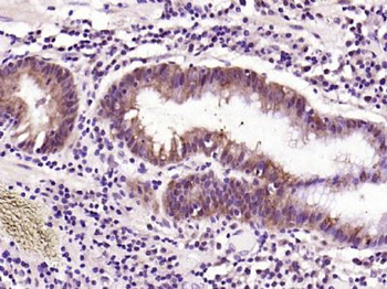
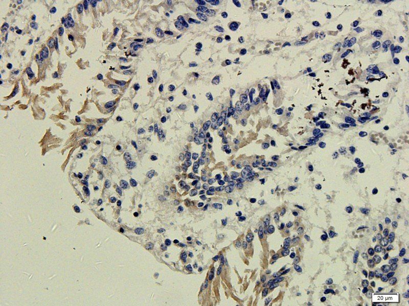
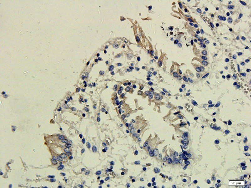
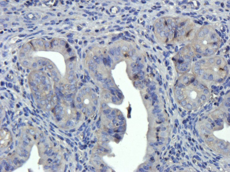
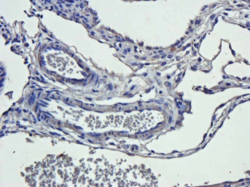
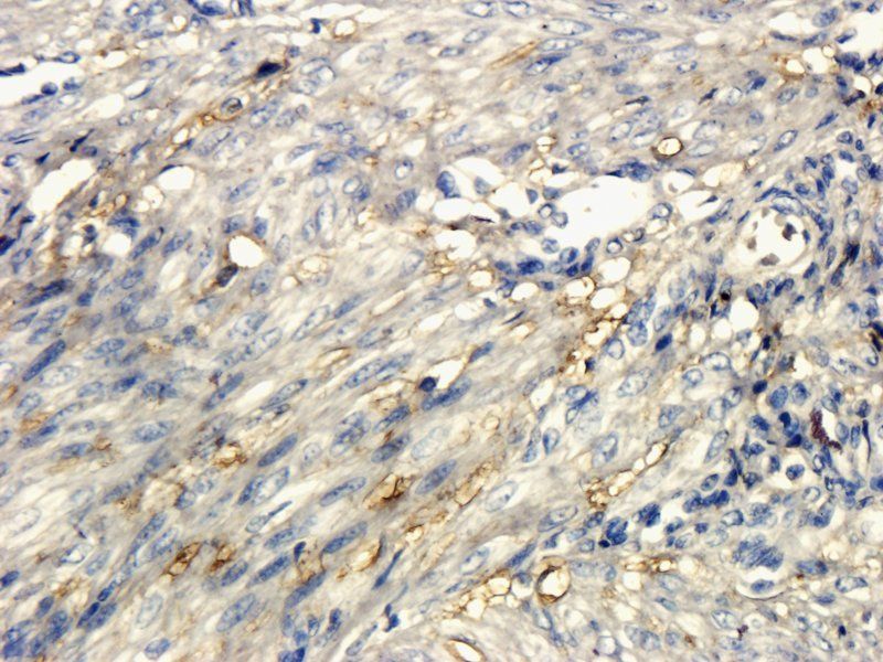
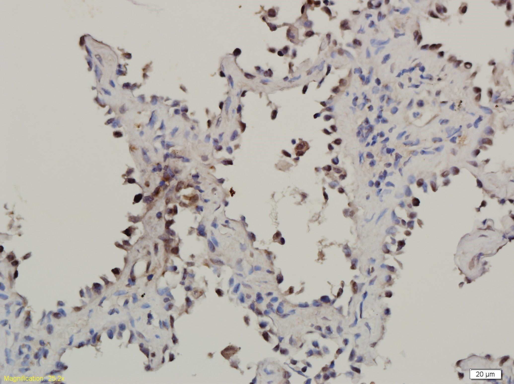
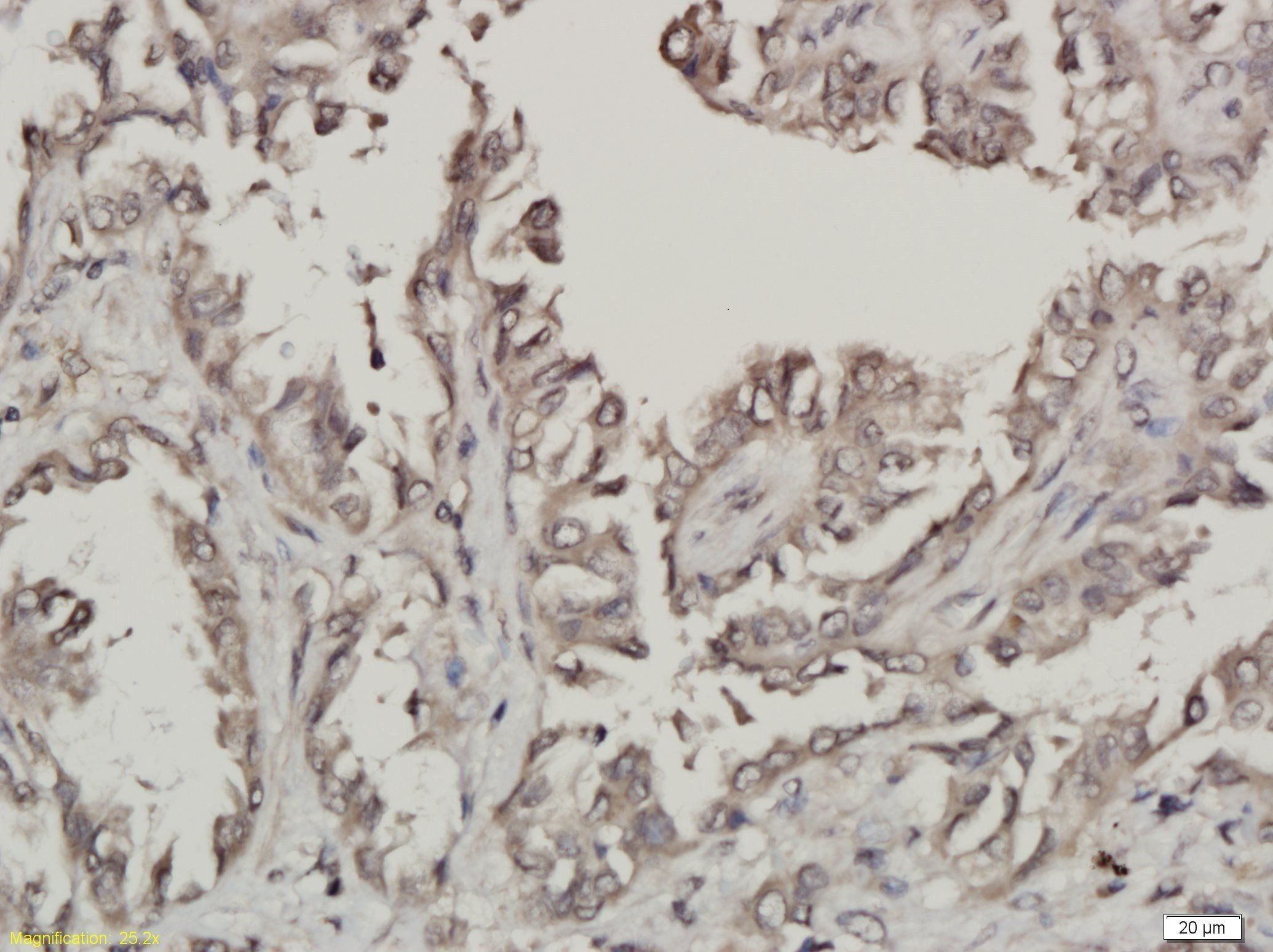
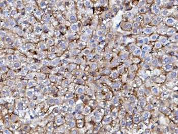
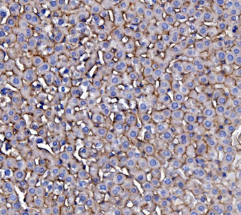
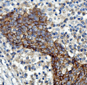
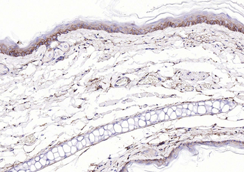
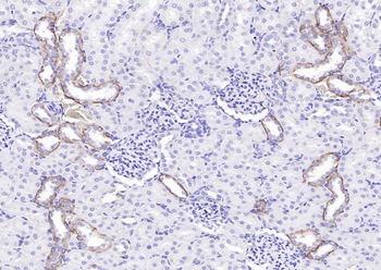
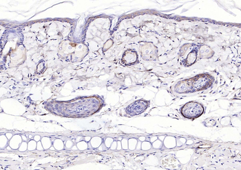
Paraformaldehyde-fixed, paraffin embedded (human gastric carcinoma), Antigen retrieval by boiling in sodium citrate buffer (pH6.0) for 15 min, Block endogenous peroxidase by 3% hydrogen peroxide for 20 minutes, Blocking buffer (normal goat serum) at 37°C for 30 min, Antibody incubation with (EGFR) Polyclonal Antibody, Unconjugated (orb10580) at 1:200 overnight at 4°C, followed by operating according to SP Kit (Rabbit) instructionsand DAB staining.
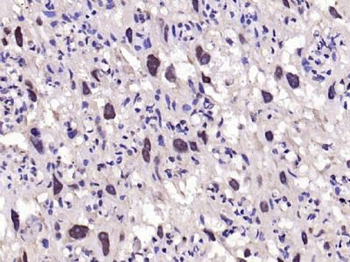
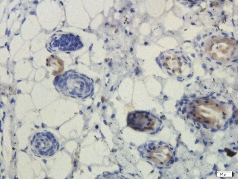
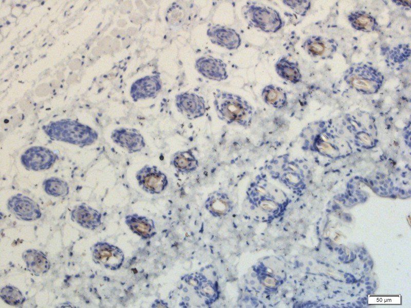
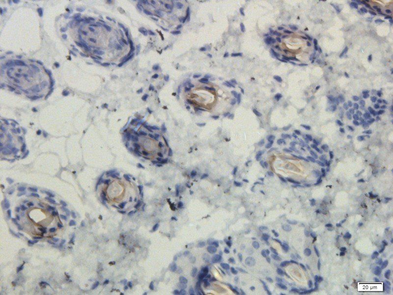
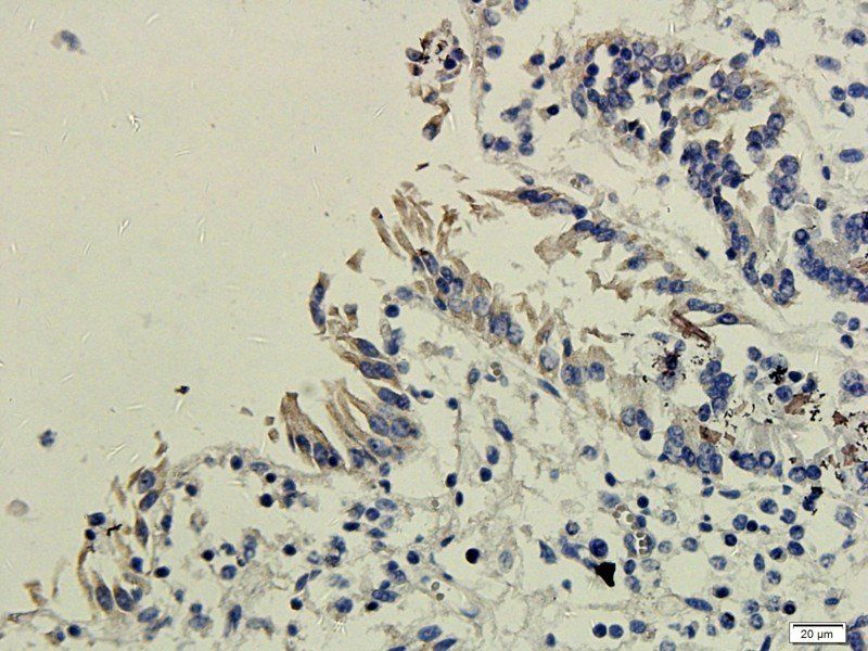
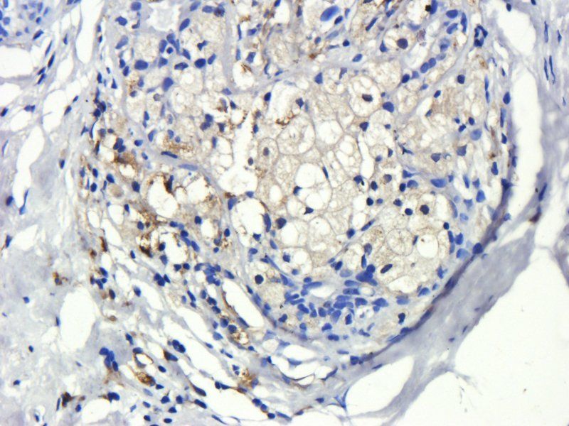
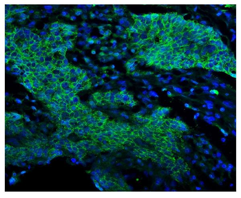
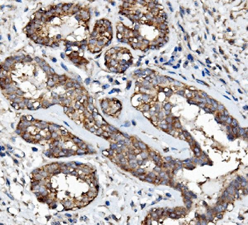
Paraformaldehyde-fixed, paraffin embedded (rat placenta), Antigen retrieval by boiling in sodium citrate buffer (pH6.0) for 15 min, Block endogenous peroxidase by 3% hydrogen peroxide for 20 minutes, Blocking buffer (normal goat serum) at 37°C for 30 min, Antibody incubation with (EGFR) Polyclonal Antibody, Unconjugated (orb10580) at 1:200 overnight at 4°C, followed by operating according to SP Kit (Rabbit) instructionsand DAB staining.
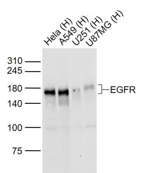
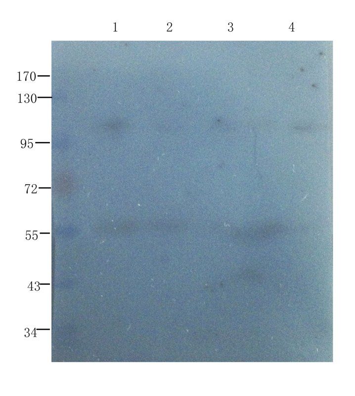
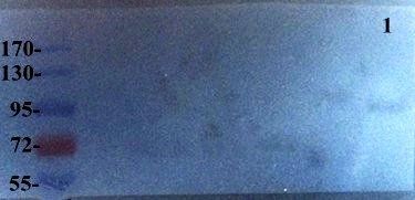
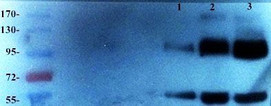
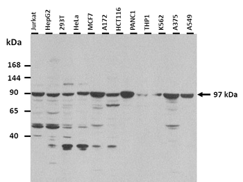
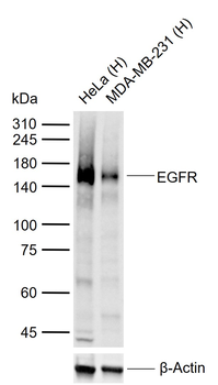
Sample: Lane 1: Hela (Human) Cell Lysate at 30 ug, Lane 2: A549 (Human) Cell Lysate at 30 ug, Lane 3: U251 (Human) Cell Lysate at 30 ug, Lane 4: U87MG (Human) Cell Lysate at 30 ug, Primary: Anti-EGFR at 1/1000 dilution, Secondary: IRDye800CW Goat Anti-Rabbit IgG at 1/20000 dilution, Predicted band size: 170 kD, Observed band size: 170 kD.
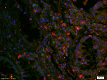
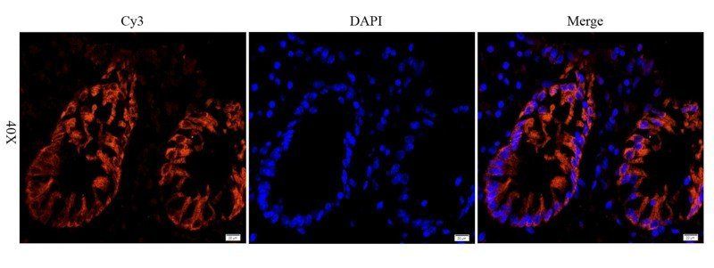
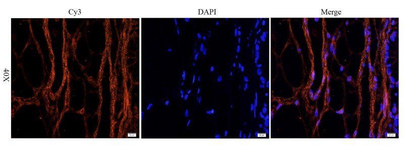
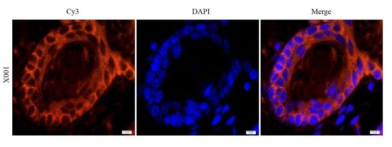
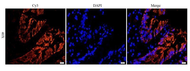
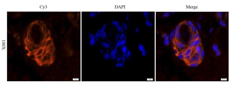
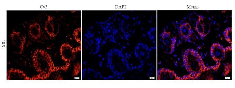
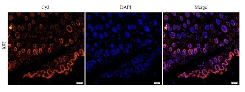
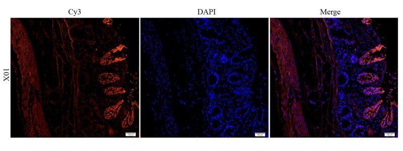
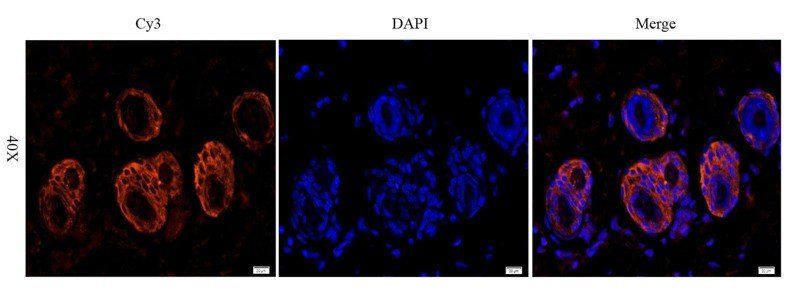
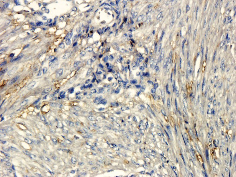
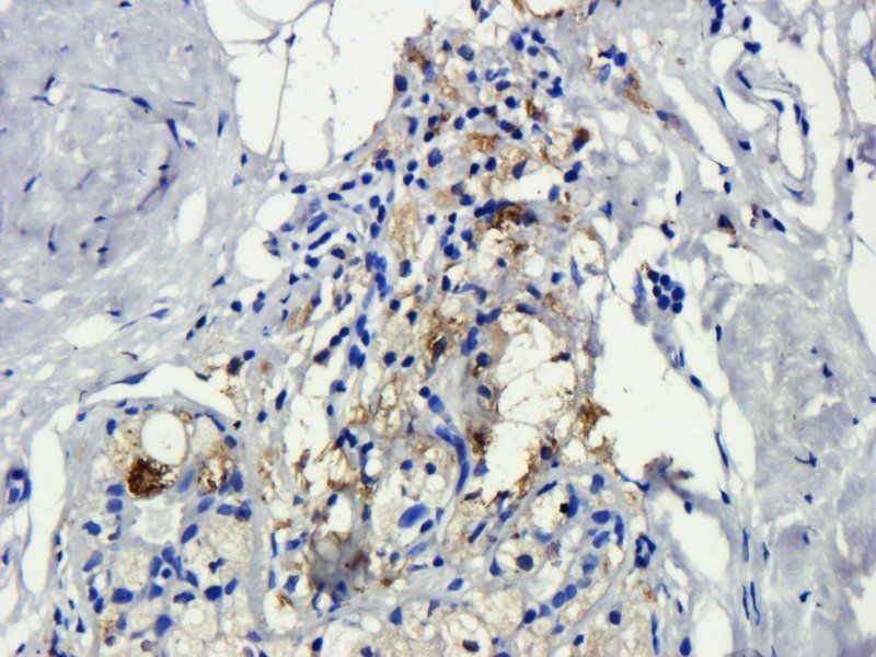
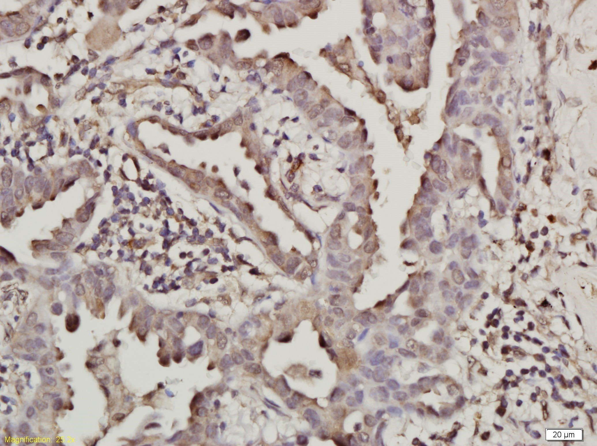
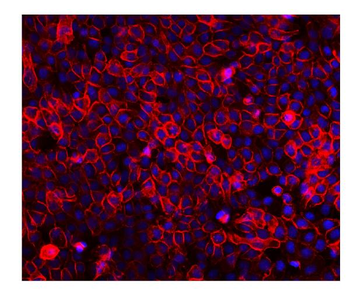
Tissue/Cell: human rectal carcinoma, 4% Paraformaldehyde-fixed and paraffin-embedded, Antigen retrieval: citrate buffer (0.01M, pH6.0), Boiling bathing for 15 min, Blocking buffer (normal goat serum) at 37°C for 20 min, Incubation: Anti-EGFR Polyclonal Antibody, Unconjugated (orb10580) 1:200, overnight at 4°C, The secondary antibody was Goat Anti-Rabbit IgG, Cy3 conjugated (orb868589) used at 1:200 dilution for 40 minutes at 37°C. DAPI (5 ug/ml, blue) was used to stain the cell nuclei.
Quick Database Links
Gene Symbol
EGFR
UniProt
UniProt Details
− No UniProt data available
Documents Download
Datasheet
Product Information
Request a Document
Protocol Information
WB
Western Blot (IB, immunoblot)
FC
Flow Cytometry
ICC
Immunocytochemistry
ELISA
Enzyme-linked Immunosorbent Assay (EIA)
EGFR Rabbit Polyclonal Antibody (orb10580)

- 0.0
Based on 0 reviews
Participating in our Biorbyt product reviews program enables you to support fellow scientists by sharing your firsthand experience with our products.
Login to Submit a ReviewAvailable Sizes
Select a size below
Free Secondary Antibody (20 ul)0/0
Please add an antibody product to your cart first.