
You have no items in your shopping cart.
Description
Research Area
Cell Biology
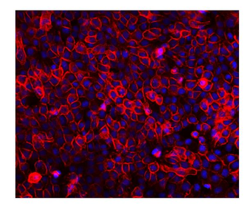
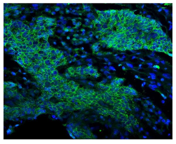
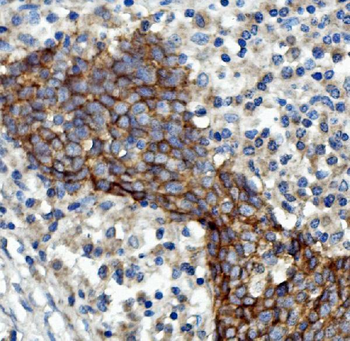
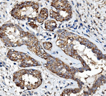
Images & Validation
−
Item 1 of 3
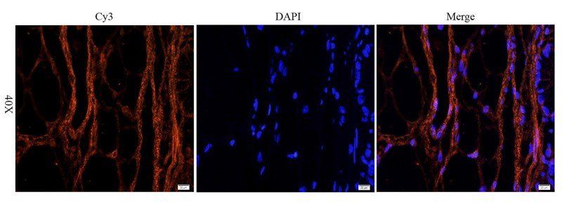
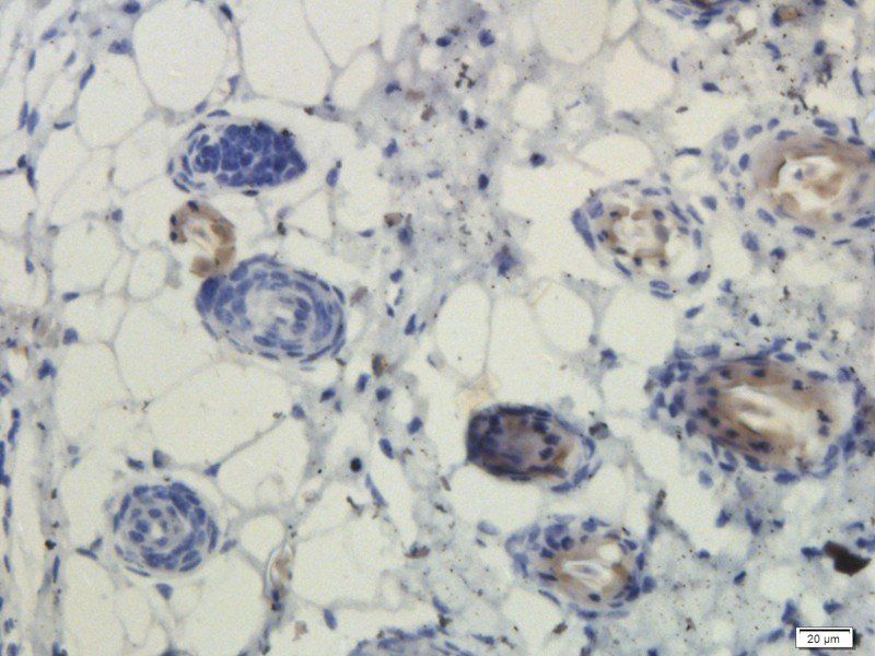
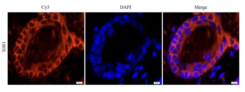
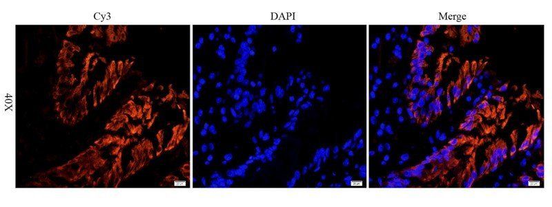
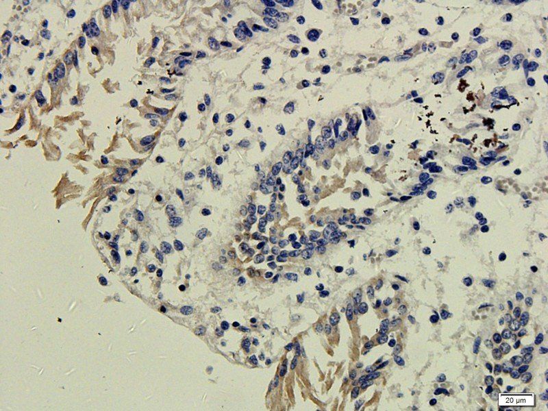
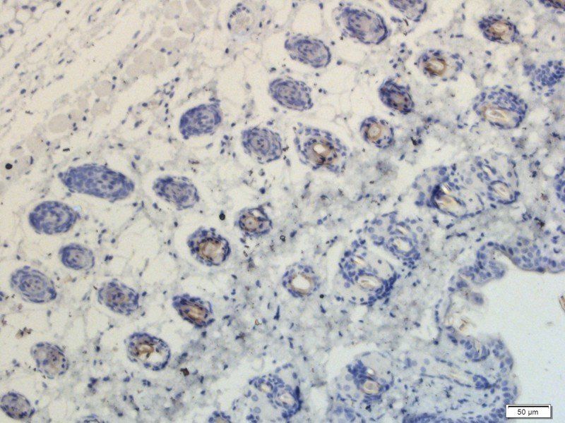
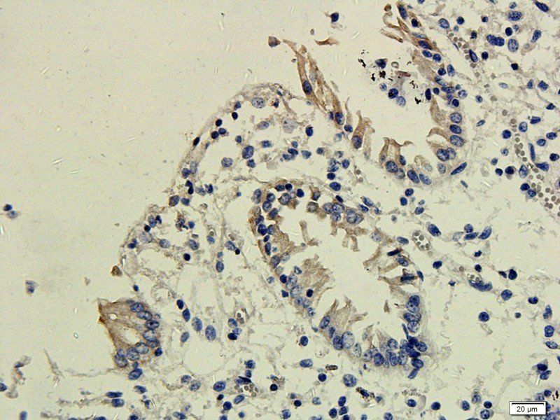
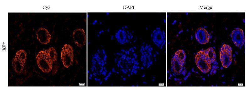
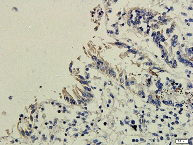
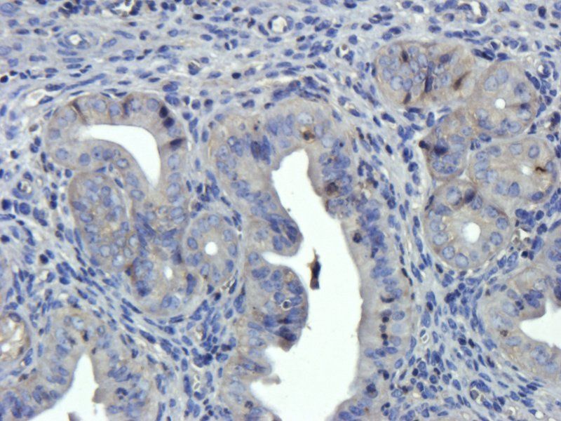
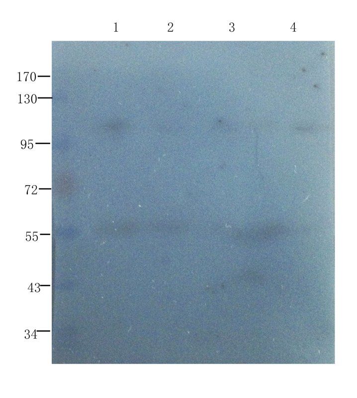
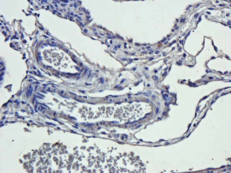
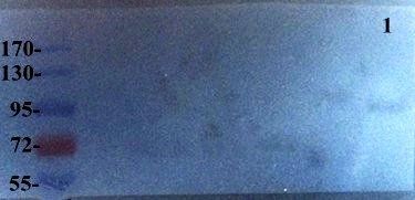
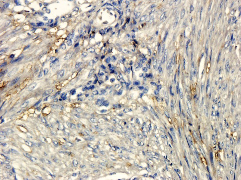
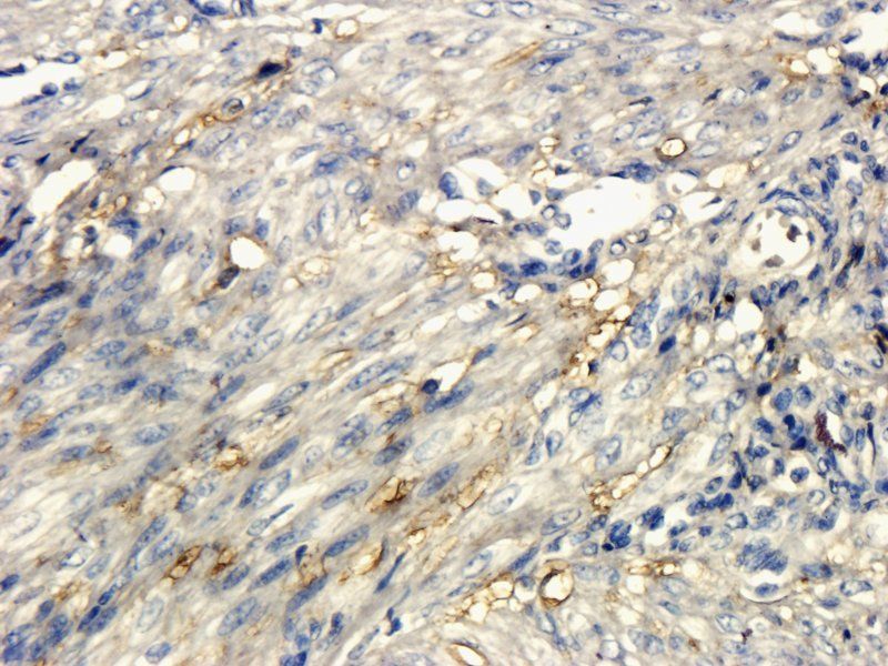
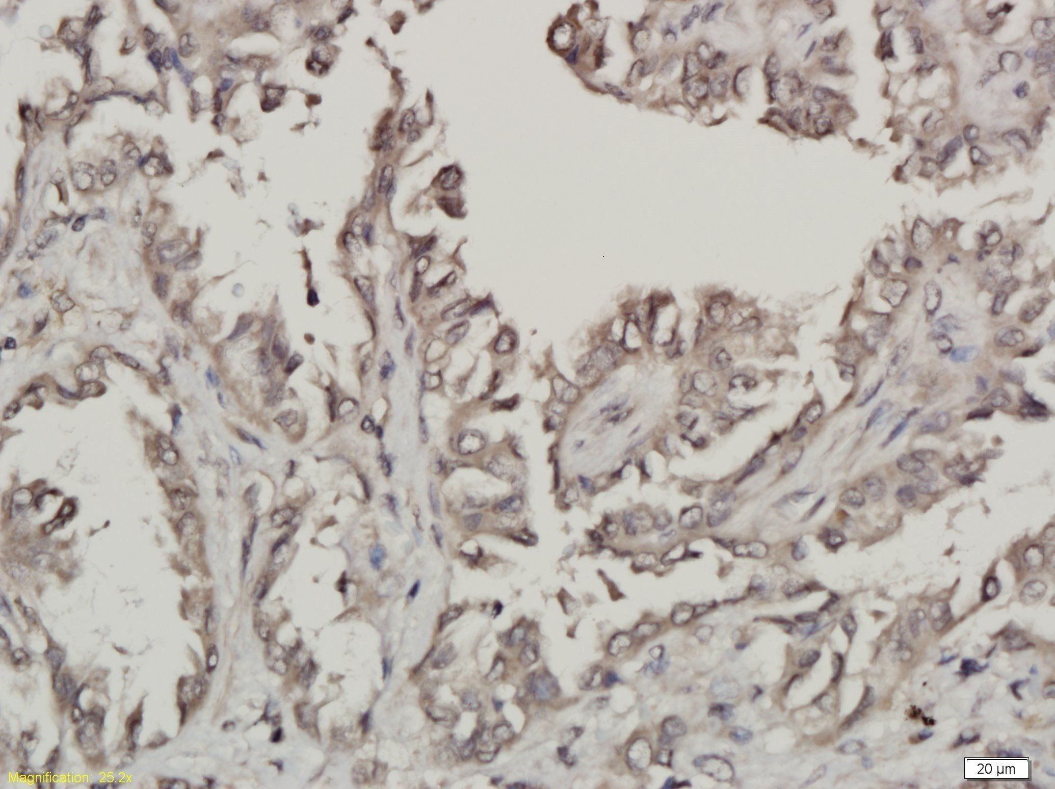
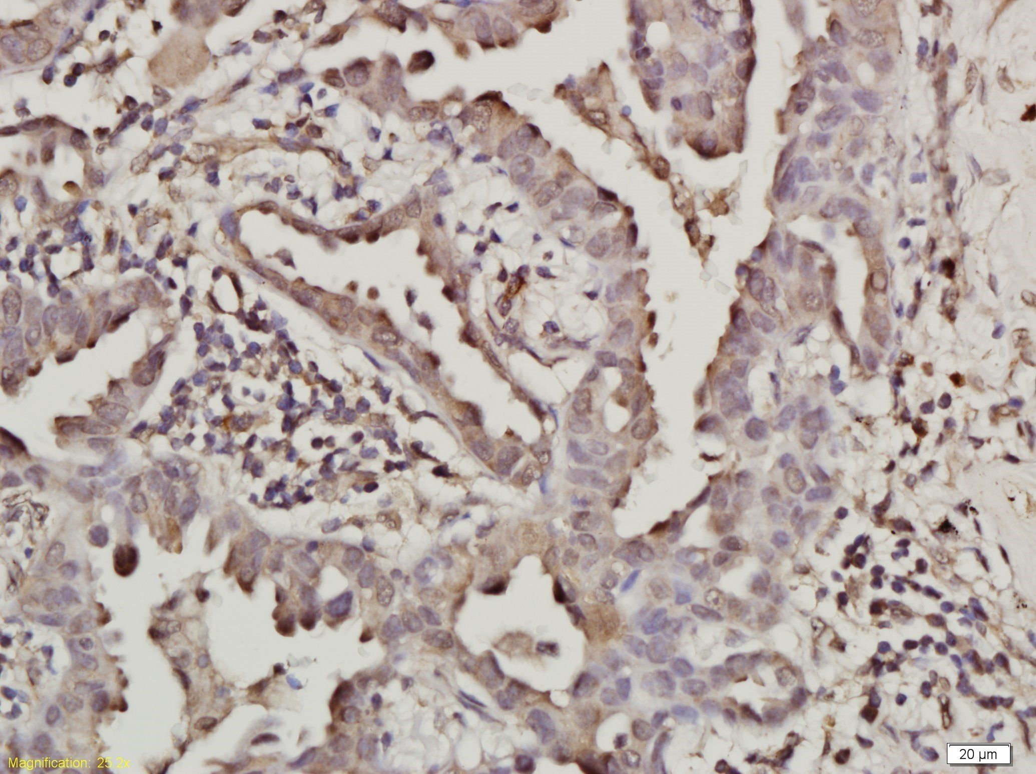
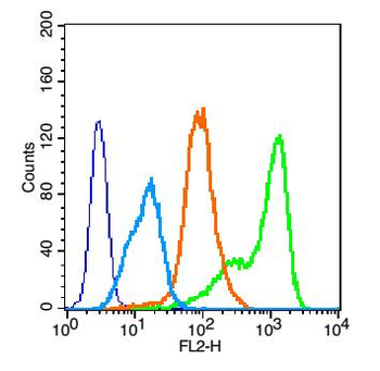
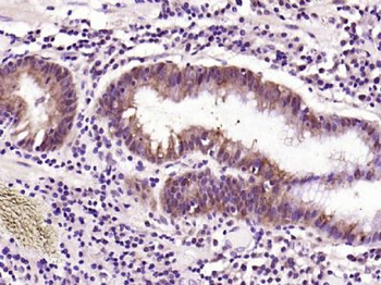
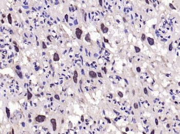
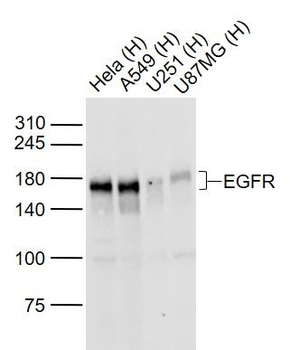
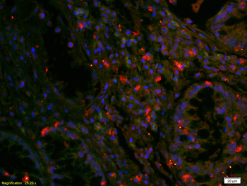
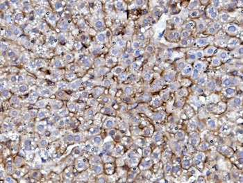
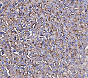
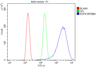
| Tested Applications | ELISA, IHC, IP, WB |
|---|---|
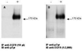
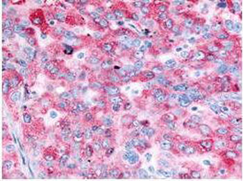
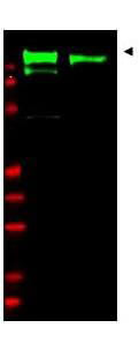
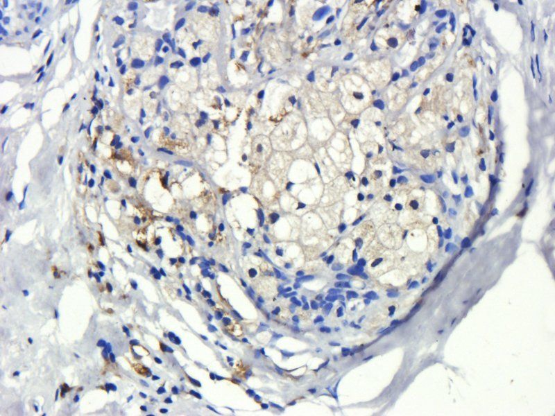
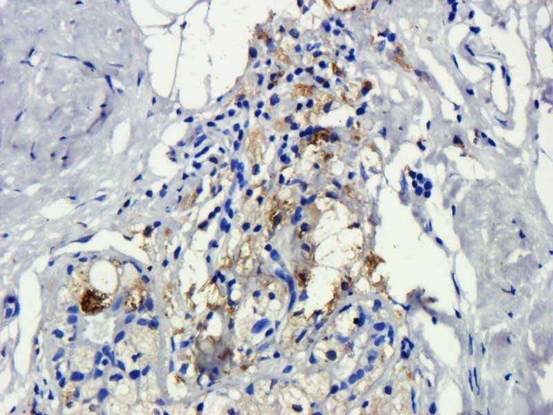
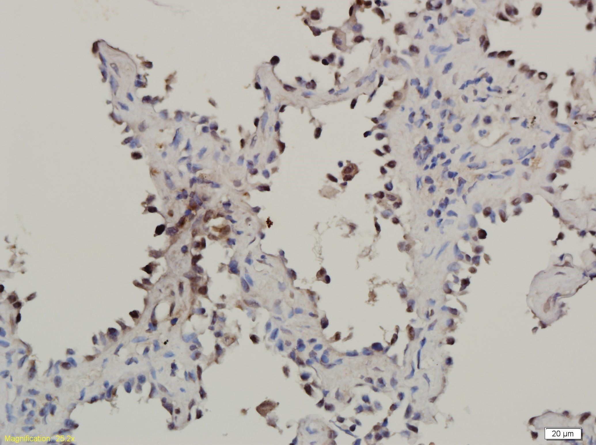
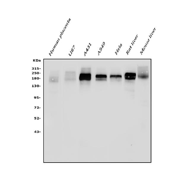
| Dilution Range | ELISA: 1:10,000 - 1:50,000, IHC: 2.5 µg/mL, IP: 10 µl, WB: 1:1,000 - 1:10,000 |
| Reactivity | Human, Rat |
| Application Notes |
Key Properties
−| Antibody Type | Primary Antibody |
|---|---|
| Host | Rabbit |
| Clonality | Polyclonal |
| Isotype | Antiserum |
| Immunogen | This whole rabbit serum was prepared by repeated immunizations with a peptide synthesized using conventional technology. The sequence of the epitope maps to a region near the carboxy terminus which is identical in human, mouse and rat EGFR. |
| Target | EGFR |
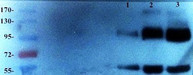
| Purity | This antiserum is directed against human epidermal growth factor receptor (EGFR) and is useful in determining its presence in western blotting and immunoprecipitation experiments. This antibody can detect EGFR from human, mouse and rat sources. Reactivity of this antibody with EGFR from other species is unknown. No reaction is observed against ErbB-2, ErbB-3 or ErbB-4. |
| Conjugation | Unconjugated |
Storage & Handling
−| Storage | Store vial at -20° C prior to opening. Aliquot contents and freeze at -20° C or below for extended storage. Avoid cycles of freezing and thawing. Centrifuge product if not completely clear after standing at room temperature. This product is stable for several weeks at 4° C as an undiluted liquid. Dilute only prior to immediate use. |
|---|---|
| Form/Appearance | Liquid (sterile filtered) |
| Buffer/Preservatives | 0.01% (w/v) Sodium Azide |
| Concentration | 85 mg/mL |
| Expiration Date | 12 months from date of receipt. |
| Dry Ice Shipping | Please note: This product requires shipment on dry ice. A dry ice surcharge will apply. |
| Disclaimer | For research use only |
Alternative Names
−rabbit anti-EGFR Antibody, rabbit anti-epidermal growth factor receptor antibody, Receptor tyrosine-protein kinase erbB-1 antibody, c-erbB-1 antibody
Similar Products
−- Item 1 of 18
EGFR isoform a variant Rabbit Polyclonal Antibody [orb308736]
ELISA, ICC, IF, IHC-P, WB
Human, Mouse, Porcine, Rat
Rabbit
Polyclonal
Unconjugated
100 μg - Item 1 of 8
Amivantamab Biosimilar Antibody, Research Grade (EGFR/c-Met) [orb2977888]
ELISA, FA, FACS, In vivo
Human
Human
Monoclonal
Unconjugated
100 μg, 1 mg - Item 1 of 9
EGFRvIII Rabbit Polyclonal Antibody [orb191506]
IHC-P, WB
Human, Mouse
Rabbit
Polyclonal
Unconjugated
200 μg, 100 μg - Item 1 of 7
EGFR Rabbit Polyclonal Antibody [orb10580]
ELISA, FC, ICC, WB
Canine, Mouse, Porcine
Human
Rabbit
Polyclonal
Unconjugated
50 μl, 100 μl, 200 μl - Item 1 of 8
EGFR Rabbit Polyclonal Antibody [orb654422]
ELISA, FC, ICC, IF, IHC, WB
Human, Mouse, Rat
Rabbit
Polyclonal
Unconjugated
100 μg
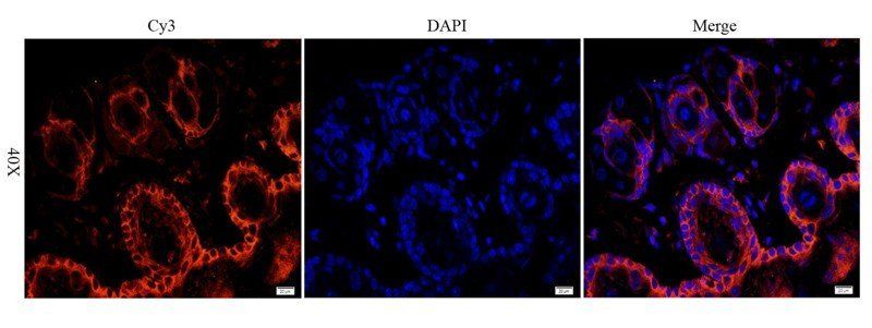
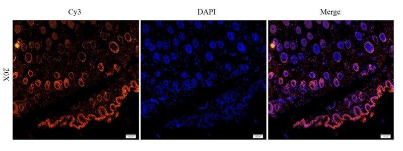
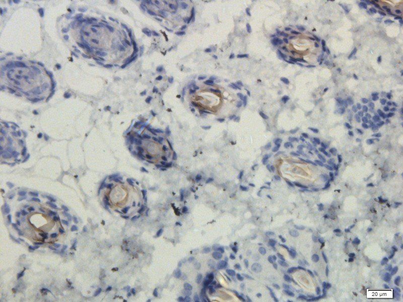
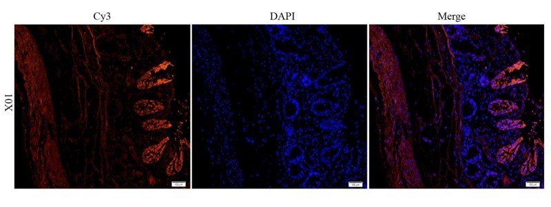



Quality Guarantee
Explore bioreagents carefree to elevate your research. All our products are rigorously tested for performance. If a product does not perform as described on its datasheet, our scientific support team will provide expert troubleshooting, a prompt replacement, or a refund. For full details, please see our Terms & Conditions and Buying Guide. Contact us at [email protected].
Protocol Information
WB
Western Blot (IB, immunoblot)
IHC
Immunohistochemistry
ELISA
Enzyme-linked Immunosorbent Assay (EIA)
IP
Immunoprecipitation
Available Sizes
Select a size below
Free Secondary Antibody (20 ul)0/0
Please add an antibody product to your cart first.
 Usually dispatched within 5-10 working days