You have no items in your shopping cart.
Description
Research Area
Cell Biology
Images & Validation
−Item 1 of 4
| Tested Applications | FACS, IF |
|---|---|
| Dilution Range | Flow cytometry: 1-2ug/10^6 cells,Immunofluorescence: 1-2ug/ml |
| Reactivity | Human |
| Application Notes |
Key Properties
−| Antibody Type | Primary Antibody |
|---|---|
| Host | Mouse |
| Clonality | Monoclonal |
| Isotype | Mouse IgG2a, kappa |
| Clone No. | GFR450 |
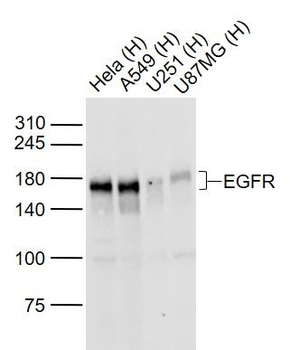
| Immunogen | The immunizing protein for this antibody was recombinant human EGFR. |
| Purification | Protein G affinity chromatography |
| Conjugation | Unconjugated |
Storage & Handling
−| Storage | Maintain refrigerated at 2-8°C for up to 2 weeks. For long term storage store at -20°C in small aliquots to prevent freeze-thaw cycles. |
|---|---|
| Buffer/Preservatives | 0.2 mg/ml in 1X PBS with 0.1 mg/ml rAlbumin and 0.05% sodium azide |
| Expiration Date | 12 months from date of receipt. |
| Disclaimer | For research use only |
Similar Products
−- Item 1 of 18
EGFR isoform a variant Rabbit Polyclonal Antibody [orb308736]
ELISA, ICC, IF, IHC-P, WB
Human, Mouse, Porcine, Rat
Rabbit
Polyclonal
Unconjugated
100 μg - Item 1 of 8
Amivantamab Biosimilar Antibody, Research Grade (EGFR/c-Met) [orb2977888]
ELISA, FA, FACS, In vivo
Human
Human
Monoclonal
Unconjugated
100 μg, 1 mg - Item 1 of 9
EGFRvIII Rabbit Polyclonal Antibody [orb191506]
IHC-P, WB
Human, Mouse
Rabbit
Polyclonal
Unconjugated
200 μg, 100 μg - Item 1 of 7
EGFR Rabbit Polyclonal Antibody [orb10580]
ELISA, FC, ICC, WB
Canine, Mouse, Porcine
Human
Rabbit
Polyclonal
Unconjugated
50 μl, 100 μl, 200 μl - Item 1 of 8
EGFR Rabbit Polyclonal Antibody [orb654422]
ELISA, FC, ICC, IF, IHC, WB
Human, Mouse, Rat
Rabbit
Polyclonal
Unconjugated
100 μg


Quality Guarantee
Explore bioreagents carefree to elevate your research. All our products are rigorously tested for performance. If a product does not perform as described on its datasheet, our scientific support team will provide expert troubleshooting, a prompt replacement, or a refund. For full details, please see our Terms & Conditions and Buying Guide. Contact us at [email protected].
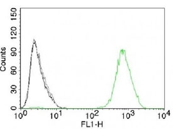
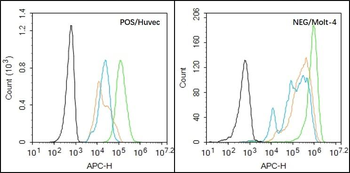
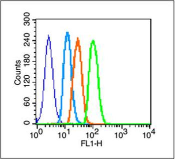
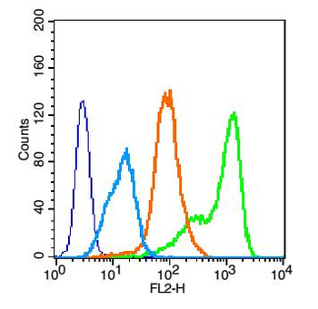
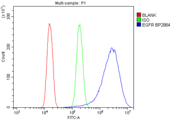
FACS testing of A431 cells with isotype control (gray), without primary antibody (black) and EGFR antibody (green, clone GFR450).
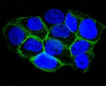
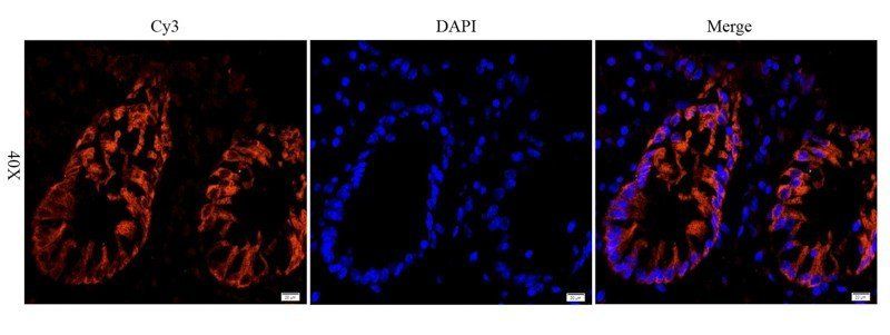
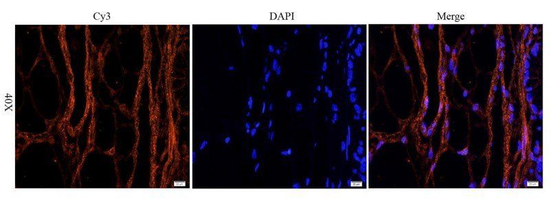
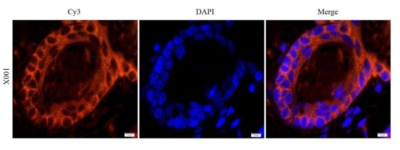
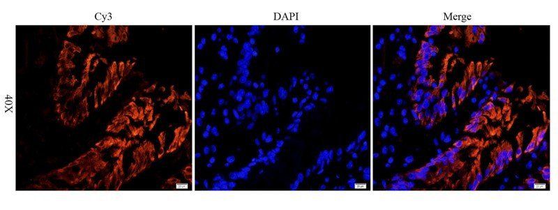
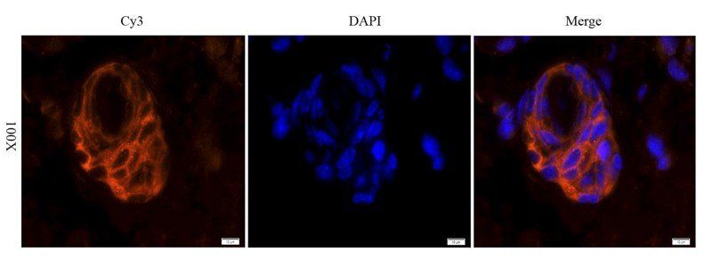
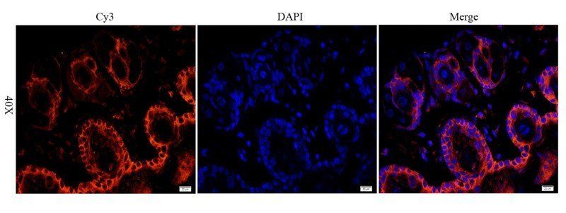
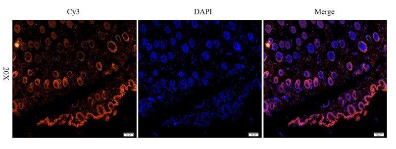
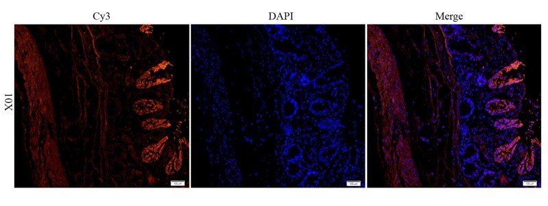
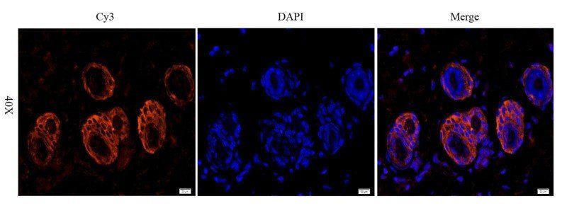
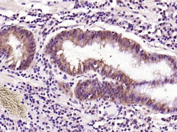
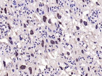
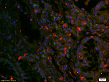
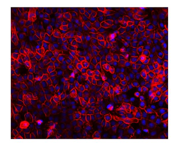
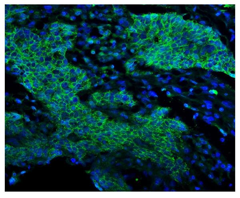
Immunofluorescent staining of A431 cells with Alexa Fluor conjugated EGFR antibody (green, clone GFR450) and DAPI nuclear stain (blue).
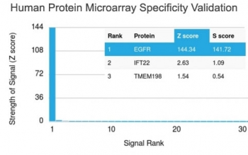
Analysis of HuProt (TM) microarray containing more than 19, 000 full-length human proteins using EGFR antibody (clone GFR450). These results demonstrate the foremost specificity of the GFR450 mAb. Z- and S- score: The Z-score represents the strength of a signal that an antibody (in combination with a fluorescently-tagged anti-IgG secondary Ab) produces when binding to a particular protein on the HuProt (TM) array. Z-scores are described in units of standard deviations (SD's) above the mean value of all signals generated on that array. If the targets on the HuProt (TM) are arranged in descending order of the Z-score, the S-score is the difference (also in units of SD's) between the Z-scores. The S-score therefore represents the relative target specificity of an Ab to its intended target.
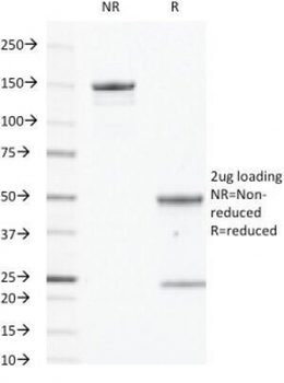
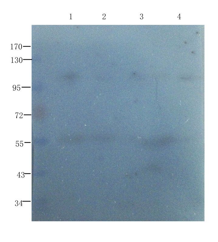
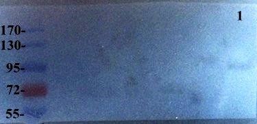
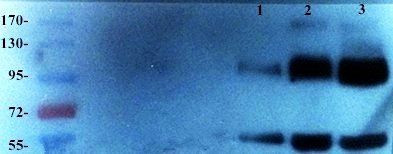
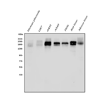
SDS-PAGE analysis of purified, BSA-free EGFR antibody (clone GFR450) as confirmation of integrity and purity.
Documents Download
Datasheet
Product Information
Request a Document
Protocol Information
FACS
Fluorescence-Activated Cell Sorting (FC, Flow cytometry)
IF
Immunofluorescence
EGFR Antibody (orb749349)

- 0.0
Based on 0 reviews
Participating in our Biorbyt product reviews program enables you to support fellow scientists by sharing your firsthand experience with our products.
Login to Submit a ReviewAvailable Sizes
Select a size below
Choose Conjugation or Carrier Free Version
Free Secondary Antibody (20 ul)0/0
Please add an antibody product to your cart first.