Queen's Award Received in 2021 ISO 9001 Certified Delivered over 1,000,000 bio-reagents to life science researchers Trusted by Life Science Communities
Cart summary

You have no items in your shopping cart.

Microtubule Associated Protein 2 (MAP2) Antibody

SKU: orb319566

Description

Our Anti-Microtubule Associated Protein 2 (MAP2) primary antibody from Biorbyt is chicken polyclonal. It detects bovine, human, mouse, and rat Microtubule Associated Protein 2 (MAP2) and is total IgY fraction.

Research Area

Disease Biomarkers, Neuroscience, Signal Transduction

Images & Validation

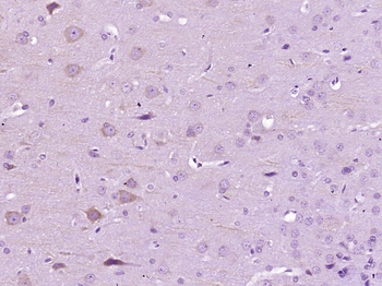
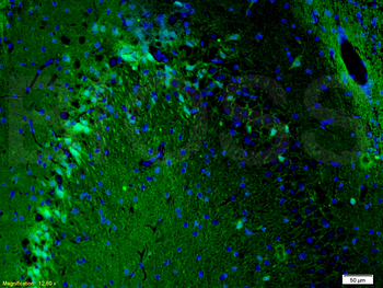
Tested ApplicationsELISA, ICC, IHC, WB
Dilution RangeWB: 1:20000, IHC: 1:2500-1:10,000, ICC: 1:1000-1:20,000
ReactivityBovine, Human, Mouse, Rat

Key Properties

Antibody TypePrimary Antibody
HostGallus
ClonalityPolyclonal
IsotypeIgY
ImmunogenRecombinant bovine MAP2 protein expressed in and purified from E. Coli.
TargetMicrotubule Associated Protein 2 (MAP2)
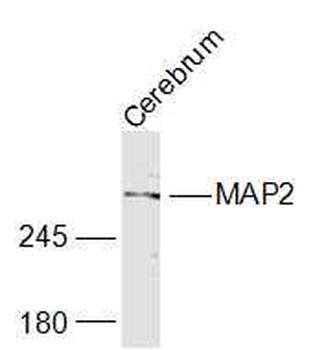
Molecular Weight280
PurificationTotal IgY fraction
ConjugationUnconjugated

Storage & Handling

StorageMaintain refrigerated at 2-8°C for up to 2 weeks. For long term storage store at -20°C in small aliquots to prevent freeze-thaw cycles.
Form/AppearanceLiquid
Buffer/PreservativesPBS + 10 mM NaN3.
Concentration19 mg/mL
Expiration Date12 months from date of receipt.
DisclaimerFor research use only

Alternative Names

DKFZp686I2148 antibody, MAP 2 antibody, MAP dendrite specific antibody, MAP-2 antibody, MAP2 antibody, MAP2A antibody, MAP2B antibody, MAP2C antibody, Microtubule associated protein 2 antibody, Microtubule-associated protein 2 antibody, MTAP2_HUMAN antibody

Similar Products

  • Phospho-ERK1/2 (Thr202 + Tyr204) Rabbit Polyclonal Antibody [orb5178]

    FC,  ICC

    Human

    Mouse

    Rabbit

    Polyclonal

    Unconjugated

    100 μl, 200 μl, 50 μl
  • Phospho-ERK1 (Thr202/Tyr204) + ERK2 (Thr183/Tyr185) Rabbit Polyclonal Antibody [orb783430]

    FC,  IF,  IHC

    Bovine, Canine, Equine, Gallus, Guinea pig, Porcine, Rabbit

    Human, Mouse, Rat

    Rabbit

    Polyclonal

    Unconjugated

    50 μl, 100 μl, 200 μl
  • ERK1/2 Recombinant Rabbit Monoclonal Antibody [orb704524]

    FC,  ICC,  IF,  IHC-Fr,  IHC-P,  WB

    Zebrafish

    Human, Mouse, Rat

    Rabbit

    Recombinant

    Unconjugated

    50 μl, 100 μl, 25 μl
  • ERK1 + ERK2 Rabbit Polyclonal Antibody [orb6018]

    FC,  ICC,  IF,  IHC-Fr,  IHC-P,  WB

    Bovine, Canine, Equine, Gallus, Goat, Porcine, Rabbit, Sheep

    Human, Mouse, Rat

    Rabbit

    Polyclonal

    Unconjugated

    50 μl, 100 μl, 200 μl
  • ERK1/2 Mouse Monoclonal Antibody [orb500888]

    FC,  KO/KD Validated,  WB

    Mouse, Rat

    Human

    Mouse

    Monoclonal

    Unconjugated

    200 μg, 50 μl, 100 μl, 200 μl
Quality Guarantee

Quality Guarantee

Explore bioreagents carefree to elevate your research. All our products are rigorously tested for performance. If a product does not perform as described on its datasheet, our scientific support team will provide expert troubleshooting, a prompt replacement, or a refund. For full details, please see our Terms & Conditions and Buying Guide. Contact us at [email protected].

Microtubule Associated Protein 2 (MAP2) Antibody

(A-B). Primary hippocampal neurons were stained with MAP2 (Cat. orb319566, 1:1000) as a negative control under basal conditions (CTRL) and with picrotoxin treatment (PTX).

Microtubule Associated Protein 2 (MAP2) Antibody

Costaining of the primary brain cells isolated from 18-day embryonic rats (E18). Neurons stained with α-MAP2 (green). Copurified astrocytes stained with α-GFAP (red).

Microtubule Associated Protein 2 (MAP2) Antibody

Day 14 (D14) RGC-iNs stained with a DAPI, TAU, and MAP2 (Cat. orb319566, 1:1000).

Microtubule Associated Protein 2 (MAP2) Antibody

Immunofluorescence of a section of rat brain stem showing colabeled with Anti-MAP2 (Cat. orb319566, red, 1:5000) and Anti-FOX3 (Cat. orb613671 , green, 1:1000). The Anti-FOX3 specifically labels the nuclei and the proximal cytoplasm of neuronal cells while the Anti-MAP2 labels dendrites and overlaps with FOX3 labeling the perikarya of neurons. The blue is DAPI staining of nuclear DNA.

Microtubule Associated Protein 2 (MAP2) Antibody

Immunofluorescence of cortical neuron-glial cell culture from E20 rat labeled with Anti- MAP2 (Cat. orb319566, red, 1:10,000), and Anti-Tau (green). The blue is DAPI staining of nuclear DNA. The anti-MAP2 antibody stains dendrites and perikarya of neurons, while the anti-TAU antibody labels neuronal perikarya, dendrites and also axonal process. As a result perikarya and dendrites appears orange-yellow, since they contain both MAP2 and tau, while axons are green.

Microtubule Associated Protein 2 (MAP2) Antibody

Immunofluorescence of rat cerebellum showing strong synaptic staining of VSNL1 (Cat. : orb319709, green) in the molecular layer (ML) and MAP2 (Cat. orb319566, red, 1:2500). The blue stain is nuclear DNA.

Microtubule Associated Protein 2 (MAP2) Antibody

Immunolabeling of mouse cortical cultures labeled with Anti-GFAP (Cat. orb319549, 1:1000, green) and anti-MAP2 (orb319566, 1:1000, red).

Microtubule Associated Protein 2 (MAP2) Antibody

Immunolabeling of mouse hippocampal cultures labeled with Anti-GFAP (Cat. orb319549, 1:1000, green) and anti-MAP2 (orb319566, 1:1000, red).

Microtubule Associated Protein 2 (MAP2) Antibody

Immunolabeling of mouse hippocampal cultures labeled with anti-MAP2 (Cat. orb319566, 1:1000, red) and anti-synapsin (Cat. orb319666 , 1:1000, green).

Microtubule Associated Protein 2 (MAP2) Antibody

Immunostaining of cultured E20 rat cortical neurons and glia stained with anti-nestin antibody (Cat. orb319583, red, 1:500) and anti-MAP2 antibody (Cat. orb319566, green, 1:500). The blue is Hoechst staining for nuclear DNA. The nestin antibody labels developing astrocytes and neuronal stem cells in a clearly filamentous fashion, while the MAP2 antibody stains dendrites and perikarya of mature neurons.

Microtubule Associated Protein 2 (MAP2) Antibody

Immunostaining of rat hippocampus showing specific immunolabeling of MAP2 (Cat. orb319566, red, 1:1000), Tau-1 (green), and nuclear DNA labeled with DAPI (blue).

Microtubule Associated Protein 2 (MAP2) Antibody

Representative 3D view images of paired helical filament (PHF)-tau-treated human brain organoid fractions. The organoids were labeled for MAP2 (Cat. orb319566, 1:1000, green), NR3A (red), PSD95 (magenta), and DAPI (blue). White arrows show the positions colocalized between PSD95 and NR3A. The dotted circles show the magnified positions. Scale bar = 10 or 3 μm.

Microtubule Associated Protein 2 (MAP2) Antibody

Representative images of anti-MAP2 (ca. orb319566) immunolabeled rat neurons at 5 DIV of T30 treatment.

Microtubule Associated Protein 2 (MAP2) Antibody

Western blot of rat cortical lysate showing specific immunolabeling of the ~280 kDa MAP2 protein.

UniProt Details

No UniProt data available

Documents Download

Datasheet
Product Information
Download

Request a Document

Protocol Information

WB
Western Blot (IB, immunoblot)
View Protocol
IHC
Immunohistochemistry
View Protocol
ICC
Immunocytochemistry
View Protocol
ELISA
Enzyme-linked Immunosorbent Assay (EIA)
View Protocol

Microtubule Associated Protein 2 (MAP2) Antibody (orb319566)

  • Star
  • Star
  • Star
  • Star
  • Star
  • 0.0
Based on 0 reviews

Participating in our Biorbyt product reviews program enables you to support fellow scientists by sharing your firsthand experience with our products.

Login to Submit a Review

No reviews yet

Available Sizes

Select a size below

100 μl
$ 560.00
Choose Conjugation or Carrier Free Version
Free Secondary Antibody (20 ul)0/0

Please add an antibody product to your cart first.

DispatchUsually dispatched within 5-10 working days
Bulk Enquiry