You have no items in your shopping cart.
Description
Research Area
Immunology & Inflammation
Images & Validation
−Item 1 of 4
| Tested Applications | FC, ICC, IHC-P, IP, WB |
|---|---|
| Reactivity | Human |
| Application Notes |
Key Properties
−| Antibody Type | Primary Antibody |
|---|---|
| Clonality | Monoclonal |
| Isotype | Mouse IgG1 |
| Clone No. | MEM-28 |
| Immunogen | Human thymocytes and T lymphocytes. |
| Target | CD45 |
| Purification | Purified by protein-A affinity chromatography. |
| Conjugation | Unconjugated |
Storage & Handling
−| Storage | Maintain refrigerated at 2-8°C for up to 2 weeks. For long term storage store at -20°C in small aliquots to prevent freeze-thaw cycles. |
|---|---|
| Buffer/Preservatives | Phosphate buffered saline (PBS), pH 7.4, 15 mM sodium azide |
| Concentration | 1 mg/ml |
| Expiration Date | 12 months from date of receipt. |
| Disclaimer | For research use only |
Alternative Names
−LCA, T200, LY5, B220, GP180, TPC
Similar Products
−- Item 1 of 13
CD45 (Intracellular) Rabbit Polyclonal Antibody [orb1294366]
FC, IF, IHC, WB
Human
Rabbit
Polyclonal
Unconjugated
100 μl, 25 μl - Item 1 of 11
- Item 1 of 6
CD45 Rabbit Polyclonal Antibody [orb1179]
FC
Mouse, Rat
Human
Rabbit
Polyclonal
Unconjugated
50 μl, 100 μl, 200 μl - Item 1 of 8
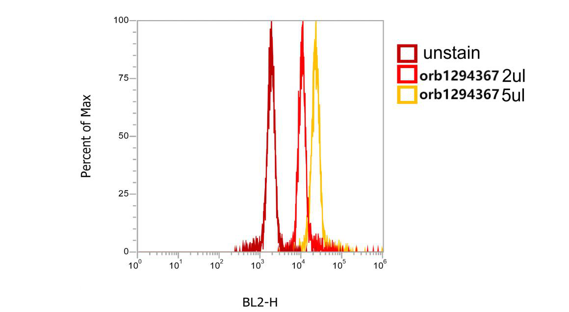
CD45 (Extracellular) Rabbit Polyclonal Antibody [orb1294367]
FC, IF, IHC, WB
Human, Mouse
Rabbit
Polyclonal
Unconjugated
100 μl, 25 μl - Item 1 of 5
CD45 Rabbit Polyclonal Antibody [orb312177]
ICC, IF, IHC-Fr, IHC-P, WB
Bovine
Human
Rabbit
Polyclonal
Unconjugated
50 μl, 100 μl, 200 μl

Quality Guarantee
Explore bioreagents carefree to elevate your research. All our products are rigorously tested for performance. If a product does not perform as described on its datasheet, our scientific support team will provide expert troubleshooting, a prompt replacement, or a refund. For full details, please see our Terms & Conditions and Buying Guide. Contact us at [email protected].
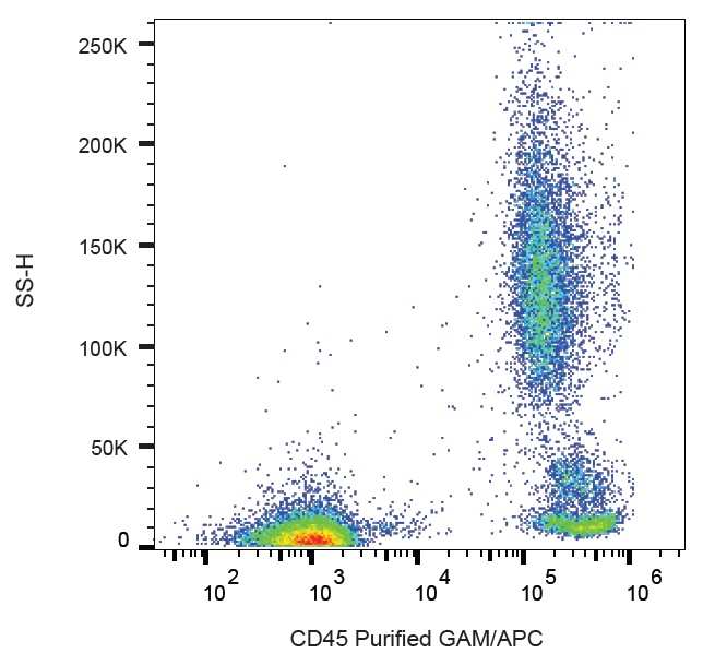
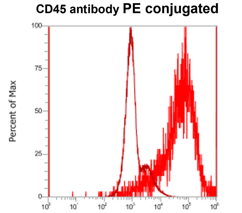
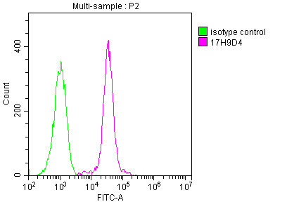
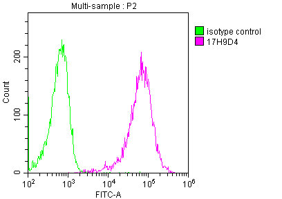
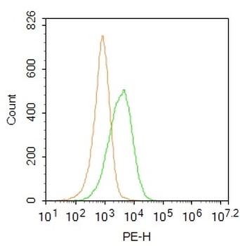
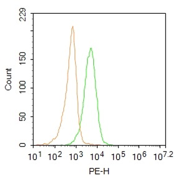
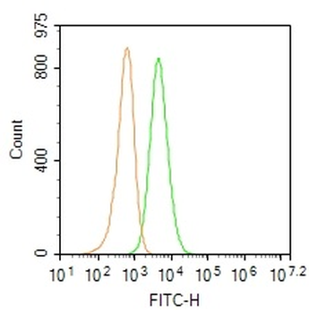
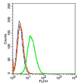
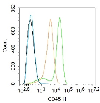
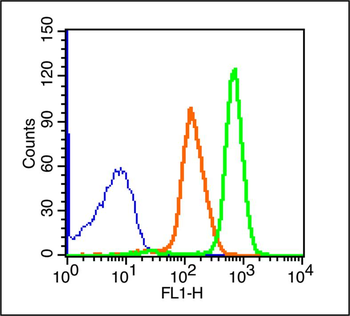
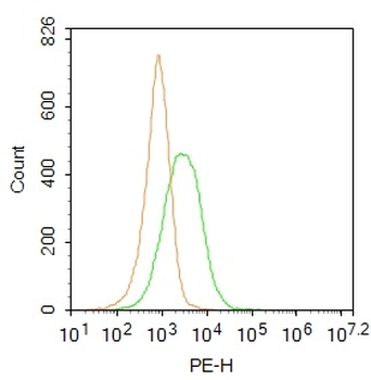
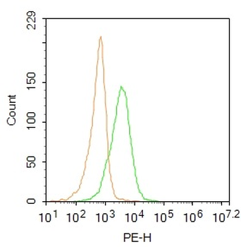
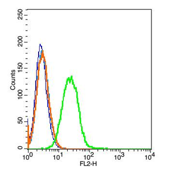
Flow cytometry analysis (surface staining) of human peripheral blood cells with anti-human CD45 (MEM-28) purified, GAM-APC.
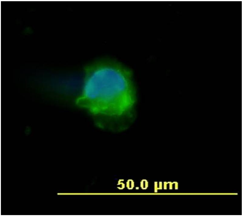
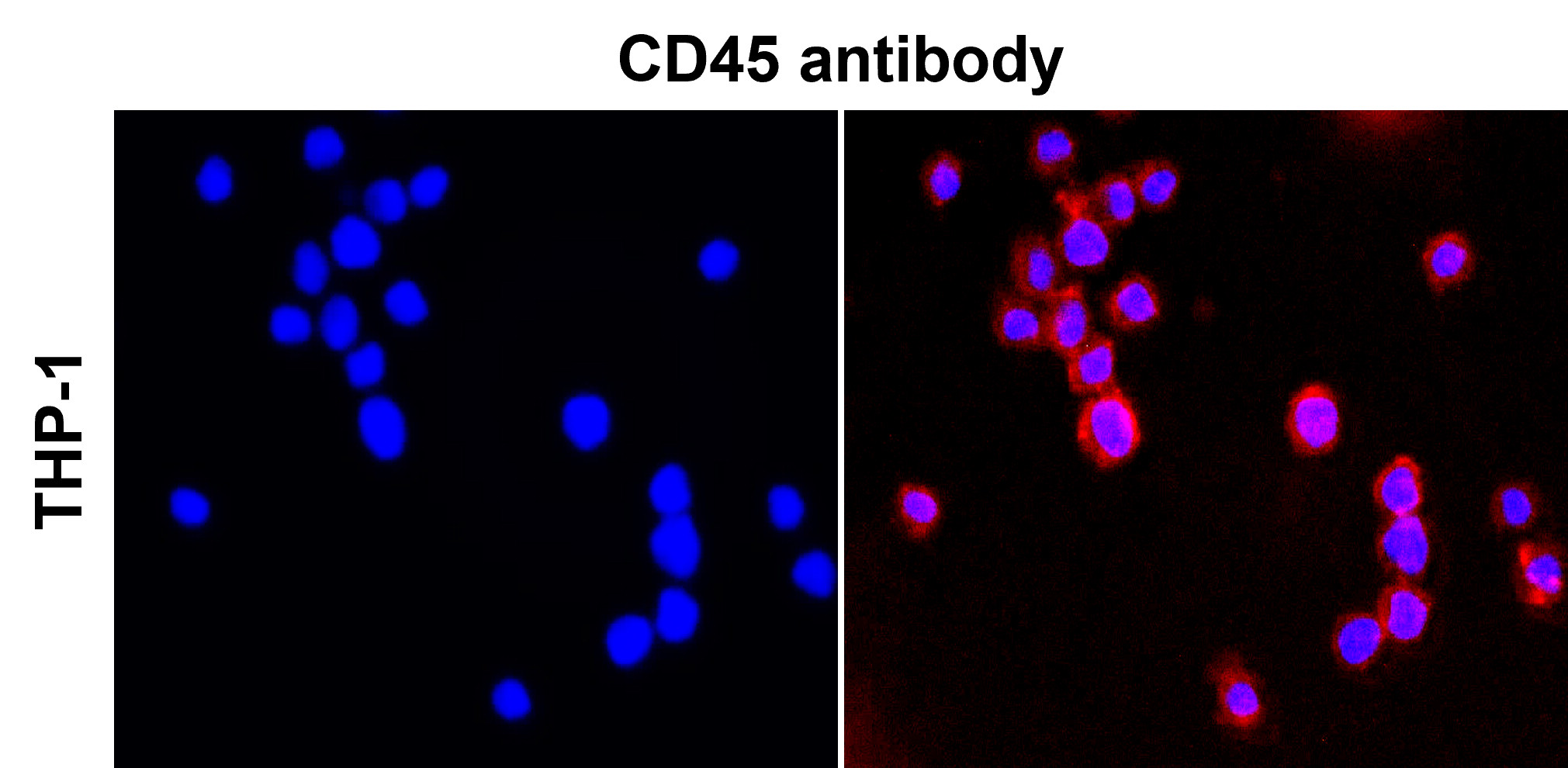
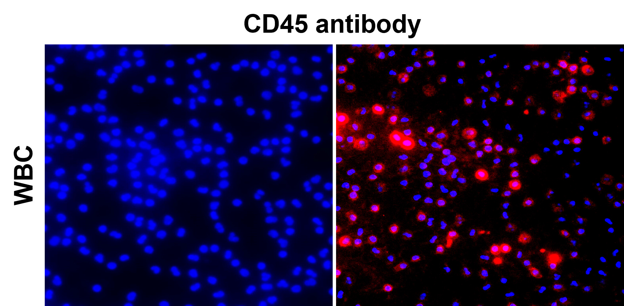
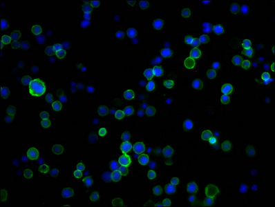
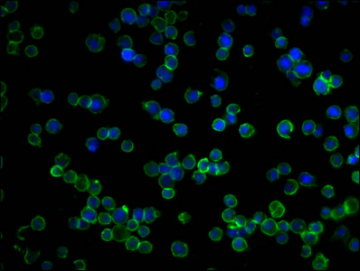
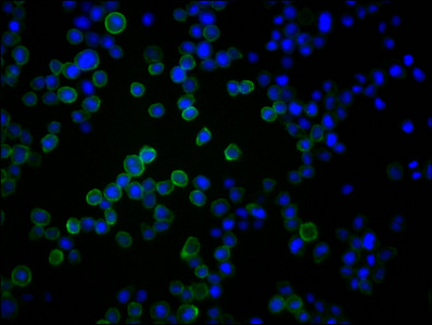
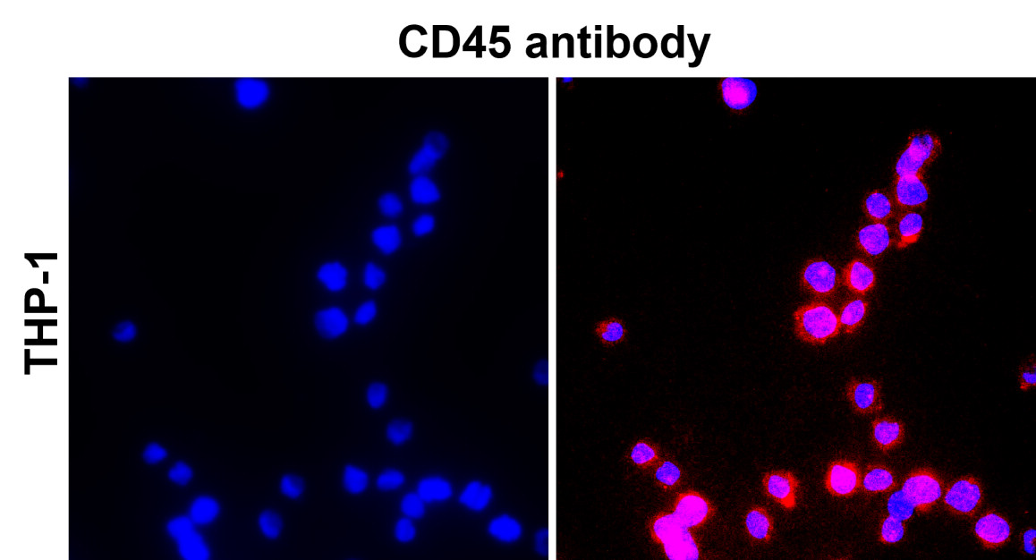
Immunocytochemistry staining of human peripheral blood mononuclear cell using anti-human CD45 (MEM-28, green). DNA visualized by DAPI (blue)
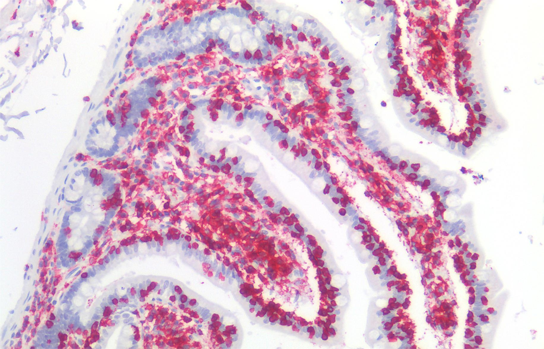
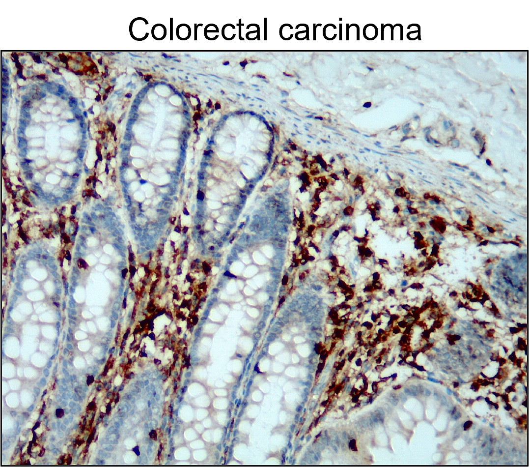
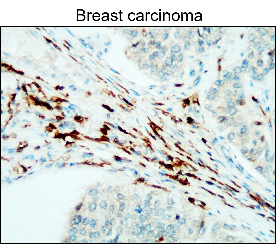
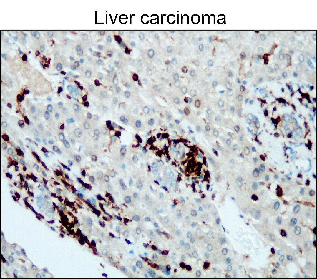
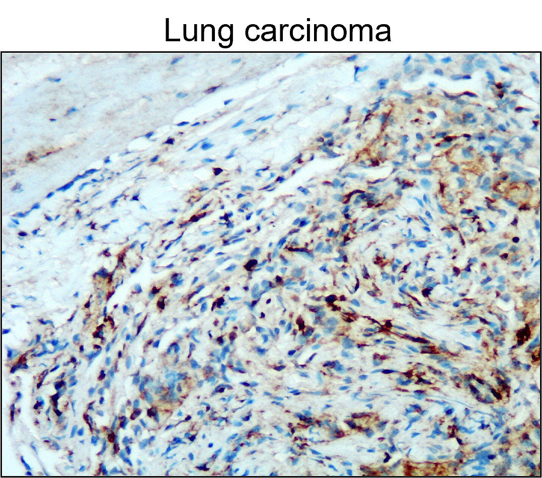
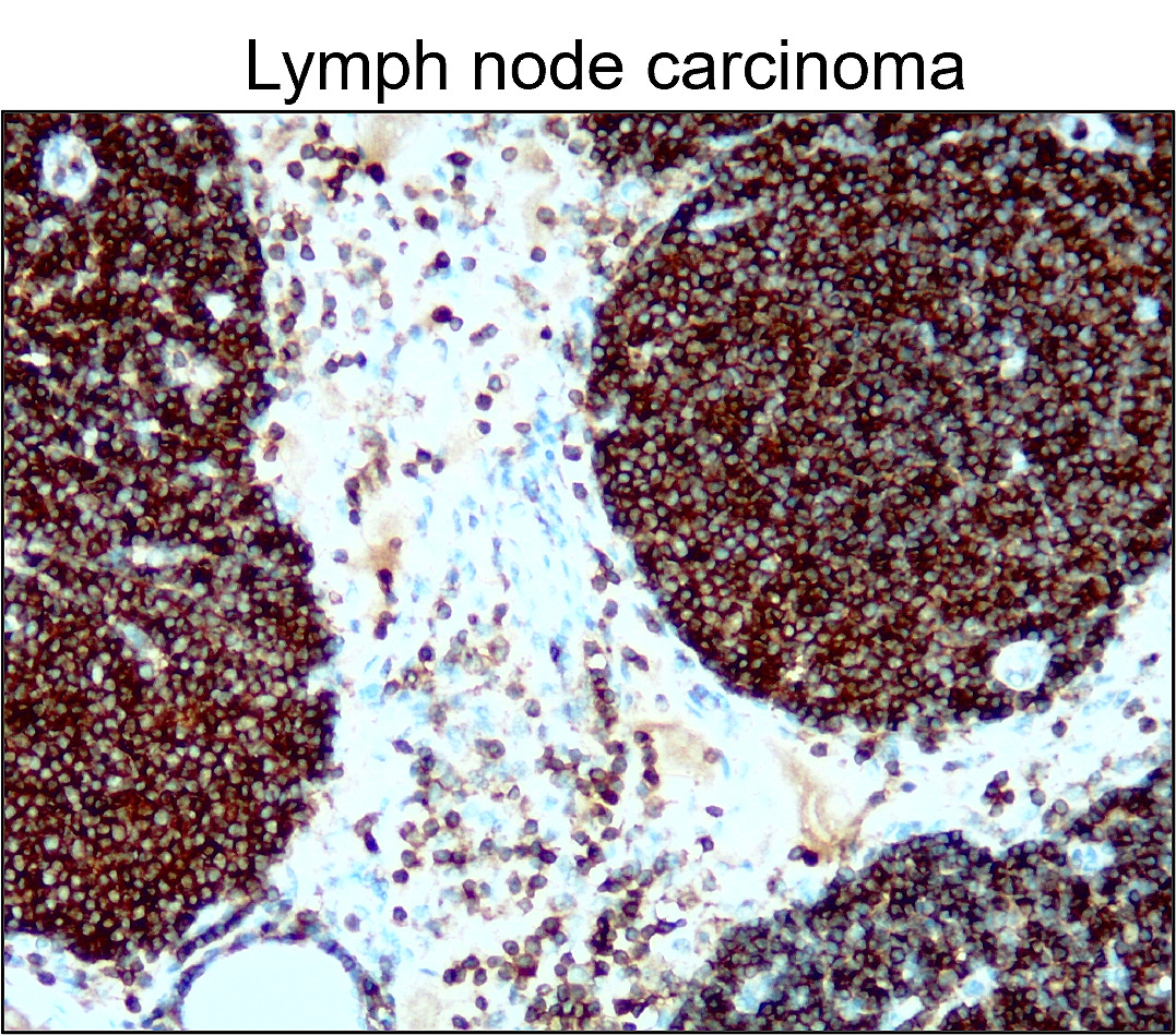
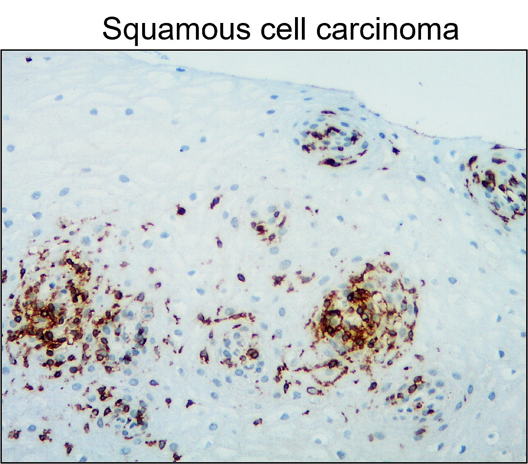
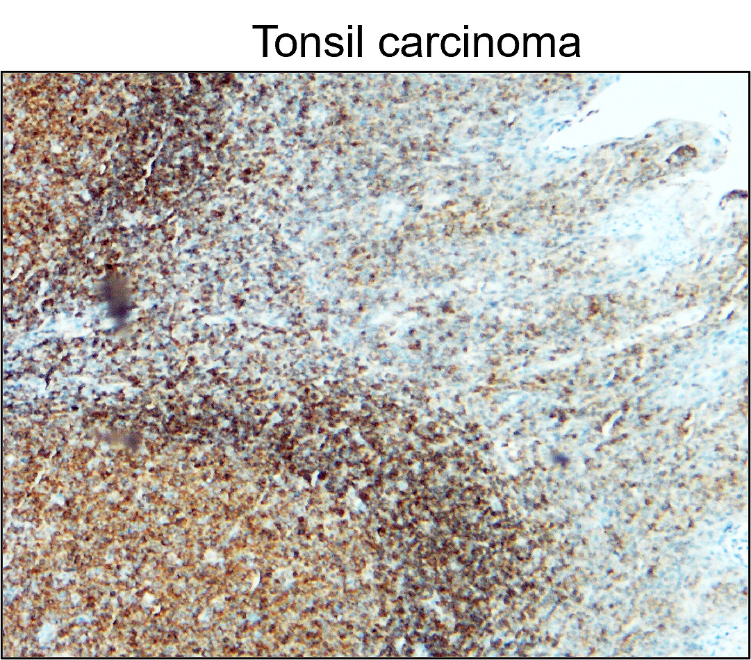
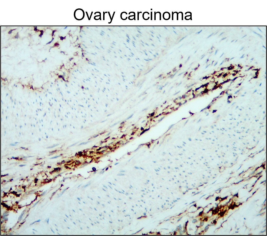
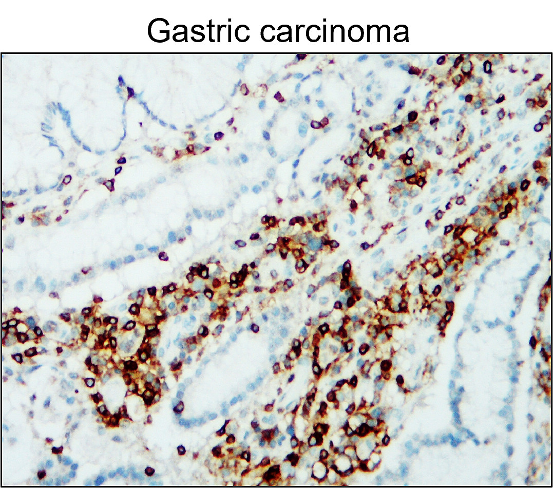
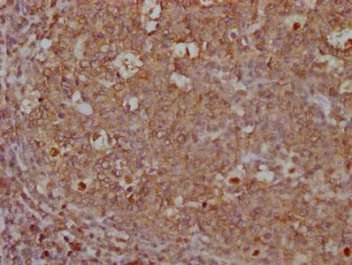
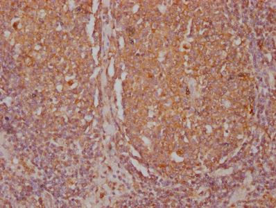
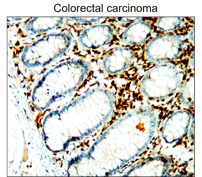
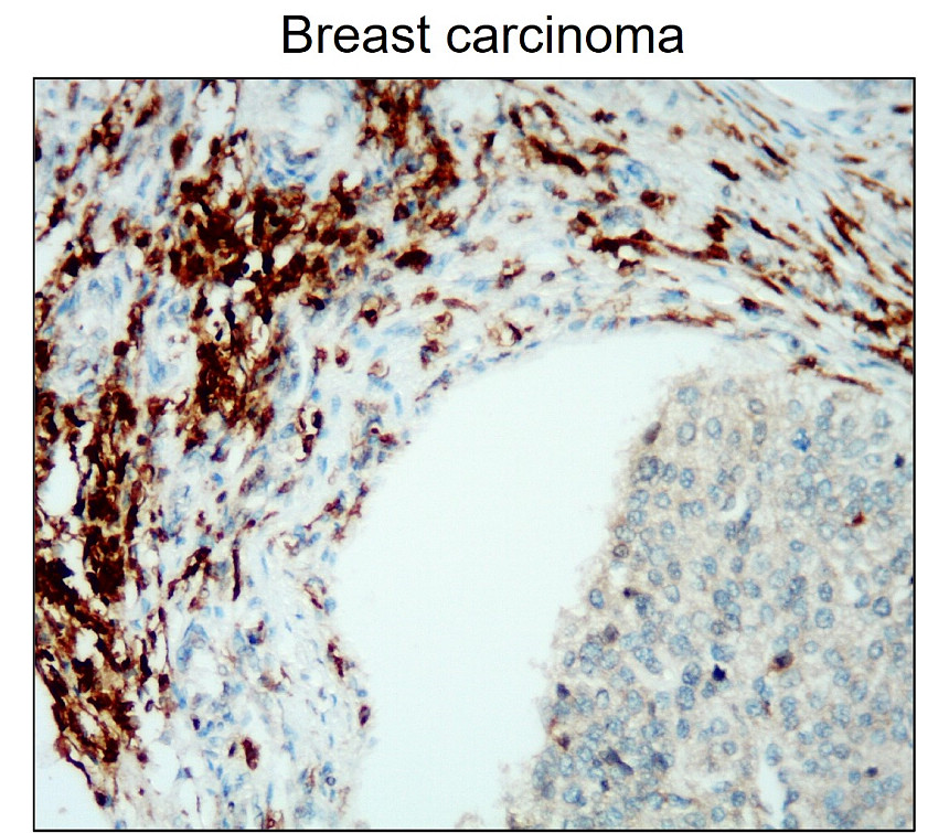
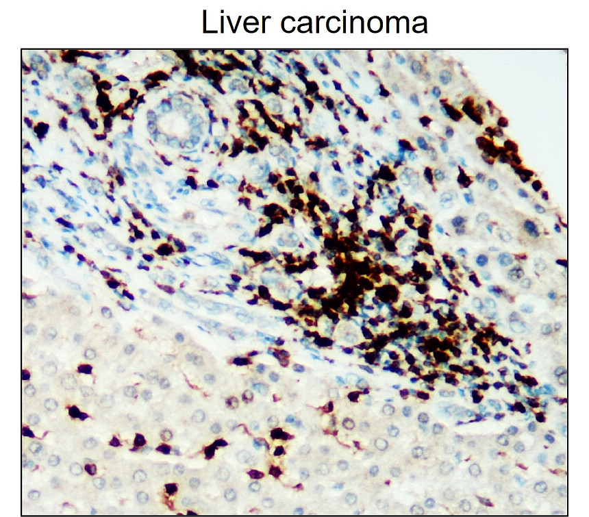
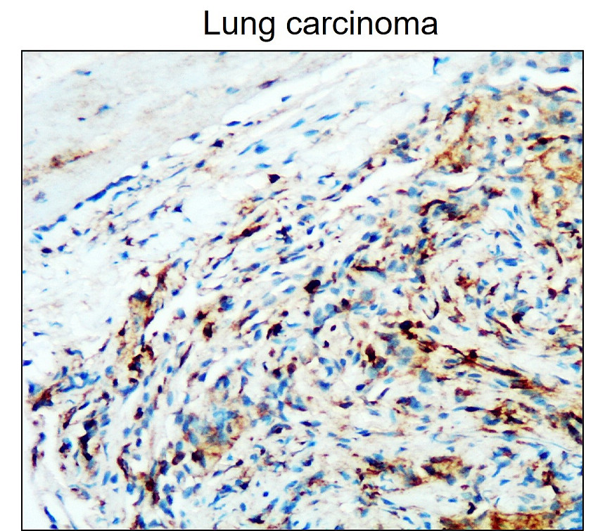
Immunohistochemistry staining of human small intestine (paraffin-embedded sections) with anti-CD45 (MEM-28), 10 µg/ml.
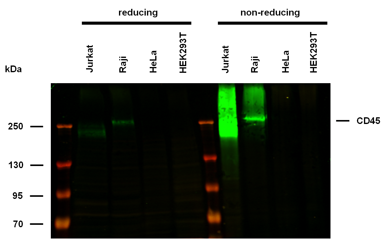
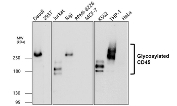
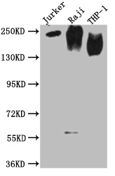
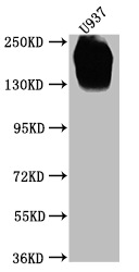
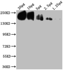
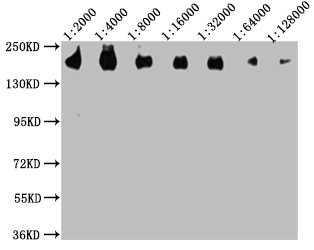
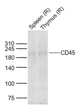
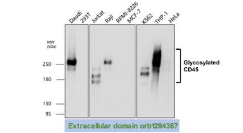
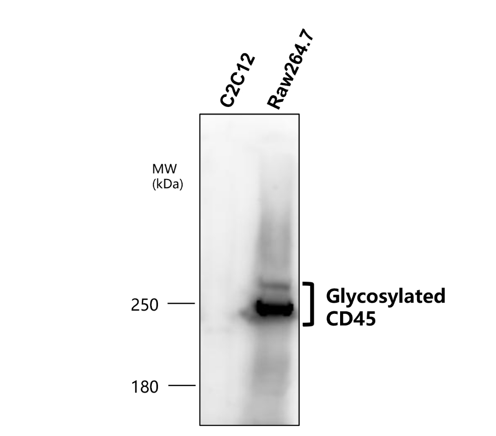
Anti-Hu CD45 Purified (clone MEM-28) works in WB application under non-reducing conditions. Western blotting analysis was performed on whole cell extracts (RIPA lysis buffer) of Jurkat, Raji, HeLa, and HEK293T cell lines, mixed and heated (100°C, 5 min) with reducing and non-reducing SDS-loading buffer. Samples were resolved using 7% Tris-glycine SDS gel electrophoresis. Nitrocellulose membrane blot was probed with mouse IgG1 monoclonal antibody MEM-28 (1 µg/ml), followed by IRDye 800CW Goat-anti-Mouse IgG (green). Multiplex fluorescent Western blot detection was performed. CD45 molecules were detected at ~180-250 kDa in Jurkat and Raji cell lines.
Documents Download
Datasheet
Product Information
Request a Document
Protocol Information
WB
Western Blot (IB, immunoblot)
IHC-P
Immunohistochemistry Paraffin
FC
Flow Cytometry
ICC
Immunocytochemistry
IP
Immunoprecipitation
CD45 Antibody (orb44266)

- 0.0
Based on 0 reviews
Participating in our Biorbyt product reviews program enables you to support fellow scientists by sharing your firsthand experience with our products.
Login to Submit a ReviewAvailable Sizes
Select a size below
Free Secondary Antibody (20 ul)0/0
Please add an antibody product to your cart first.