You have no items in your shopping cart.
Description
Research Area
Cancer Biology, Epigenetics & Chromatin, Immunology & Inflammation, Molecular Biology
Images & Validation
−Item 1 of 2
| Tested Applications | ELISA, FC, IHC |
|---|---|
| Dilution Range | Immunohistochemistry(Paraffin-embedded Section), 2-5 μg/ml, Human Flow Cytometry (Fixed), 1-3 μg/1x10^6 cells, Human ELISA, 0.1-0.5 μg/ml |
| Reactivity | Human |
Related Conjugates & Formulations
−Key Properties
−| Antibody Type | Primary Antibody |
|---|---|
| Host | Rabbit |
| Clonality | Polyclonal |
| Isotype | Rabbit IgG |
| Immunogen | E.coli-derived human CCR7 recombinant protein (Position: K332-E371). |
| Target | C-C chemokine receptor type 7 |
| Molecular Weight | 38 kDa |
| Purification | Immunogen affinity purified. |
| Conjugation | Unconjugated |
Storage & Handling
−| Storage | Maintain refrigerated at 2-8°C for up to 2 weeks. For long term storage store at -20°C in small aliquots to prevent freeze-thaw cycles. |
|---|---|
| Form/Appearance | Lyophilized |
| Buffer/Preservatives | Each vial contains 4 mg Trehalose, 0.9 mg NaCl, 0.2 mg Na2HPO4. |
| Concentration | Adding 0.2 ml of distilled water will yield a concentration of 500 μg/ml. |
| Expiration Date | 12 months from date of receipt. |
| Disclaimer | For research use only |
Alternative Names
−BLR2; C C chemokine receptor type 7; C C CKR 7; CC CKR 7; CCR 7; CCR7; CD197; CDw197; CMKBR7; EBI1; EVI1; MIP 3 beta receptor
Similar Products
−- Item 1 of 9
CCR7 Rabbit Polyclonal Antibody [orb10276]
FC, ICC, WB
Canine
Human, Mouse, Rat
Rabbit
Polyclonal
Unconjugated
50 μl, 100 μl, 200 μl - Item 1 of 6
CCR7 Antibody (N-term) [orb1930722]
FC, IF, IHC-P, WB
Human, Mouse
Rabbit
Polyclonal
Unconjugated
50 μl, 100 μl - Item 1 of 4
CKR-7 rabbit pAb Antibody [orb767586]
ELISA, IF, WB
Human, Monkey
Polyclonal
Unconjugated
50 μl, 100 μl - Item 1 of 3
CCR7 Rabbit Polyclonal Antibody [orb1040883]
IF, IHC-Fr, IHC-P
Bovine, Human, Mouse
Rat
Rabbit
Polyclonal
Unconjugated
50 μl, 100 μl, 200 μl 
CCR7 Rabbit Polyclonal Antibody (FITC) [orb15268]
FC, ICC, IF
Canine
Human, Mouse, Rat
Rabbit
Polyclonal
FITC
100 μl

Quality Guarantee
Explore bioreagents carefree to elevate your research. All our products are rigorously tested for performance. If a product does not perform as described on its datasheet, our scientific support team will provide expert troubleshooting, a prompt replacement, or a refund. For full details, please see our Terms & Conditions and Buying Guide. Contact us at [email protected].
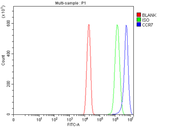
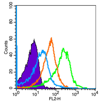
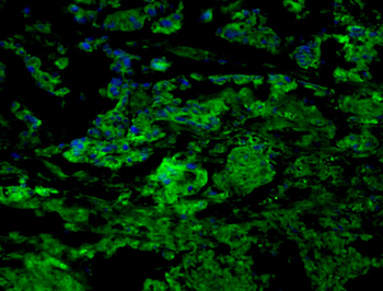
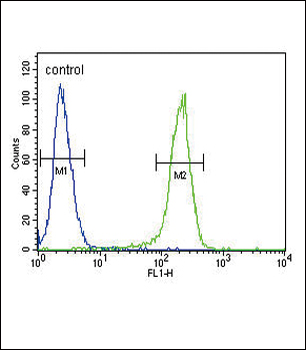
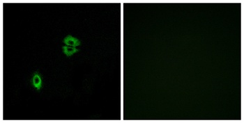
Flow Cytometry analysis of U20S cells using anti-CCR7 antibody. Overlay histogram showing U20S cells (Blue line). To facilitate intracellular staining, cells were fixed with 4% paraformaldehyde and permeabilized with permeabilization buffer. The cells were blocked with 10% normal goat serum. And then incubated with rabbit anti-CCR7 Antibody (1 µg/1x10^6 cells) for 30 min at 20°C. DyLight®488 conjugated goat anti-rabbit IgG (5-10 µg/1x10^6 cells) was used as secondary antibody for 30 minutes at 20°C. Isotype control antibody (Green line) was rabbit IgG (1 µg/1x10^6) used under the same conditions. Unlabelled sample without incubation with primary antibody and secondary antibody (Red line) was used as a blank control.
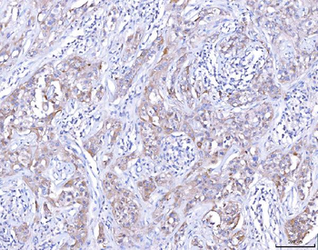
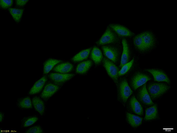
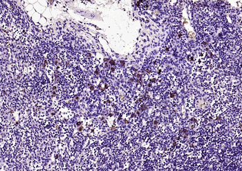
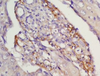
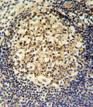
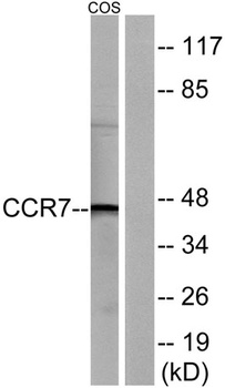
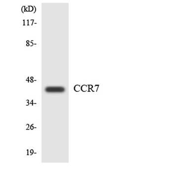
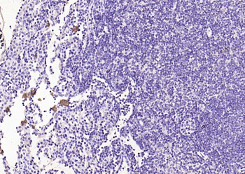
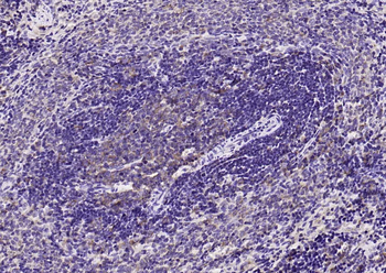
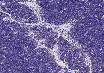
IHC analysis of CCR7 using anti-CCR7 antibody. CCR7 was detected in a paraffin-embedded section of human lymphoma tissue. Heat mediated antigen retrieval was performed in EDTA buffer (pH8.0, epitope retrieval solution). The tissue section was blocked with 10% goat serum. The tissue section was then incubated with 2 µg/ml rabbit anti-CCR7 Antibody overnight at 4°C. Peroxidase Conjugated Goat Anti-rabbit IgG was used as secondary antibody and incubated for 30 minutes at 37°C. The tissue section was developed using HRP Conjugated Rabbit IgG Super Vision Assay Kit with DAB as the chromogen.
Quick Database Links
Gene Symbol
C-C chemokine receptor type 7
UniProt
UniProt Details
− No UniProt data available
Documents Download
Datasheet
Product Information
Request a Document
Protocol Information
IHC
Immunohistochemistry
FC
Flow Cytometry
ELISA
Enzyme-linked Immunosorbent Assay (EIA)
CCR7 Rabbit Polyclonal Antibody (orb1145762)

- 0.0
Based on 0 reviews
Participating in our Biorbyt product reviews program enables you to support fellow scientists by sharing your firsthand experience with our products.
Login to Submit a ReviewAvailable Sizes
Select a size below
Free Secondary Antibody (20 ul)0/0
Please add an antibody product to your cart first.