You have no items in your shopping cart.
Featured
Description
Research Area
Immunology & Inflammation
Images & Validation
−Item 1 of 9
| Tested Applications | FC, ICC, WB |
|---|---|
| Dilution Range | WB=1:500-2000, ICC/IF=1:100-500, Flow-Cyt=1μg/Test |
| Reactivity | Human, Mouse, Rat |
| Predicted Reactivity | Canine |
Related Conjugates & Formulations
−Key Properties
−| Antibody Type | Primary Antibody |
|---|---|
| Host | Rabbit |
| Clonality | Polyclonal |
| Isotype | IgG |
| Immunogen | KLH conjugated synthetic peptide derived from human CCR7 (25-59/379aa) |
| Target | CCR7 |
| Molecular Weight | 43 kDa |
| Purification | Affinity purified by Protein A |
| Conjugation | Unconjugated |
Storage & Handling
−| Storage | Maintain refrigerated at 2-8°C for up to 2 weeks. For long term storage store at -20°C in small aliquots to prevent freeze-thaw cycles. |
|---|---|
| Form/Appearance | Liquid |
| Buffer/Preservatives | 0.01M TBS (pH7.4) with 1% rAlbumin, 0.02% Proclin300 and 50% Glycerol. |
| Concentration | 1mg/ml |
| Expiration Date | 12 months from date of receipt. |
| Disclaimer | For research use only |
Alternative Names
−BLR2; CC-CKR-7; CCR-7; CD197; CDw197; CMKBR7; EBI1; Ebi1h; CCR7_HUMAN; CCR7; C-C CKR-7; Epstein-Barr virus-induced G-protein coupled receptor 1 (EBI1 | EBV-induced G-protein coupled receptor 1); MIP-3 beta receptor; EVI1; CCR7_MOUSE; C-C motif chemokine receptor 7; chemokine (C-C motif) receptor 7
Similar Products
−- Item 1 of 6
CCR7 Antibody (N-term) [orb1930722]
FC, IF, IHC-P, WB
Human, Mouse
Rabbit
Polyclonal
Unconjugated
50 μl, 100 μl - Item 1 of 4
CKR-7 rabbit pAb Antibody [orb767586]
ELISA, IF, WB
Human, Monkey
Polyclonal
Unconjugated
50 μl, 100 μl - Item 1 of 3
CCR7 Rabbit Polyclonal Antibody [orb1040883]
IF, IHC-Fr, IHC-P
Bovine, Human, Mouse
Rat
Rabbit
Polyclonal
Unconjugated
50 μl, 100 μl, 200 μl 
CCR7 Rabbit Polyclonal Antibody (FITC) [orb15268]
FC, ICC, IF
Canine
Human, Mouse, Rat
Rabbit
Polyclonal
FITC
100 μl- Item 1 of 2
CCR7 Rabbit Polyclonal Antibody [orb625528]
ELISA, IHC, WB
Human
Rabbit
Polyclonal
Unconjugated
50 μg, 100 μg
Quality Guarantee
Explore bioreagents carefree to elevate your research. All our products are rigorously tested for performance. If a product does not perform as described on its datasheet, our scientific support team will provide expert troubleshooting, a prompt replacement, or a refund. For full details, please see our Terms & Conditions and Buying Guide. Contact us at [email protected].
Blank control (Black line): Mouse spleen (Black). Primary Antibody (green line): Rabbit Anti-CD4 antibody, Dilution: 3 µg/10^6 cells, Isotype Control Antibody (orange line): Rabbit IgG-PE. Secondary Antibody (white blue line): Goat anti-rabbit IgG-PE, Dilution: 1 µg/test. Protocol, The cells were fixed with 4% PFA (10 min at room temperature) and then permeabilized with 90% ice-cold methanol for 20 min at room temperature. The cells were then incubated in 5% BSA to block non-specific protein-protein interactions for 30 min at room temperature. Cells stained with Primary Antibody for 30 min at room temperature. The secondary antibody used for 40 min at room temperature. Acquisition of 20000 events was performed.
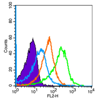
Blank control: Raji (blue). Primary Antibody: Rabbit Anti-CCR7 antibody (orb10276), Dilution: 1 µg in 100 µL 1X PBS containing 0.5% BSA, Isotype Control Antibody: Rabbit IgG (orange), used under the same conditions, Secondary Antibody: Goat anti-rabbit IgG-PE (white blue), Dilution: 1:200 in 1 X PBS containing 0.5% BSA. Protocol, The cells were fixed with 2% paraformaldehyde (10 min). Primary antibody (orb10276, 1 µg/1x10^6 cells) were incubated for 30 min on the ice, followed by 1 X PBS containing 0.5% BSA + 10% goat serum (15 min) to block non-specific protein-protein interactions. Then the Goat Anti-rabbit IgG/PE antibody was added into the blocking buffer mentioned above to react with the primary antibody at 1/200 dilution for 30 min on ice. Acquisition of 20000 events was performed.

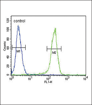
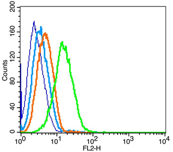
Blank control: THP-1. Primary Antibody (green line): Rabbit Anti-CCR7 antibody (orb10276), Dilution: 1 µg/10^6 cells, Isotype Control Antibody (orange line): Rabbit IgG. Secondary Antibody: Goat anti-rabbit IgG-FITC, Dilution: 0.5 µg/test. Protocol, The cells were fixed with 4% PFA (10 min at room temperature) and then permeabilized with 0.1% PBST for 20 min at room temperature. The cells were then incubated in 5% BSA to block non-specific protein-protein interactions for 30 min at room temperature. Cells stained with Primary Antibody for 30 min at room temperature. The secondary antibody used for 40 min at room temperature. Acquisition of 20000 events was performed.
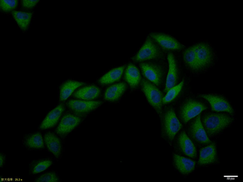
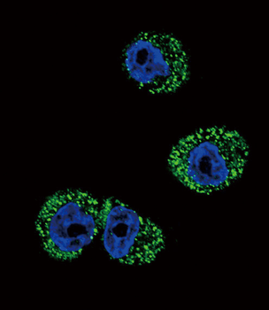
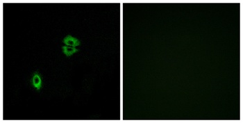
Hela cell, 4% Paraformaldehyde-fixed, Triton X-100 at room temperature for 20 min, Blocking buffer (normal goat serum) at 37°C for 20 min, Antibody incubation with (CCR7) polyclonal Antibody, Unconjugated (orb10276) 1:100, 90 minutes at 37°C, followed by a conjugated Goat Anti-Rabbit IgG antibody at 37°C for 90 minutes, DAPI (blue) was used to stain the cell nuclei.
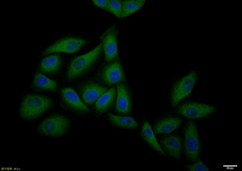
Hela cell, 4% Paraformaldehyde-fixed, Triton X-100 at room temperature for 20 min, Blocking buffer (normal goat serum) at 37°C for 20 min, Antibody incubation with (CCR7) polyclonal Antibody, Unconjugated (orb10276) 1:100, 90 minutes at 37°C, followed by a conjugated Goat Anti-Rabbit IgG antibody at 37°C for 90 minutes, DAPI (blue) was used to stain the cell nuclei.
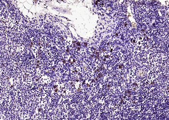
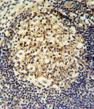
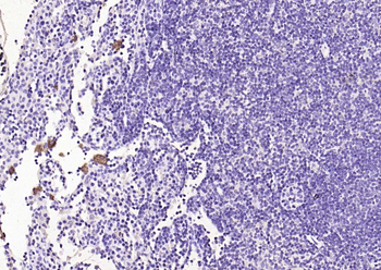
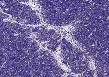
Paraformaldehyde-fixed, paraffin embedded (RAT lymphoid), Antigen retrieval by boiling in sodium citrate buffer (pH6.0) for 15 min, Block endogenous peroxidase by 3% hydrogen peroxide for 20 minutes, Blocking buffer (normal goat serum) at 37°C for 30 min, Antibody incubation with (CCR7) Polyclonal Antibody, Unconjugated (orb10276) at 1:200 overnight at 4°C, followed by operating according to SP Kit (Rabbit) instructionsand DAB staining.
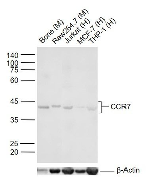
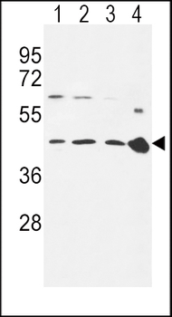
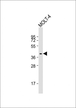
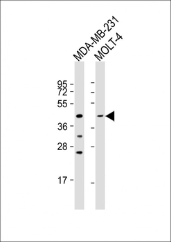
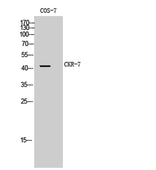
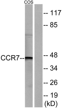
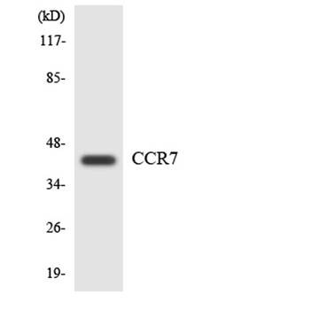
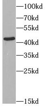
Sample: Lane 1: Mouse Bone tissue lysates, Lane 2: Mouse Raw264.7 cell lysates, Lane 3: Human Jurkat cell lysates, Lane 4: Human MCF-7 cell lysates, Lane 5: Human THP-1 cell lysates, Primary: Anti-CCR7 (orb10276) at 1/1000 dilution, Secondary: IRDye800CW Goat Anti-Rabbit IgG at 1/20000 dilution, Predicted band size: 42 kDa, Observed band size: 42 kDa.
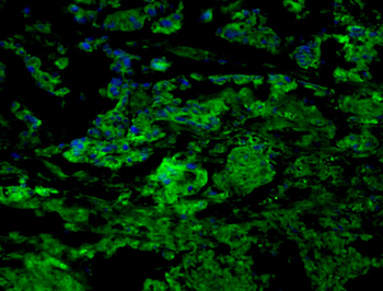
Tissue/cell: human gastric tissue, 4% Paraformaldehyde-fixed and paraffin-embedded, Antigen retrieval: citrate buffer (0.01M, pH 6.0), Boiling bathing for 15 min, Blocking buffer (normal goat serum) at 37℃ for 20 min, Incubation: Anti-CCR7 Polyclonal Antibody, Unconjugated (orb10276) 1:200, overnight at 4°C, The secondary antibody was Goat Anti-Rabbit IgG, FITC conjugated (orb868805) used at 1:200 dilution for 40 minutes at 37°C. DAPI (5 ug/ml, blue) was used to stain the cell nuclei.
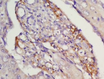
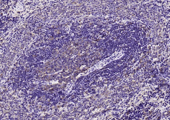
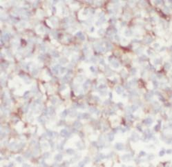
Tissue/cell: human laryngocarcinoma, 4% Paraformaldehyde-fixed and paraffin-embedded, Antigen retrieval: citrate buffer (0.01M, pH 6.0), Boiling bathing for 15 min, Block endogenous peroxidase by 3% Hydrogen peroxide for 30 min, Blocking buffer (normal goat serum) at 37℃ for 20 min, Incubation: Anti-CCR7 Polyclonal Antibody, Unconjugated (orb10276) 1:200, overnight at 4°C, followed by conjugation to the secondary antibody and DAB staining.
Quick Database Links
Gene Symbol
CCR7
UniProt
UniProt Details
− No UniProt data available
Documents Download
Datasheet
Product Information
Request a Document
Protocol Information
WB
Western Blot (IB, immunoblot)
FC
Flow Cytometry
ICC
Immunocytochemistry
Hao Xu 1 2, Xin Li 1 2, Wenxue Wang 1 2, Li Zhen 1 2, Baodong Zhao Strontium-Doped Marine Collagen Membranes Promote Osteogenesis by Inducing M2 Macrophage Polarization Tissue Eng Regen Med, (2025)
Li, Xuelu et al. High Expression of CCR7 Predicts Lymph Node Metastasis and Good Prognosis in Triple Negative Breast Cancer Cell Physiol Biochem, 43, 531-539 (2017)
CCR7 Rabbit Polyclonal Antibody (orb10276)

- 0.0
Based on 0 reviews
Participating in our Biorbyt product reviews program enables you to support fellow scientists by sharing your firsthand experience with our products.
Login to Submit a ReviewAvailable Sizes
Select a size below
Free Secondary Antibody (20 ul)0/0
Please add an antibody product to your cart first.