You have no items in your shopping cart.
Description
Research Area
Immunology & Inflammation
Images & Validation
−Item 1 of 5
| Tested Applications | ELISA, IF, IHC, WB |
|---|---|
| Dilution Range | ELISA: 1:1,000 - 1:5,000, IHC: 1:100 - 1:500, WB: 1:500 - 1:2,000 |
| Reactivity | Human |
| Application Notes |
Key Properties
−| Antibody Type | Primary Antibody |
|---|---|
| Host | Rabbit |
| Clonality | Polyclonal |
| Isotype | IgG |
| Immunogen | TNF alpha antibody used to produce this IgG fraction antibody was prepared by repeated immunizations with recombinant human TNFα. |
| Target | TNF |
| Purity | This antibody is primarily directed against mature 17,000 MW human TNFα and is useful in determining its presence in various assays. In general, this antibody also detects primate TNFα in the same formats using similar dilutions. The antibody does not recognize human TNFß (lymphotoxin). This IgG fraction antibody will recognize the cell-bound precursor of TNFα as a 26,000 protein in immunoblots, particularly in denatured samples. This antibody is also useful for neutralization of human and primate TNFα activity in bioassays. It does not neutralize the biological activity of lymphotoxin. For neutralization, it is recommended to incubate the sample with a 1:200 dilution of the antibody for at least 4 hours before being tested. A control of similarly diluted normal rabbit IgG is recommended. |
| Conjugation | Unconjugated |
Storage & Handling
−| Storage | Store anti-TNF alpha at -20° C prior to opening. Aliquot contents and freeze at -20° C or below for extended storage. Avoid cycles of freezing and thawing. Centrifuge product if not completely clear after standing at room temperature. This product is stable for several weeks at 4° C as an undiluted liquid. Dilute only prior to immediate use. |
|---|---|
| Form/Appearance | Liquid (sterile filtered) |
| Buffer/Preservatives | Preservative: None. Stabilizer: None; Buffer: 0.02 M Potassium Phosphate, 0.15 M Sodium Chloride, pH 7.2 |
| Concentration | 1.0 mg/ml |
| Expiration Date | 12 months from date of receipt. |
| Dry Ice Shipping | Please note: This product requires shipment on dry ice. A dry ice surcharge will apply. |
| Disclaimer | For research use only |
Alternative Names
−APC1 antibody, Cachectin antibody, DIF antibody, Differentiation inducing factor antibody, Macrophage cytotoxic factor antibody, MCF antibody, Necrosin antibody, Tumour Necrosis Factor Alpha antibody, rabbit anti-Tumor Necrosis Factor Alpha Antibody, rabbit anti-TNF Alpha Antibody, Tumor necrosis factor, TNF-alpha, Tumor necrosis factor ligand superfamily member 2, TNF-a, Tumor necrosis factor membrane form, N-terminal fragment, NTF, Intracellular domain 1, ICD1, Intracellular domain 2, ICD2, C-domain 1, C-domain 2, Tumor necrosis factor, soluble form, TNF, TNFA, TNFSF2
Similar Products
−- Item 1 of 24
Osteoprotegerin/TNFRSF11B Rabbit Polyclonal Antibody [orb570415]
ELISA, FC, IHC, WB
Human, Monkey, Mouse, Rat
Rabbit
Polyclonal
Unconjugated
100 μg - Item 1 of 7
TNF alpha Rabbit Polyclonal Antibody [orb11495]
ELISA, ICC, IF, IHC-P, WB
Human, Porcine, Rat
Rabbit
Polyclonal
Unconjugated
200 μg, 100 μg - Item 1 of 5
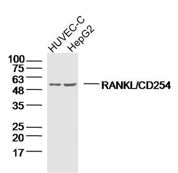
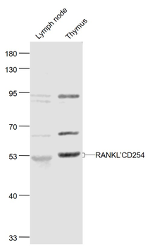
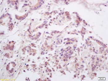
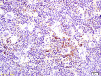
RANKL/CD254 Rabbit Polyclonal Antibody [orb11190]
FC, IF, IHC-Fr, IHC-P, WB
Canine, Rat
Human, Mouse
Rabbit
Polyclonal
Unconjugated
50 μl, 100 μl, 200 μl - Item 1 of 9
TNFR2 Recombinant Rabbit Monoclonal Antibody [orb612211]
IF, IHC-Fr, IHC-P
Mouse, Rat
Human
Rabbit
Recombinant
Unconjugated
50 μl, 100 μl - Item 1 of 7
TNF alpha Rabbit Polyclonal Antibody [orb371962]
ICC, IF, IHC-P
Human, Mouse, Rat
Rabbit
Polyclonal
Unconjugated
100 μg


Quality Guarantee
Explore bioreagents carefree to elevate your research. All our products are rigorously tested for performance. If a product does not perform as described on its datasheet, our scientific support team will provide expert troubleshooting, a prompt replacement, or a refund. For full details, please see our Terms & Conditions and Buying Guide. Contact us at [email protected].
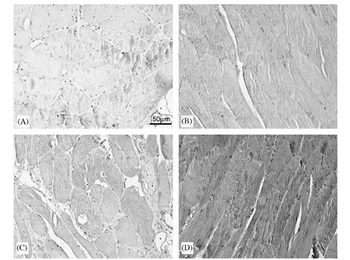
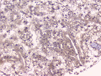
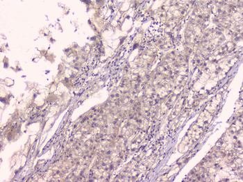
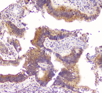
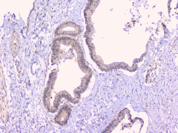
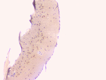
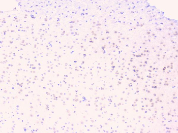
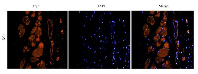
(I) Immunostaining for TNF-a: (A) negative control; (B) muscle from subject 465 yr (positive control); (C) muscle from weight maintainer; and (D) muscle from weight gainer. Anti-human TNF-a antibody (p/n orb345125) at 1:50. The sites of peroxidase binding were demonstrated with diamonobenzidine.
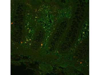
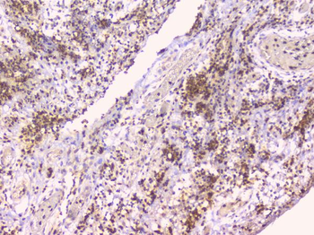
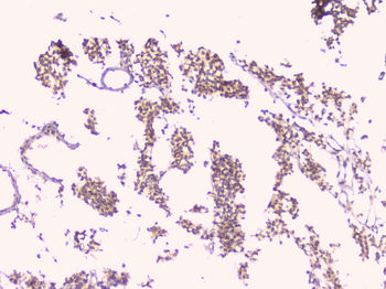
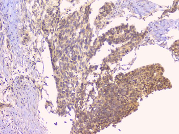
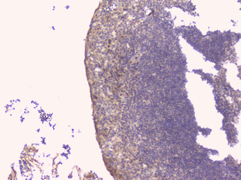
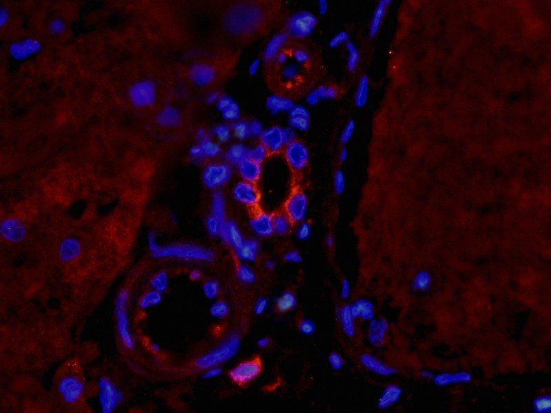
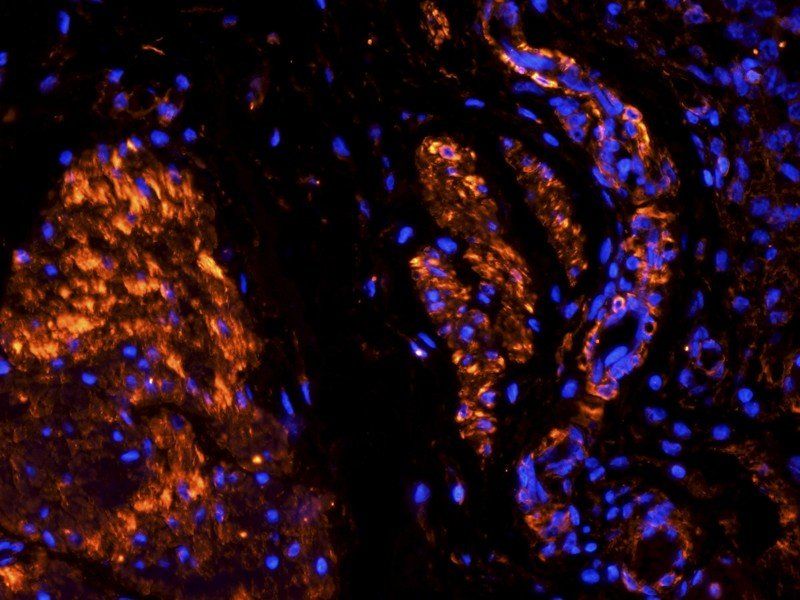
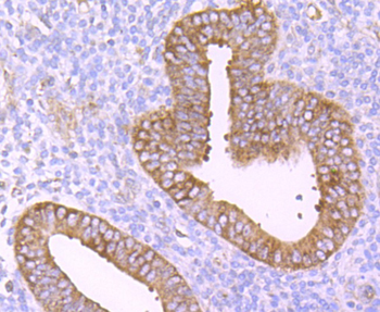
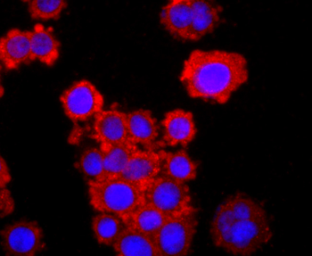
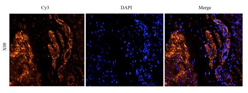
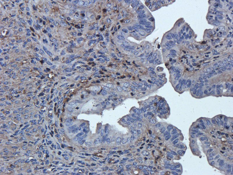
Fluorescent immunohistochemistry showing staining of human colon by Biorbyt's anti-TNF alpha (formalin/PFA-fixed paraffin-embedded sections). Samples were formaldehyde-fixed, then blocked in 10% serum for 2 hours at 20°C. The primary antibody was diluted 1:100 and incubated with the sample for 2 hours at 20°C. Alexa Fluor® 680 goat polyclonal secondary antibody was used diluted 1:5000.
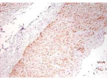
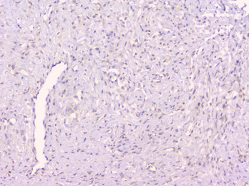
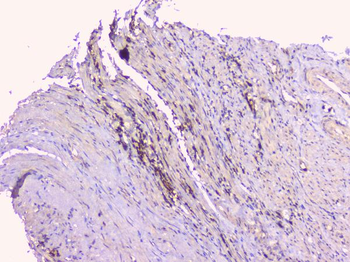
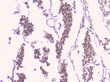
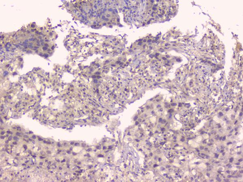
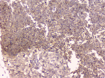
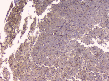
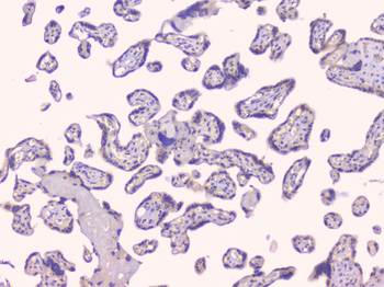
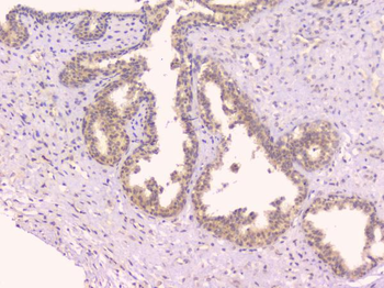
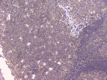
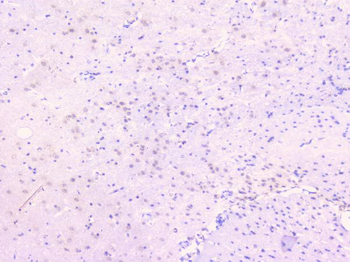
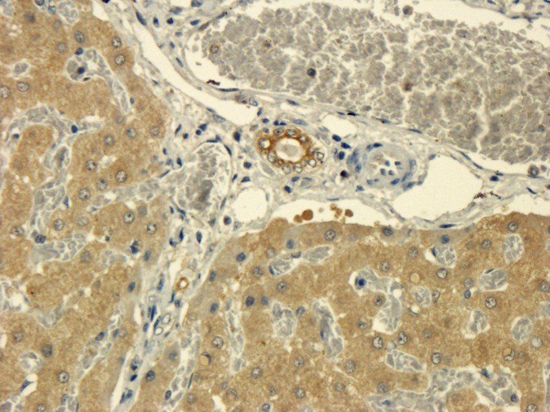
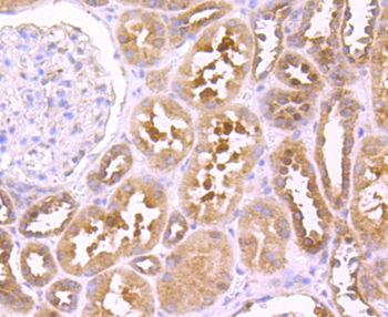
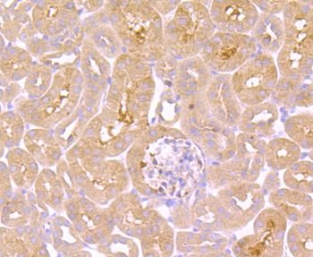
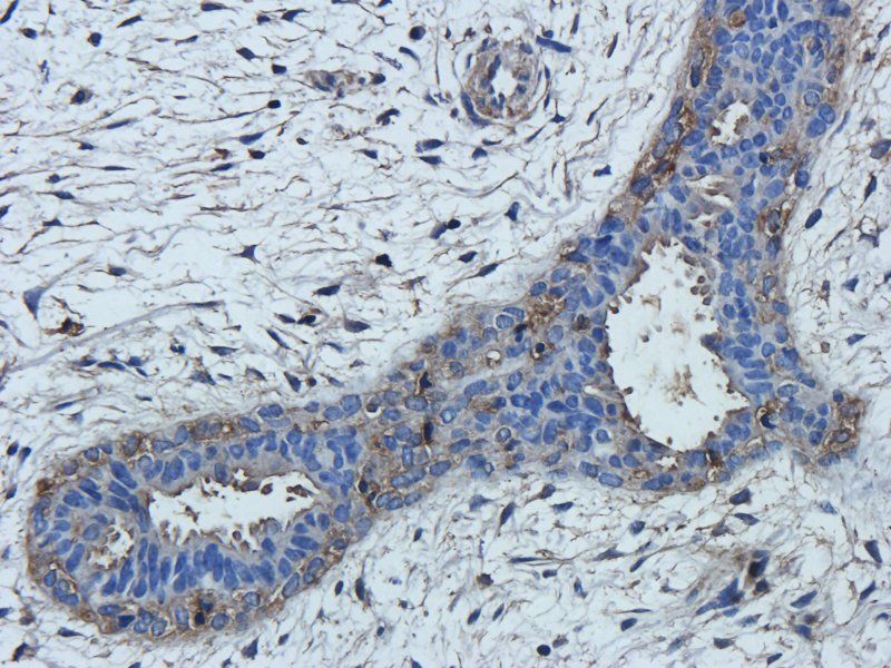
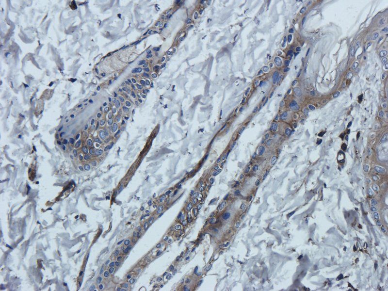
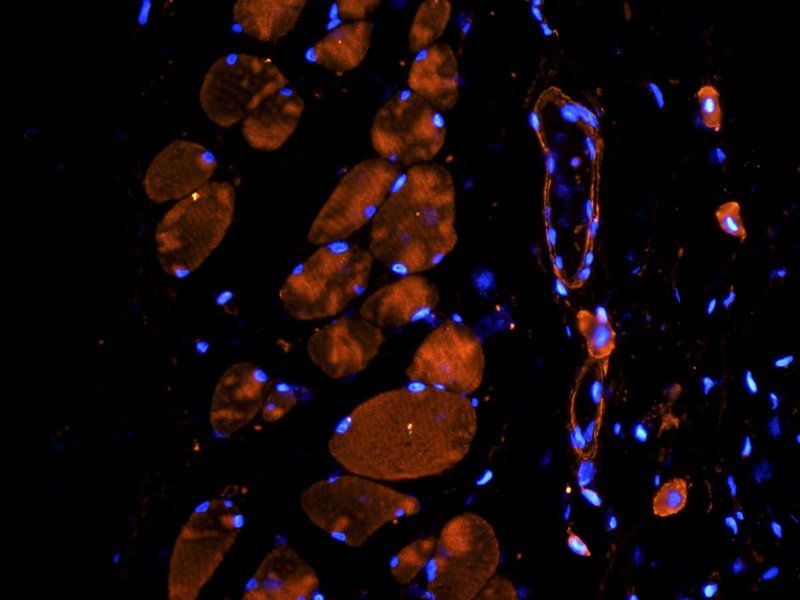
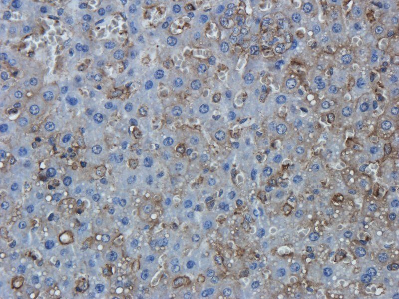
Immunohistochemistry using Biorbyt's polyclonal TNFa antibody showing staining of formalin/PFA-fixed paraffin-embedded sections of human artery tissue sections. Sections were fixed in formaldehyde and subjected to heat mediated antigen retrieval in citrate buffer (pH6.0). Slides were blocked for ten minutes with 1.5% serum. Primary antibody was diluted 1:100 and incubated with samples for 24 hours at 4°C. HRP-conjugated goat anti-rabbit antibody was used as the secondary antibody.
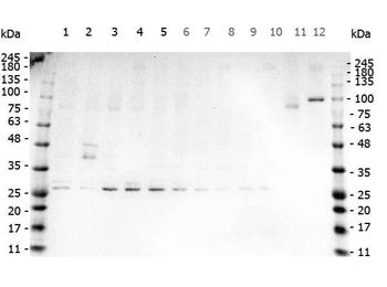
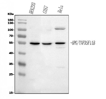
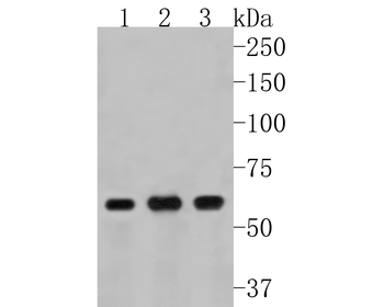
Western Blot of Rabbit anti-TNF Alpha antibody. Marker: Opal Pre-stained ladder. Lane 1: HEK293 lysate (p/n orb348669). Lane 2: HeLa Lysate (p/n orb348668). Lane 3: MCF-7 Lysate (p/n orb348664). Lane 4: Jurkat Lysate. Lane 5: A431 Lysate (p/n orb348665). Lane 6: A549 Lysate (p/n orb348675). Lane 7: LNCap Lysate (p/n orb348694). Lane 8: MOLT-4 Lysate (p/n orb348696). Lane 9: Ramos Lysate. Lane 10: Raji Lysate (p/n orb348672). Lane 11: A-172 Lysate (p/n orb348708). Lane 12: NIH/3T3 Lysate (p/n orb348714). Load: 35 µg per lane. Primary antibody: TNF Alpha antibody at 1 ug/ml overnight at 4C. Secondary antibody: Peroxidase rabbit secondary antibody (p/n orb347654) at 1:30000 for 60 min at RT. Blocking Buffer: 1% Casein-TTBS for 30 min at RT. Predicted/Observed size: 26kDa for TNF Alpha.
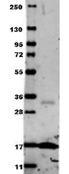
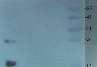
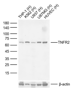
Western blot using Biorbyt's Anti-Human TNF-a (RABBIT) Antibody. Membrane blocked in 1% BSA-TBS-T for 30 min at RT, Rb-a-TNF alpha added at 1:1000 in 1% BSA-TBS-T o/n 4°C, DyLight 649 Gt-a-Rb added at 1:20000 in buffer (p/n orb348637) for 30 min at RT.
Documents Download
Datasheet
Product Information
Request a Document
Protocol Information
WB
Western Blot (IB, immunoblot)
IHC
Immunohistochemistry
IF
Immunofluorescence
ELISA
Enzyme-linked Immunosorbent Assay (EIA)
TNF Antibody (orb420293)

- 0.0
Based on 0 reviews
Participating in our Biorbyt product reviews program enables you to support fellow scientists by sharing your firsthand experience with our products.
Login to Submit a ReviewAvailable Sizes
Select a size below
Choose Conjugation or Carrier Free Version
Free Secondary Antibody (20 ul)0/0
Please add an antibody product to your cart first.