You have no items in your shopping cart.
Description
Research Area
Immunology & Inflammation
Images & Validation
−Item 1 of 6
| Tested Applications | FACS, FC, ICC, IF, IHC, WB |
|---|---|
| Dilution Range | WB (1:500), IHC-P (1:50) |
| Reactivity | Human, Mouse |
| Application Notes |
Key Properties
−| Host | Rabbit |
|---|---|
| Clonality | Polyclonal |
| Immunogen | Developed against a synthetic peptide corresponding to amino acids 420-435 of human TLR4 |
| Target | TLR4 |
| Molecular Weight | 75-80kDa |
| Purification | Protein A Purified |
| Conjugation | Unconjugated |
Storage & Handling
−| Storage | Maintain refrigerated at 2-8°C for up to 2 weeks. For long term storage store at -20°C in small aliquots to prevent freeze-thaw cycles. |
|---|---|
| Buffer/Preservatives | PBS, 50% glycerol, 0.09% sodium azide. Storage buffer changes when conjugated. |
| Concentration | 1 mg/ml |
| Expiration Date | 12 months from date of receipt. |
| Disclaimer | For research use only |
Alternative Names
−ARMD10, CD284, TLR 4, TOLL, Toll like receptor 4
Similar Products
−- Item 1 of 5
TLR4 Rabbit Polyclonal Antibody [orb11489]
FC, ICC, IF, IHC-Fr, IHC-P, WB
Bovine, Canine, Mouse, Porcine, Sheep
Human, Rat
Rabbit
Polyclonal
Unconjugated
50 μl, 100 μl, 200 μl - Item 1 of 5
TLR4 Rabbit Polyclonal Antibody [orb371961]
ICC, IF, IHC-P
Human, Mouse, Porcine, Rat
Rabbit
Polyclonal
Unconjugated
100 μg, 200 μg - Item 1 of 5
- Item 1 of 6
- Item 1 of 6
TLR4 Antibody (Biotin) [orb152202]
FACS, FC, ICC, IF, IHC, WB
Human, Mouse
Rabbit
Polyclonal
Biotin
100 μg


Quality Guarantee
Explore bioreagents carefree to elevate your research. All our products are rigorously tested for performance. If a product does not perform as described on its datasheet, our scientific support team will provide expert troubleshooting, a prompt replacement, or a refund. For full details, please see our Terms & Conditions and Buying Guide. Contact us at [email protected].

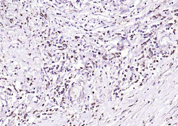
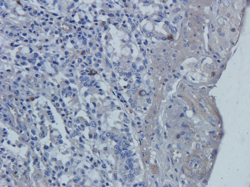
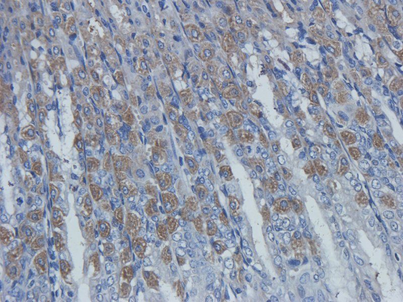
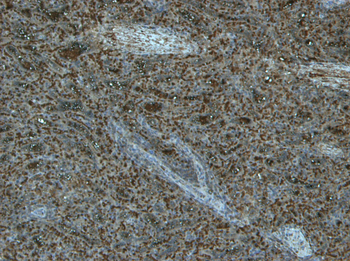
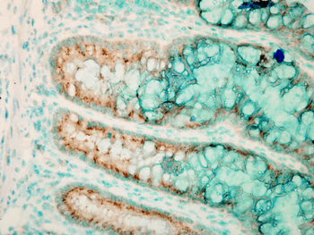
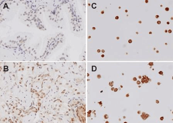
Immunohistochemistry analysis using Rabbit Anti-TLR4 Polyclonal Antibody. Tissue: colon colitis. Species: Mouse. Fixation: Formalin. Primary Antibody: Rabbit Anti-TLR4 Polyclonal Antibody at 1:100000 for 12 hours at 4°C. Secondary Antibody: Biotin Goat Anti-Rabbit at 1:2000 for 1 hour at RT. Counterstain: Methyl Green at 200uL for 2 min at RT. Localization: Inflammatory cells.
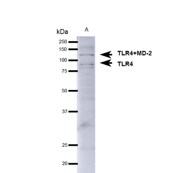
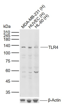
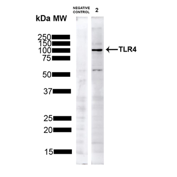
Western blot analysis of Mouse brain showing detection of ~75-80kDa TLR4 protein using Rabbit Anti-TLR4 Polyclonal Antibody. Lane 1: Molecular Weight Ladder. Lane 2: Mouse Brain. Load: 15 μg. Block: 5% Skim Milk in 1X TBST. Primary Antibody: Rabbit Anti-TLR4 Polyclonal Antibody at 1:1000 for 1 hour at RT. Secondary Antibody: Goat Anti-Rabbit IgG: HRP at 1:4000 for 1 hour at RT. Predicted/Observed Size: ~75-80kDa. Other Band (s): ~130kDa (TLR4+MD-2).

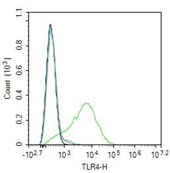
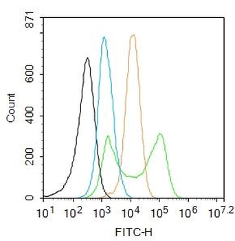
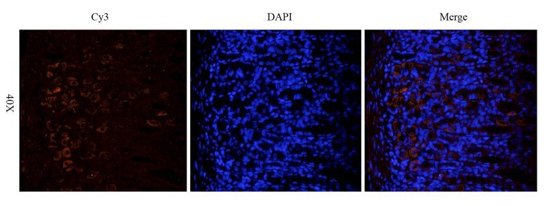
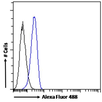
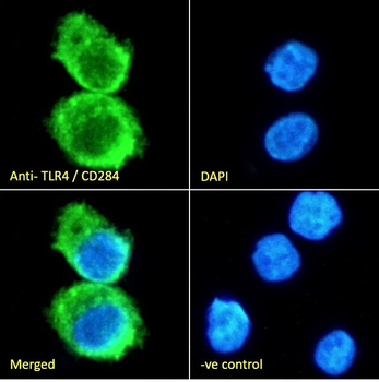
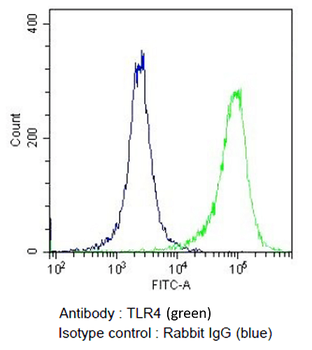
Flow Cytometry analysis using Rabbit Anti-TLR4 Polyclonal Antibody. Tissue: Monocytic Leukemia cells (THP-1). Species: Human. Fixation: 2% Formaldehyde for 10 min at RT. Primary Antibody: Rabbit Anti-TLR4 Polyclonal Antibody at 2 μg/106 cells for 60 min at 37°C. Secondary Antibody: Goat Anti-Rabbit Dylight 488 at 1:200 for 40 min at 37°C.
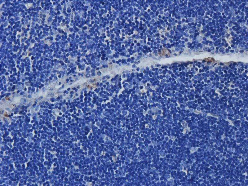
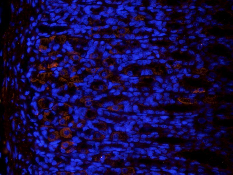
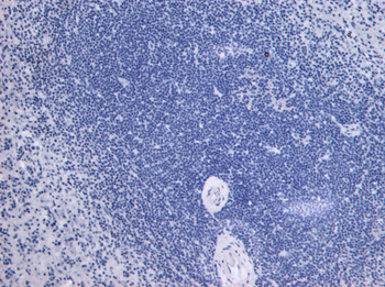
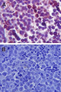
Immunohistochemistry analysis using Rabbit Anti-TLR4 Polyclonal Antibody. Tissue: spleen tissue. Species: Mouse. Primary Antibody: Rabbit Anti-TLR4 Polyclonal Antibody at 1:100.

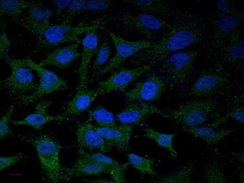
Western blot analysis of Human HeLa showing detection of ~75-80kDa TLR4 protein using Rabbit Anti-TLR4 Polyclonal Antibody. Lane 1: Molecular Weight Ladder. Lane 2: Human HeLa. Load: 10 μg. Block: 5% Skim Milk in 1X TBST. Primary Antibody: Rabbit Anti-TLR4 Polyclonal Antibody at 1:1000 for 1 hour at RT. Secondary Antibody: Goat Anti-Rabbit IgG: HRP at 1:4000 for 1 hour at RT. Predicted/Observed Size: ~75-80kDa. Other Band (s): ~60kDa (observed degradation bands (PMID: 16885150).

Immunohistochemistry analysis using Rabbit Anti-TLR4 Polyclonal Antibody. Tissue: Spleen tissue. Species: Mouse. Primary Antibody: Rabbit Anti-TLR4 Polyclonal Antibody at 1:100.
Quick Database Links
UniProt Details
− No UniProt data available
NCBI Gene Details
− No NCBI Gene data available
NCBI Reference Sequences
−Associated Accession Numbers
Curated reference sequences for the gene transcript and protein product| Protein | NP_612564.1 |
|---|
Documents Download
Datasheet
Product Information
Request a Document
Protocol Information
WB
Western Blot (IB, immunoblot)
IHC
Immunohistochemistry
FACS
Fluorescence-Activated Cell Sorting (FC, Flow cytometry)
FC
Flow Cytometry
IF
Immunofluorescence
ICC
Immunocytochemistry
Filter by Applications
Filter by Species
Shaokang Cai et al. Saikosaponin D Attenuates Postherpetic Neuralgia and Reduces Inflammation by Regulating Gut Microbiota in a Rodent Model Biochem Genet, (2026)
Applications
WB
Reactivity
Mouse
TLR4 Antibody (orb67563)

- 0.0
Based on 0 reviews
Participating in our Biorbyt product reviews program enables you to support fellow scientists by sharing your firsthand experience with our products.
Login to Submit a ReviewAvailable Sizes
Select a size below
Choose Conjugation or Carrier Free Version
Free Secondary Antibody (20 ul)0/0
Please add an antibody product to your cart first.