You have no items in your shopping cart.
Featured
KO/KD
Validated
Validated
Description
Research Area
Immunology & Inflammation
Images & Validation
−Item 1 of 11
| Tested Applications | ELISA, ICC, IF, KO/KD Validated, WB |
|---|---|
| Reactivity | Human, Rat |
| Predicted Reactivity | Mouse |
Key Properties
−| Antibody Type | Primary Antibody |
|---|---|
| Host | Rabbit |
| Clonality | Polyclonal |
| Isotype | IgG |
| Immunogen | Anti-TACE antibody (orb1240074) was raised against a peptide corresponding to 17 amino acids near the carboxy terminus of human TACE. The immunogen is located within the last 50 amino acids of TACE. |
| Target | ADAM17 |
| Molecular Weight | Predicted: 93kDObserved: 93-125kD (Post-modification: 9 N-linked glycosylation) |
| Purification | TACE Antibody is affinity chromatography purified via peptide column. |
| Conjugation | Unconjugated |
Storage & Handling
−| Storage | Maintain refrigerated at 2-8°C for up to 2 weeks. For long term storage store at -20°C in small aliquots to prevent freeze-thaw cycles. |
|---|---|
| Form/Appearance | Liquid |
| Buffer/Preservatives | TACE Antibody is supplied in PBS containing 0.02% sodium azide. |
| Concentration | 1 mg/mL |
| Expiration Date | 12 months from date of receipt. |
| Disclaimer | For research use only |
Alternative Names
−TACE Antibody: CSVP, TACE, NISBD, ADAM18, CD156B, CSVP, Disintegrin and metalloproteinase domain-containing protein 17, Snake venom-like protease, ADAM 17
Similar Products
−- Item 1 of 3
ADAM17 Rabbit Polyclonal Antibody [orb4374]
FC, ICC, IF, IHC-Fr, IHC-P, WB
Bovine, Canine, Porcine, Rabbit
Human, Mouse, Rat
Rabbit
Polyclonal
Unconjugated
50 μl, 100 μl, 200 μl - Item 1 of 1
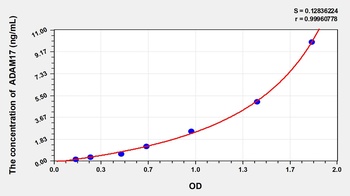
Human A Disintegrin and Metalloprotease 17 (ADAM17) ELISA Kit [orb775182]
Human
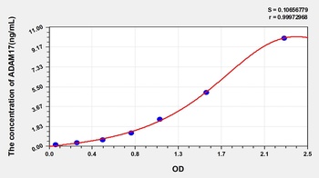
0.16-10 ng/mL
0.052 ng/mL
48 T, 96 T - Item 1 of 1
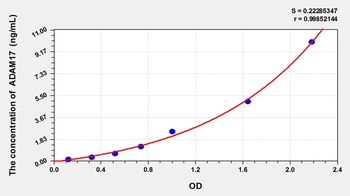
Rat A Disintegrin and Metalloprotease 17 (ADAM17) ELISA Kit [orb780602]
Rat
0.16-10 ng/mL
0.06 ng/mL
48 T, 96 T - Item 1 of 1
Mouse A Disintegrin and Metalloprotease 17 (ADAM17) ELISA Kit [orb779324]
Mouse
0.16-10 ng/mL
0.054 ng/mL
96 T, 48 T - Item 1 of 3
LEF1 Rabbit Polyclonal Antibody [orb763052]
ELISA, FC, ICC, IF, IHC, WB
Human, Mouse, Rat
Rabbit
Polyclonal
Unconjugated
100 μg

Quality Guarantee
Explore bioreagents carefree to elevate your research. All our products are rigorously tested for performance. If a product does not perform as described on its datasheet, our scientific support team will provide expert troubleshooting, a prompt replacement, or a refund. For full details, please see our Terms & Conditions and Buying Guide. Contact us at [email protected].
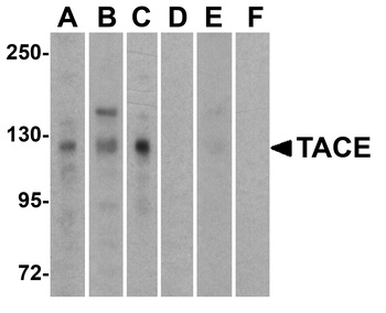
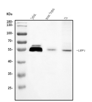
Western Blot Validation of TACE in Human Cell Lines. Loading: 15 µg of lysates per lane. Antibodies: TACE (1 µg/mL), 1h incubation at RT in 5% NFDM/TBST. Secondary: Goat anti-rabbit IgG HRP conjugate at 1:10000 dilution. Lanes: HeLa (A, D), Jurkat (B, E), Raji (C, F) in the absence (A-C) or presence (E-F) of blocking peptide.
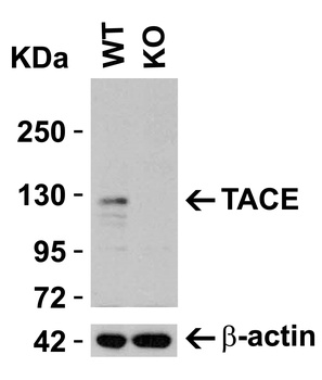
KO Validation in HeLa Cells. Loading: 10 µg of HeLa WT cell lysates or TACE KO cell lysates. Antibodies: TACE orb1240074 (0.25 µg/mL) and beta-actin orb1240312 (1 µg/mL), 1 h incubation at RT in 5% NFDM/TBST. Secondary: Goat Anti-Rabbit IgG HRP conjugate at 1:10000 dilution.
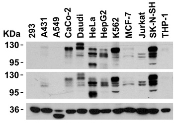
Independent Antibody Validation (IAV) via Protein Expression Profile in Cell Lines. Loading: 15 µg of lysates per lane. Antibodies: TACE orb1240074 (0.5 µg/mL), TACE orb1257204 (2 µg/mL), and GAPDH (0.02 µg/mL), 1h incubation at RT in 5% NFDM/TBST. Secondary: Goat anti-rabbit IgG HRP conjugate at 1:10000 dilution.
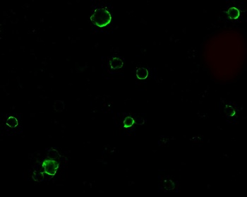
Immunofluorescence Validation of TACE in HeLa Cells. Immunofluorescent analysis of 4% paraformaldehyde-fixed HeLa cells labeling TACE with orb1240074 at 10 µg/mL, followed by goat anti-rabbit IgG secondary antibody at 1/500 dilution (green).
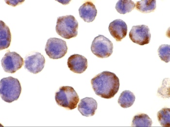
Immunocytochemistry Validation of TACE in HeLa Cells. Immunohistochemical analysis of HeLa cells using anti-TACE antibody (orb1240074) at 10 µg/ml. Cells was fixed with formaldehyde and blocked with 10% serum for 1 h at RT; antigen retrieval was by heat mediation with a citrate buffer (pH6). Samples were incubated with primary antibody overnight at 4°C. A goat anti-rabbit IgG H&L (HRP) at 1/250 was used as secondary. Counter stained with Hematoxylin.
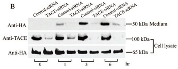
KD Validation of TACE in Monkey COS Cells. (Wang et al., 2006). COS cells stably expressing Pref-1A were transfected with control siRNA or TACE siRNA. TACE was detected in lysates by using the anti-TACE antibody (orb1240074). TACE expression levels were markedly reduced in TACE knockdown cell lysate.
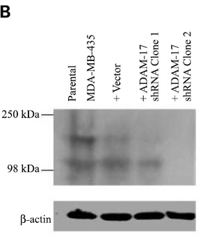
KD Validation of TACE in MDA-MB-435 Cells. (McGowan et al., 2007). ADAM-17 protein expression, following transfection with ADAM-17 shRNA (two clones) or neomycin-resistant negative control vector, was examined by immunoblot analysis with anti-ADAM-17 antibodies (orb1240074).
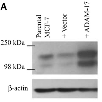
Overexpression Validation of TACE in MCF-7 Cells. (McGowan et al., 2007). ADAM-17 (TACE) protein expression, following transfection of vector and ADAM-17 cDNA, was examined by immunoblot analysis with anti-ADAM-17 (orb1240074) antibodies in MCF-7 cells. Increased ADAM-17 was detected in ADAM-17 transfected cells.
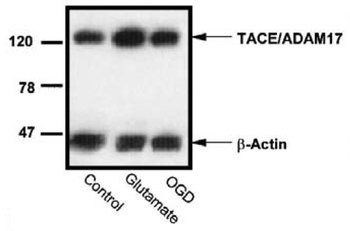
Induced Expression Validation of TACE in Rat Cortical Neurons (Hurtado et al., 2002). Effect of oxygen–glucose deprivation (OGD) or glutamate on the levels of TACE/ADAM17 in rat cortical cultures. Western blot analysis of TACE in homogenates from control, glutamate, and OGD-exposed cultures from a representative experiment.
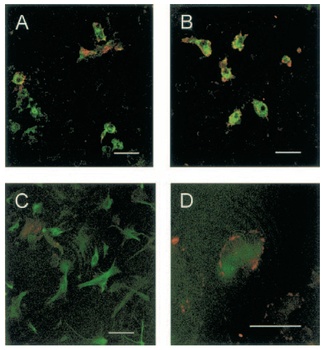
Immunofluorescence Validation of TACE in Rat Cortical Neurons (Hurtado et al., 2002). Double immunostaining of control and glutamate-exposed rat cortical cultures. (A) Control cultures show TACE immunoreactivity at the cellular membrane of some microglial cells (B) Glutamate-exposed cultures show that most microglial cells express TACE immunoreactivity. (C) Control cultures show that TACE immunostaining does not colocalize with astrocytes [glial fibrillary acidic protein (GFAP) -positive cells]. (D) Astrocyte (GFAP-positive cell) showing TACE immunoreactivity in its surface after treatment with glutamate.
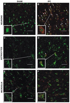
Immunofluorescence Validation of TACE in Rat Brain (Pradillo et al, 2005). Cellular localization of TACE. Double immunofluorescence staining of brain sections from sham-operated (SHAM; A, C, E) and IPC-exposed animals (IPC; B, D, F) of TACE (red) and the cellular markers (green) NeuN (neurons; A, B), GFAP (astrocytes; C, D) and L.esculentum lectin (microglia and endothelium; E, F). White arrows indicate TACE-positive cells.
Documents Download
Datasheet
Product Information
Request a Document
Protocol Information
WB
Western Blot (IB, immunoblot)
IF
Immunofluorescence
ICC
Immunocytochemistry
ELISA
Enzyme-linked Immunosorbent Assay (EIA)
ADAM17 Antibody (orb1240074)

- 0.0
Based on 0 reviews
Participating in our Biorbyt product reviews program enables you to support fellow scientists by sharing your firsthand experience with our products.
Login to Submit a ReviewAvailable Sizes
Select a size below
Free Secondary Antibody (20 ul)0/0
Please add an antibody product to your cart first.