You have no items in your shopping cart.
Featured
KO/KD
Validated
Validated
Description
Research Area
Cell Biology
Images & Validation
−Item 1 of 10
| Tested Applications | ELISA, IF, IHC-P, IP, KO/KD Validated, WB |
|---|---|
| Reactivity | Human, Mouse, Rat |
Key Properties
−| Antibody Type | Primary Antibody |
|---|---|
| Host | Rabbit |
| Clonality | Polyclonal |
| Isotype | IgG |
| Immunogen | Anti-Smac antibody (orb1240005) was raised against a peptide corresponding to 16 amino acids near the carboxy terminus of murine Smac. The immunogen is located within the last 50 amino acids of Smac. |
| Target | Diablo |
| Molecular Weight | Predicted: 27kDObserved: 20 kD |
| Purification | Smac Antibody is DEAE purified. |
| Conjugation | Unconjugated |
Storage & Handling
−| Storage | Maintain refrigerated at 2-8°C for up to 2 weeks. For long term storage store at -20°C in small aliquots to prevent freeze-thaw cycles. |
|---|---|
| Form/Appearance | Liquid |
| Buffer/Preservatives | Smac Antibody is supplied in PBS containing 0.02% sodium azide. |
| Concentration | 1 mg/ml |
| Expiration Date | 12 months from date of receipt. |
| Disclaimer | For research use only |
Alternative Names
−Smac Antibody: Smac, AU040403, 0610041G12Rik, 1700006L01Rik, Smac, Diablo homolog, mitochondrial, Direct IAP-binding protein with low pI
Similar Products
−- Item 1 of 10
Diablo Antibody [orb1240004]
ELISA, IF, IHC-P, IP, KO/KD Validated, WB
Human, Mouse, Rat
Rabbit
Polyclonal
Unconjugated
0.02 mg, 0.1 mg - Item 1 of 6
Smac/Diablo Rabbit Polyclonal Antibody [orb334593]
ELISA, FC, ICC, IF, IHC, IP, WB
Human, Mouse, Rat
Rabbit
Polyclonal
Unconjugated
100 μg - Item 1 of 1
- Item 1 of 1
- Item 1 of 4


Quality Guarantee
Explore bioreagents carefree to elevate your research. All our products are rigorously tested for performance. If a product does not perform as described on its datasheet, our scientific support team will provide expert troubleshooting, a prompt replacement, or a refund. For full details, please see our Terms & Conditions and Buying Guide. Contact us at [email protected].
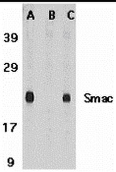
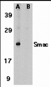
Western Blot Validation in (A and B) Mouse Heart Tissue Lysate and (C) Rat Heart Tissue Lysate. Loading: 15 µg of lysates per lane. Antibodies: Smac orb1240005 (1 µg/mL), 1h incubation at RT in 5% NFDM/TBST. Secondary: Goat anti-rabbit IgG HRP conjugate at 1:10000 dilution. A Mouse heartB Mouse heart and blocking peptideC Rat heart.
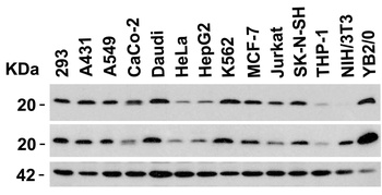
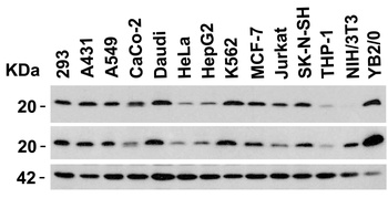
Independent Antibody Validation (IAV) via Protein Expression Profile in Cell Lines. Loading: 15 µg of lysates per lane. Antibodies: Smac orb1240004 (1 µg/mL), Smac orb1240005 (1 µg/mL), and beta-actin (1 µg/mL), 1h incubation at RT in 5% NFDM/TBST. Secondary: Goat anti-rabbit IgG HRP conjugate at 1:10000 dilution.
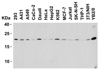
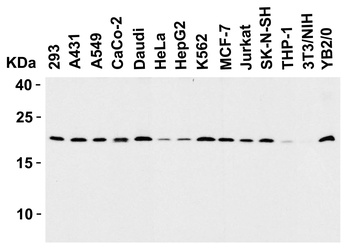
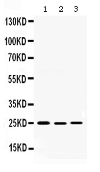
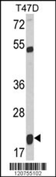
Western Blot Validation in Human, Mouse and Rat Cell Lines. Loading: 15 µg of lysates per lane. Antibodies: Smac orb1240005 (1 µg/mL), 1h incubation at RT in 5% NFDM/TBST. Secondary: Goat anti-rabbit IgG HRP conjugate at 1:10000 dilution.
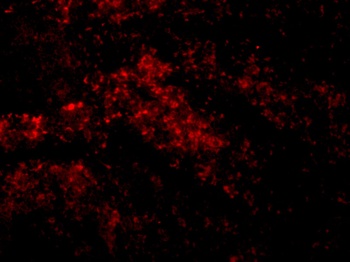
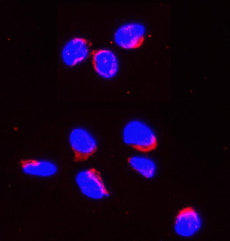
Immunofluorescence Validation of Smac in Mouse Spleen Cells. Immunofluorescent analysis of 4% paraformaldehyde-fixed Mouse Spleen Cells labeling Smac with orb1240005 at 10 µg/mL, followed by goat anti-rabbit IgG secondary antibody at 1/500 dilution (red).
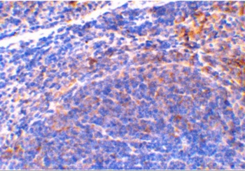
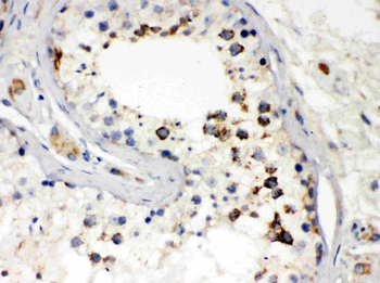
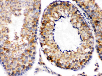
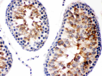
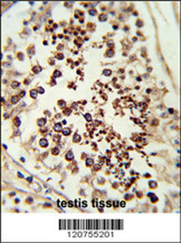
Immunohistochemistry Validation of Smac in Mouse Spleen Tissue. Immunohistochemical analysis of paraffin-embedded Mouse Spleen Tissue using anti-Smac antibody (orb1240005) at 2 µg/ml. Tissue was fixed with formaldehyde and blocked with 10% serum for 1 h at RT; antigen retrieval was by heat mediation with a citrate buffer (pH6). Samples were incubated with primary antibody overnight at 4°C. A goat anti-rabbit IgG H&L (HRP) at 1/250 was used as secondary. Counter stained with Hematoxylin.
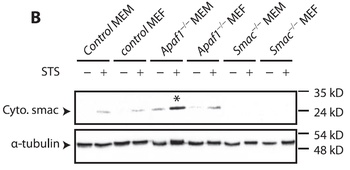
KO Validation in Mouse Fibroblasts and Myoblasts (Ho et al., 2007). The indicated MEFs or MEMs were exposed to 2 µM STS for 4 h and analyzed by Western blot. Accumulation of Smac/Diablo in mitochondrion-depleted cytosol fractions fromSTS-treated Apaf-1 KO cells were detected by anti-smac antibodies. Smac expression was not detected in smac KO mice.
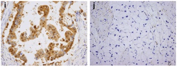
Immunohistochemistry Validation of Smac in Human gastric carcinoma (Kim et al., 2011). Smac was highly expressed in gastric mucosa of patients with gastric carcinoma.

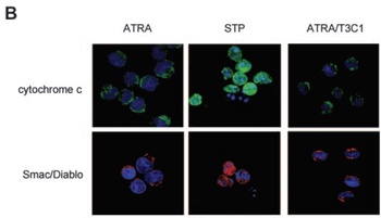
Immunofluorescence Analysis of Smac in NB4-LR1 Cells (Saumet et al., 2005). NB4-LR1 cells were either treated with ATRA (1 µM) for 3 days without or with the T3C1 recombinant fragment (3 µM) or treated with staurosporine (STP; 5 µM) for 3.5 hours. STP, but not ATRA or AYRA/T3C1 induced the release of smac.

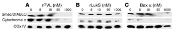
Induced Expression Validation in Rat Liver (Genestier et al., 2005). Mitochondria from rat liver were treated with increasing concentrations of rPVL (A), rLukS (B), or Bax alpha (C) for 1 hour at 30 °C. rPVL induces the release of the apoptogenic proteins cytochrome c and Smac/DIABLO from isolated mitochondria.

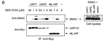
Overxpression Validation in HEK293T Cells (Flygare et al., 2012). HEK293T cells were transiently transfected with Smac and Myc-tagged cIAP1, cIAP2, ML-IAP, or empty vector. Cells were lysed, and lysates were incubated with the indicated concentrations of 1 and immunoprecipitated with anti-Myc antibody (left panels). Samples were then immunoblotted with anti-Smac and anti-Myc antibodies. Whole-cell lysates are shown in the right panel.
Documents Download
Datasheet
Product Information
Request a Document
Protocol Information
WB
Western Blot (IB, immunoblot)
IHC-P
Immunohistochemistry Paraffin
IF
Immunofluorescence
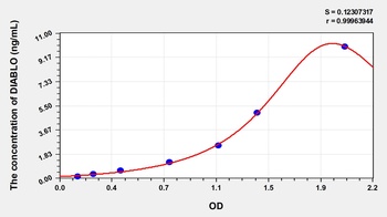
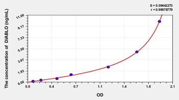
ELISA
Enzyme-linked Immunosorbent Assay (EIA)
IP
Immunoprecipitation
Diablo Antibody (orb1240005)

- 0.0
Based on 0 reviews
Participating in our Biorbyt product reviews program enables you to support fellow scientists by sharing your firsthand experience with our products.
Login to Submit a ReviewAvailable Sizes
Select a size below
Free Secondary Antibody (20 ul)0/0
Please add an antibody product to your cart first.