You have no items in your shopping cart.
Featured
Description
Research Area
Immunology & Inflammation
Images & Validation
−Item 1 of 7
| Tested Applications | FC, ICC, IHC, WB |
|---|---|
| Dilution Range | WB: 1/500 - 1/2000; IHC: 1/200 - 1/1000; IF/ICC: 1/200 - 1/1000; FCM: 1/200 - 1/400; ELISA: 1/10000 |
| Reactivity | Human, Mouse |
Key Properties
−| Antibody Type | Primary Antibody |
|---|---|
| Host | Mouse |
| Clonality | Monoclonal |
| Isotype | Mouse IgG1 |
| Clone No. | B3C5J6 |
| Immunogen | Purified recombinant fragment of human SDC1 (AA: 28-171) expressed in E. Coli. |
| Target | SDC1 |
| Molecular Weight | 32.5kDa |
| Conjugation | Unconjugated |
Storage & Handling
−| Storage | Maintain refrigerated at 2-8°C for up to 2 weeks. For long term storage store at -20°C in small aliquots to prevent freeze-thaw cycles. |
|---|---|
| Buffer/Preservatives | Purified antibody in PBS with 0.05% sodium azide. |
| Concentration | Approximately 1mg/ml (varies from batch to batch). Please inquire for precise concentration. |
| Expiration Date | 12 months from date of receipt. |
| Disclaimer | For research use only |
Alternative Names
−SDC; CD138; SYND1; syndecan
Similar Products
−- Item 1 of 7
CD138 Antibody (C-term) (Ascites) [orb652175]
FC, IF, WB
Human
Mouse
Monoclonal
Unconjugated
50 μl, 100 μl - Item 1 of 8
CD138 Recombinant Rabbit Monoclonal Antibody [orb2563454]
IF, IHC-Fr, IHC-P
Mouse
Human
Rabbit
Recombinant
Unconjugated
100 μl, 50 μl, 25 μl - Item 1 of 4
Syndecan-1 Rabbit Polyclonal Antibody [orb10289]
IF, IHC-Fr, IHC-P, WB
Bovine, Canine, Equine, Gallus, Porcine, Rabbit, Rat
Human, Mouse
Rabbit
Polyclonal
Unconjugated
50 μl, 100 μl, 200 μl - Item 1 of 1
- Item 1 of 4
SDC1 Antibody [orb1410037]
IHC
Human
Mouse
Monoclonal
Unconjugated
20 μg, 100 μg, 100 μg (without BSA and Azide)


Quality Guarantee
Explore bioreagents carefree to elevate your research. All our products are rigorously tested for performance. If a product does not perform as described on its datasheet, our scientific support team will provide expert troubleshooting, a prompt replacement, or a refund. For full details, please see our Terms & Conditions and Buying Guide. Contact us at [email protected].
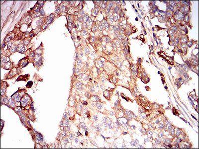
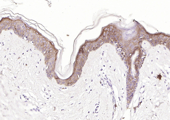
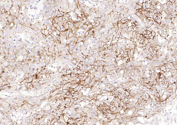
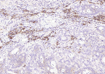
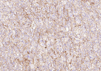
Immunohistochemical analysis of formalin-fixed and paraffin-embedded ovarian cancer tissues using Syndecan 1 antibody
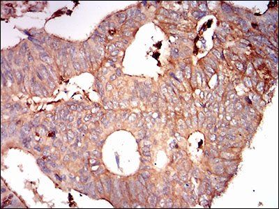
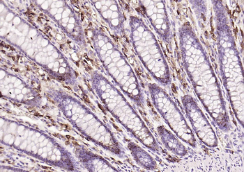
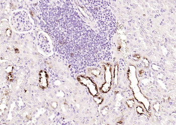
Immunohistochemical analysis of formalin-fixed and paraffin-embedded rectum cancer tissues using Syndecan 1 antibody
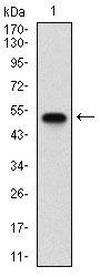
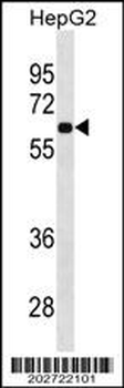
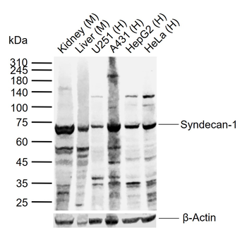
Western blot analysis of human SDC1 using Syndecan 1 antibody
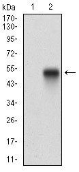
Western blot analysis of HEK293 (Lane 1) HEK293 (Lane 2) cell lysate using Syndecan 1 antibody
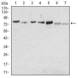
Western blot analysis of MCF-7 (Lane 1), Hela (Lane 2), HepG2 (Lane 3), T47D (Lane 4), SW620 (Lane 5), Jurkat (Lane 6) NIH/3T3 (Lane 7) cell lysate using Syndecan 1 antibody
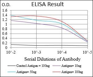
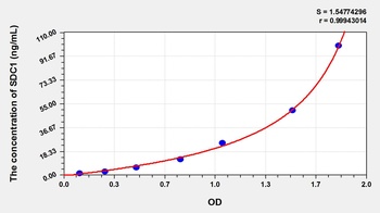
Line graph illustrates about the Ag-Ab reactions using different concentrations of antigen and serial dilutions of Syndecan 1 antibody
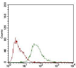
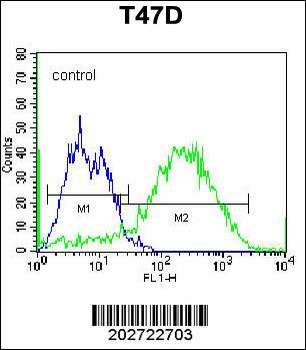
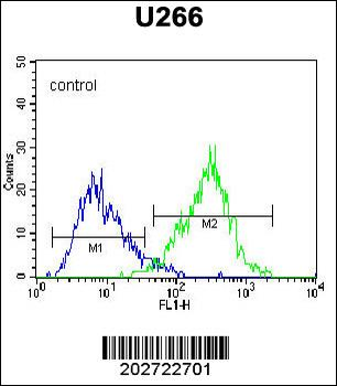
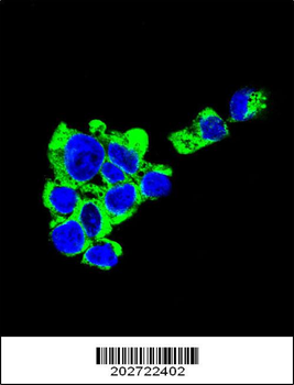
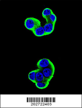
Flow cytometric analysis of HepG2 cells lysates using Syndecan 1 antibody
Documents Download
Datasheet
Product Information
Request a Document
Protocol Information
WB
Western Blot (IB, immunoblot)
IHC
Immunohistochemistry
FC
Flow Cytometry
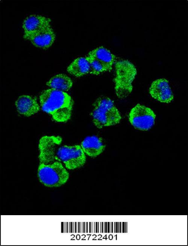
ICC
Immunocytochemistry
SDC1 Antibody (orb153547)

- 0.0
Based on 0 reviews
Participating in our Biorbyt product reviews program enables you to support fellow scientists by sharing your firsthand experience with our products.
Login to Submit a ReviewAvailable Sizes
Select a size below
Choose Conjugation or Carrier Free Version
Free Secondary Antibody (20 ul)0/0
Please add an antibody product to your cart first.