You have no items in your shopping cart.
Description
Research Area
Stem Cell & Developmental Biology
Images & Validation
−Item 1 of 5
| Tested Applications | FC, IHC-P, WB |
|---|---|
| Dilution Range | WB - 1:4000, IHC-P - 1:100, FC - 1:25 |
| Reactivity | Human, Mouse |
Key Properties
−| Host | Rabbit |
|---|---|
| Clonality | Polyclonal |
| Isotype | Rabbit IgG |
| Immunogen | This ROR1 antibody is generated from rabbits immunized with recombinant human ROR1 protein (aa region: 112 - 399). |
| Target | ROR1 |
| Molecular Weight | 104283 Da |
| Conjugation | Unconjugated |
Storage & Handling
−| Storage | Maintain refrigerated at 2-8°C for up to 2 weeks. For long term storage store at -20°C in small aliquots to prevent freeze-thaw cycles |
|---|---|
| Form/Appearance | Purified polyclonal antibody supplied in PBS with 0.09% (W/V) sodium azide. This antibody is purified through a protein A column, followed by peptide affinity purification. |
| Expiration Date | 12 months from date of receipt. |
| Disclaimer | For research use only |
Alternative Names
−Tyrosine-protein kinase transmembrane receptor ROR1, Neurotrophic tyrosine kinase, receptor-related 1, ROR1, NTRKR1
Similar Products
−- Item 1 of 6
ROR1 Rabbit Polyclonal Antibody [orb11664]
IHC-P, WB
Human, Mouse, Rat
Rabbit
Polyclonal
Unconjugated
100 μg - Item 1 of 5
- Item 1 of 5
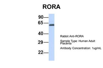
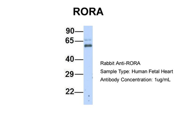
RORA Rabbit Polyclonal Antibody [orb329792]
WB
Bovine, Canine, Equine, Goat, Guinea pig, Rabbit, Rat, Zebrafish
Human, Mouse
Rabbit
Polyclonal
Unconjugated
100 μl - Item 1 of 1
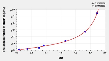
Human Receptor Tyrosine Kinase Like Orphan Receptor 1 (ROR1) ELISA Kit [orb778708]
Human
0.16-10 ng/mL
0.057 ng/mL
48 T, 96 T - Item 1 of 1
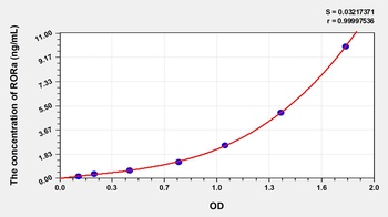
Human RAR Related Orphan Receptor Alpha (RORa) ELISA Kit [orb779150]
Human
0.16-10 ng/mL
0.057 ng/mL
96 T, 48 T

Quality Guarantee
Explore bioreagents carefree to elevate your research. All our products are rigorously tested for performance. If a product does not perform as described on its datasheet, our scientific support team will provide expert troubleshooting, a prompt replacement, or a refund. For full details, please see our Terms & Conditions and Buying Guide. Contact us at [email protected].
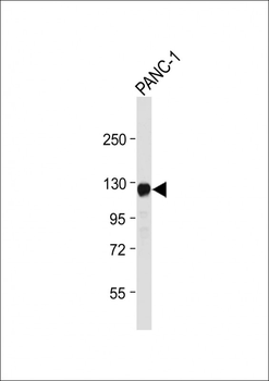
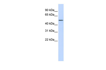
Anti-ROR1 Antibody at 1:4000 dilution + PANC-1 whole cell lysate. Lysates/proteins at 20 µg per lane. Secondary Goat Anti-Rabbit IgG, (H+L), Peroxidase conjugated at 1/10000 dilution. Predicted band size: 104 kDa. Blocking/Dilution buffer: 5% NFDM/TBST.
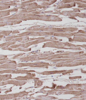
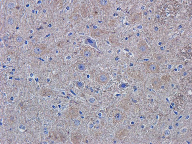
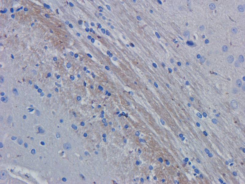
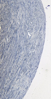
Immunohistochemical analysis on paraffin-embedded Human heart tissue. Tissue was fixed with formaldehyde at room temperature. Heat induced epitope retrieval was performed by EDTA buffer (pH9.0). Samples were incubated with primary antibody (1:100) for 1 hour at room temperature. Undiluted CRF Anti-Polyvalent HRP Polymer antibody was used as the secondary antibody.
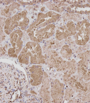
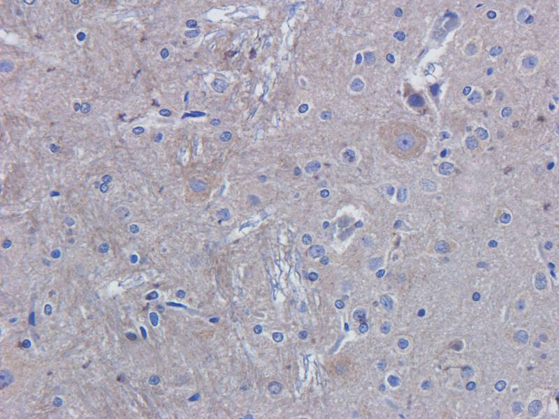
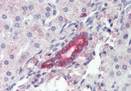
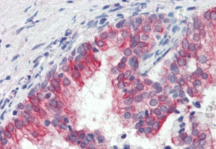
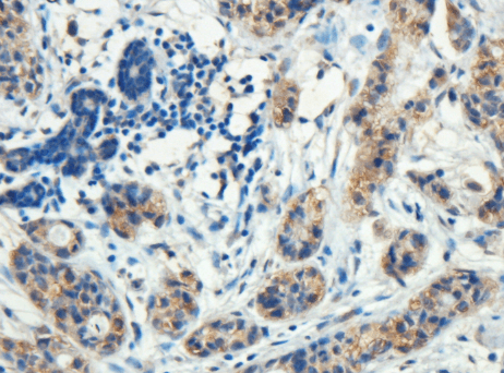
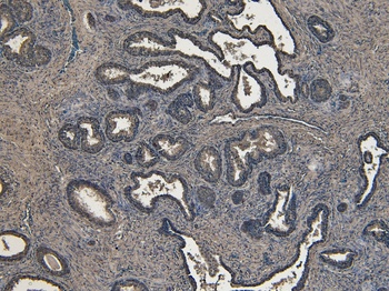
Immunohistochemical analysis on paraffin-embedded Human kidney tissue. Tissue was fixed with formaldehyde at room temperature. Heat induced epitope retrieval was performed by EDTA buffer (pH9.0). Samples were incubated with primary antibody (1:100) for 1 hour at room temperature. Undiluted CRF Anti-Polyvalent HRP Polymer antibody was used as the secondary antibody.
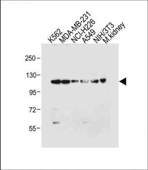
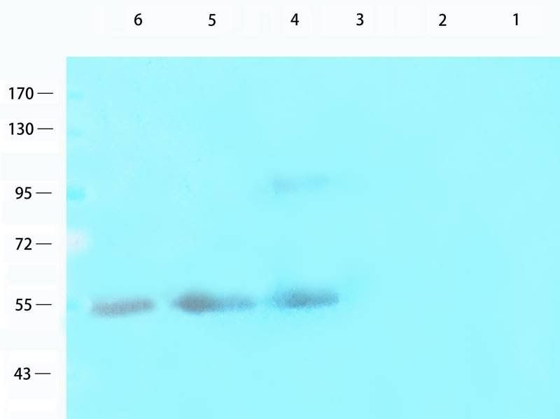
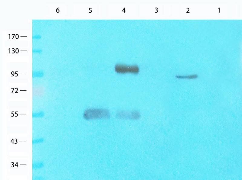
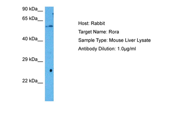
All lanes: Anti-ROR1 Antibody at 1:4000 dilution. Lane 1: K562 whole cell lysate. Lane 2: MDA-MB-231 whole cell lysate. Lane 3: NCI-H226 whole cell lysate. Lane 4: A549 whole cell lysate. Lane 5: NIH/3T3 whole cell lysate. Lane 6: Mouse kidney tissue lysate. Lysates/proteins at 20 µg per lane. Secondary Goat Anti-Rabbit IgG, (H+L), Peroxidase conjugated at 1/10000 dilution. Predicted band size: 104 kDa. Blocking/Dilution buffer: 5% NFDM/TBST.
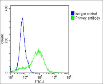
Overlay histogram showing A549 cells stained (green line). The cells were fixed with 2% paraformaldehyde (10 min). The cells were then icubated in 2% bovine serum albumin to block non-specific protein-protein interactions followed by the antibody (1:25 dilution) for 60 min at 37°C. The secondary antibody used was Goat-Anti-Rabbit IgG, DyLight 488 Conjugated Highly Cross-Adsorbed at 1/200 dilution for 40 min at 37°C. Isotype control antibody (blue line) was rabbit IgG1 (1 μg/1x10^6 cells) used under the same conditions. Acquisition of > 10000 events was performed.
Quick Database Links
UniProt Details
− No UniProt data available
NCBI Reference Sequences
−Associated Accession Numbers
Curated reference sequences for the gene transcript and protein product| Protein | NP_001077061.1, NP_005003.2 |
|---|
Documents Download
Datasheet
Product Information
Request a Document
Protocol Information
WB
Western Blot (IB, immunoblot)
IHC-P
Immunohistochemistry Paraffin
FC
Flow Cytometry
ROR1 Antibody (orb1928997)

- 0.0
Based on 0 reviews
Participating in our Biorbyt product reviews program enables you to support fellow scientists by sharing your firsthand experience with our products.
Login to Submit a ReviewAvailable Sizes
Select a size below
Choose Conjugation or Carrier Free Version
Free Secondary Antibody (20 ul)0/0
Please add an antibody product to your cart first.