You have no items in your shopping cart.
Description
Images & Validation
−Item 1 of 3
| Tested Applications | ELISA, WB |
|---|---|
| Dilution Range | ELISA: 1:2,000 - 1:10,000, WB: 1:1000-1:10,000 |
| Reactivity | Other |
| Application Notes |
Key Properties
−| Antibody Type | Primary Antibody |
|---|---|
| Host | Goat |
| Clonality | Polyclonal |
| Isotype | IgG |
| Immunogen | The immunogen is a Red Fluorescent Protein (RFP) fusion protein corresponding to the full length amino acid sequence (234aa) derived from the mushroom polyp coral Discosoma. |
| Target | DsRed |
| Purity | Anti-RFP (GOAT) is an IgG fraction purified from monospecific antiserum by a multi-step process which includes delipidation, salt fractionation and ion exchange chromatography followed by extensive dialysis against the buffer stated above. Assay by immunoelectrophoresis resulted in a single precipitin arc against anti-Goat Serum and purified and partially purified Red Fluorescent Protein (Discosoma). No reaction was observed against Human, Mouse or Rat serum proteins. Expect reactivity against RFP and its variants: mCherry, tdTomato, mBanana, mOrange, mPlum, mOrange and mStrawberry. |
| Conjugation | Unconjugated |
Storage & Handling
−| Storage | Store vial at -20° C prior to opening. Aliquot contents and freeze at -20° C or below for extended storage. Avoid cycles of freezing and thawing. Centrifuge product if not completely clear after standing at room temperature. This product is stable for several weeks at 4° C as an undiluted liquid. Dilute only prior to immediate use. |
|---|---|
| Form/Appearance | Liquid (sterile filtered) |
| Buffer/Preservatives | Preservative: 0.01% (w/v) Sodium Azide. Stabilizer: None; Buffer: 0.02 M Potassium Phosphate, 0.15 M Sodium Chloride, pH 7.2 |
| Concentration | 1 mg/mL |
| Expiration Date | 12 months from date of receipt. |
| Dry Ice Shipping | Please note: This product requires shipment on dry ice. A dry ice surcharge will apply. |
| Disclaimer | For research use only |
Alternative Names
−goat anti-RFP antibody, DsRed, rDsRed, Discosoma sp. Red Fluorescent Protein, Red fluorescent protein drFP583
Similar Products
−- Item 1 of 45
DsRed Antibody [orb345391]
ELISA, FC, IF, IHC, KO/KD Validated, WB
Other
Rabbit
Polyclonal
Unconjugated
100 μg - Item 1 of 45
DsRed Antibody [orb345392]
ELISA, FC, IF, IHC, KO/KD Validated, WB
Other
Rabbit
Polyclonal
Unconjugated
25 μl - Item 1 of 2
tdTomato Antibody [orb182397]
ELISA, FACS, IF, IHC-Fr, IHC-P, WB
Other
Goat
Polyclonal
Unconjugated
100 μg - Item 1 of 4
mCherry Goat Polyclonal Antibody [orb11618]
FC, ICC, IEM, IF, IHC-Fr, IHC-P, WB
Other
Goat
Polyclonal
Unconjugated
20 μg, 100 μg - Item 1 of 12


Quality Guarantee
Explore bioreagents carefree to elevate your research. All our products are rigorously tested for performance. If a product does not perform as described on its datasheet, our scientific support team will provide expert troubleshooting, a prompt replacement, or a refund. For full details, please see our Terms & Conditions and Buying Guide. Contact us at [email protected].

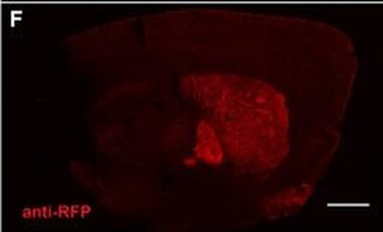
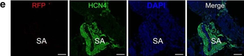
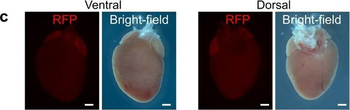
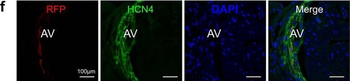
Population of CD11b + myeloid progenitor cells differentiate into SMA + stromal cells within tumors and in vitro. (A) Representative image of red fluorescent protein (RFP) + stromal cells in tumor from CCR2-RFP heterozygous SCID mouse. (B) RFP+ SMA + double positive cells within tumor stroma. (C) CD11b + SMA + double positive cells within tumor stroma. (D) CD45 + CD11b + CD34 + myeloid progenitor cells in the mammary gland at 1.5 (n = 5 empty vector (EV), 7 CCL2) and 2.5 weeks (n = 5 EV, 7 CCL2) post-transplantation were quantified by flow cytometry. (E) CD45 + CD11b + CD34 + myeloid progenitor cells in the bone marrow at 1.5 weeks (n = 6 EV, 7 CCL2) and 2.5 weeks (n = 3 mice/group) post-transplantation were quantified by flow cytometry. (F) Representative brightfield image of colony formed by CD45 + CD11b + CD34 + myeloid progenitor cells isolated using fluorescence-activated cell sorting (FACS). (G) Colonies in culture co-stained with SMA and collagen I. Statistical differences determined by Mann–Whitney U test. Magnification bars = 50 µm.

Population of CD11b + myeloid progenitor cells differentiate into SMA + stromal cells within tumors and in vitro. (A) Representative image of red fluorescent protein (RFP) + stromal cells in tumor from CCR2-RFP heterozygous SCID mouse. (B) RFP+ SMA + double positive cells within tumor stroma. (C) CD11b + SMA + double positive cells within tumor stroma. (D) CD45 + CD11b + CD34 + myeloid progenitor cells in the mammary gland at 1.5 (n = 5 empty vector (EV), 7 CCL2) and 2.5 weeks (n = 5 EV, 7 CCL2) post-transplantation were quantified by flow cytometry. (E) CD45 + CD11b + CD34 + myeloid progenitor cells in the bone marrow at 1.5 weeks (n = 6 EV, 7 CCL2) and 2.5 weeks (n = 3 mice/group) post-transplantation were quantified by flow cytometry. (F) Representative brightfield image of colony formed by CD45 + CD11b + CD34 + myeloid progenitor cells isolated using fluorescence-activated cell sorting (FACS). (G) Colonies in culture co-stained with SMA and collagen I. Statistical differences determined by Mann–Whitney U test. Magnification bars = 50 µm.

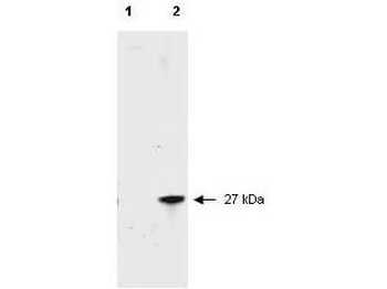
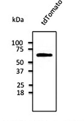
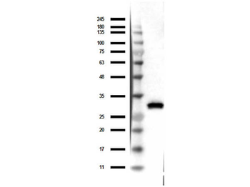
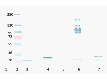
Western Blot of Goat Anti-RFP Antibody. Lane 1: 50 ng of RFP protein (p/n orb345960). Primary Antibody: Goat Anti-RFP at 1:10000 overnight at 2-8°C. Secondary Antibody: Donkey anti-Goat IgG HRP (p/n orb347032) at 1:40000 for 30 min at RT. Blocking buffer: BlockOut® (p/n orb348644). Predicted MW: ~27kDa.
Quick Database Links
Gene Symbol
DsRed
UniProt
UniProt Details
− No UniProt data available
Documents Download
Datasheet
Product Information
Request a Document
Protocol Information
WB
Western Blot (IB, immunoblot)
ELISA
Enzyme-linked Immunosorbent Assay (EIA)
DsRed Antibody (orb420218)

- 0.0
Based on 0 reviews
Participating in our Biorbyt product reviews program enables you to support fellow scientists by sharing your firsthand experience with our products.
Login to Submit a ReviewAvailable Sizes
Select a size below
Choose Conjugation or Carrier Free Version
Free Secondary Antibody (20 ul)0/0
Please add an antibody product to your cart first.