You have no items in your shopping cart.
Description
Research Area
Signal Transduction
Images & Validation
−Item 1 of 5
| Tested Applications | FC, ICC, IP, WB |
|---|---|
| Dilution Range | WB - 1:1,000; IP - 6 µl/1 mg lysate; ICC - 1:100 to 1:500. Epitope retrieval with citrate buffer pH 6.0 is recommended for FFPE cell sections. |
| Reactivity | Mouse |
| Application Notes |
Key Properties
−| Antibody Type | Primary Antibody |
|---|---|
| Host | Rabbit |
| Clonality | Recombinant |
| Isotype | IgG |
| Clone No. | BLR242L |
| Immunogen | Residues 32-530 (ECD) |
| Target | PDGFR beta |
| Purification | Purified |
| Conjugation | Unconjugated |
Storage & Handling
−| Storage | 2 - 8°C |
|---|---|
| Form/Appearance | Liquid |
| Buffer/Preservatives | Borate Buffered Saline (BBS) pH 8.2 with 0.09% Sodium Azide, rAlbumin-Free |
| Concentration | 1000 µg/ml |
| Expiration Date | 12 months from date of receipt. |
| Disclaimer | For research use only |
Alternative Names
−AI528809; beta platelet-derived growth factor receptor; beta-type platelet-derived growth factor receptor; CD antigen CD140b; CD140 antigen-like family member B; CD140b; PDGF beta chain; Pdgfr; PDGFR-1; PDGF-R-beta; PDGFR-beta; platelet-derived growth factor receptor 1; platelet-derived growth factor receptor beta; platelet-derived growth factor receptor beta variant 1
Similar Products
−- Item 1 of 4
PDGF Receptor beta Recombinant Rabbit Monoclonal Antibody [orb2563055]
IF, IHC-Fr, IHC-P, WB
Mouse, Rat
Human, Mouse, Rat
Rabbit
Recombinant
Unconjugated
50 μl, 100 μl, 25 μl - Item 1 of 1
Rabbit PDGFR beta Recombinant Monoclonal Antibody [orb1519799]
FC, ICC, IP, WB
Human
Rabbit
Recombinant
Unconjugated
100 μl, 10 μl 
PDGF Receptor beta Recombinant Rabbit Monoclonal Antibody (PE) [orb2330972]
IF
Human, Mouse, Rat
Rabbit
Recombinant
PE
100 μlPDGFR beta Rabbit Monoclonal Antibody [orb3072926]
IP, WB
Human, Mouse, Rat
Rabbit
Monoclonal
Unconjugated
200 μl, 100 μl, 50 μl, 30 μlPhospho-PDGF Receptor beta (Tyr1021) Recombinant Rabbit Monoclonal Antibody [orb2561256]
WB
Mouse
Mouse
Rabbit
Recombinant
Unconjugated
50 μl, 100 μl, 25 μl
Quality Guarantee
Explore bioreagents carefree to elevate your research. All our products are rigorously tested for performance. If a product does not perform as described on its datasheet, our scientific support team will provide expert troubleshooting, a prompt replacement, or a refund. For full details, please see our Terms & Conditions and Buying Guide. Contact us at [email protected].
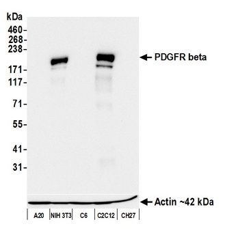
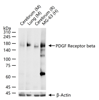
Detection of mouse PDGFR beta by western blot. Samples: Whole cell lysate (10 µg) from A20, NIH 3T3, C6, C2C12, and CH27 cells prepared using NETN lysis buffer. Antibody: Rabbit anti-PDGFR beta recombinant monoclonal antibody (orb1570943) used at 1:1000. Secondary: HRP-conjugated goat anti-rabbit IgG. Detection: Chemiluminescence with an exposure time of 10 seconds. Lower Panel: Rabbit anti-Actin recombinant monoclonal antibody.
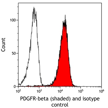
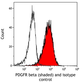
Detection of mouse PDGFR beta (shaded) in NIH3T3 cells by flow cytometry. Antibody: Rabbit anti-PDGFR beta recombinant monoclonal antibody (orb1570943) or isotype control (unshaded). Secondary: DyLight® 650-conjugated goat anti-rabbit IgG.
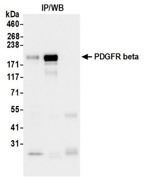
Detection of mouse PDGFR beta by western blot of immunoprecipitates. Samples: Whole cell lysate (1.0 mg per IP reaction; 5% of IP loaded) from NIH 3T3 cells prepared using NETN lysis buffer. Antibodies: Rabbit anti-PDGFR beta recombinant monoclonal antibody (orb1570943) used for IP at 6 µl/mg lysate.
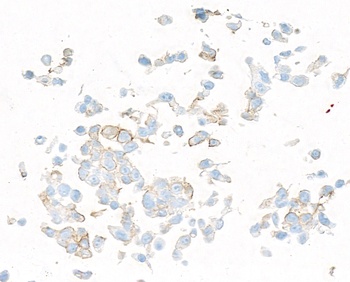
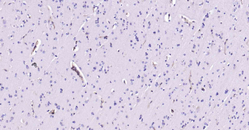
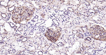
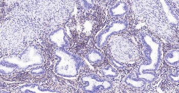
Detection of mouse PDGFR beta by immunocytochemistry. Sample: FFPE section of mouse CT26 cells. Antibody: Rabbit anti-PDGFR beta recombinant monoclonal antibody (orb1570943). Secondary: HRP-conjugated goat anti-rabbit IgG.
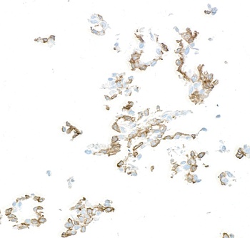
Detection of mouse PDGFR beta by immunocytochemistry. Sample: FFPE section of mouse C2C12 cells. Antibody: Rabbit anti-PDGFR beta recombinant monoclonal antibody (orb1570943). Secondary: HRP-conjugated goat anti-rabbit IgG.
Quick Database Links
UniProt Details
− No UniProt data available
NCBI Reference Sequences
−Associated Accession Numbers
Curated reference sequences for the gene transcript and protein product| Protein | NP_005745.1 |
|---|
Documents Download
Datasheet
Product Information
Request a Document
Protocol Information
WB
Western Blot (IB, immunoblot)
FC
Flow Cytometry
ICC
Immunocytochemistry
IP
Immunoprecipitation
Rabbit PDGFR beta Recombinant Monoclonal Antibody (orb1570943)

- 0.0
Based on 0 reviews
Participating in our Biorbyt product reviews program enables you to support fellow scientists by sharing your firsthand experience with our products.
Login to Submit a ReviewAvailable Sizes
Select a size below
Choose Conjugation or Carrier Free Version
Free Secondary Antibody (20 ul)0/0
Please add an antibody product to your cart first.