You have no items in your shopping cart.
Description
Research Area
Neuroscience
Images & Validation
−Item 1 of 4
| Tested Applications | IHC, IP, WB |
|---|---|
| Dilution Range | WB - 1:2,000 - 1:10,000; IP - 2 - 10 µg/mg lysate; IHC - 1:500 - 1:2,000. Epitope retrieval with citrate buffer pH 6.0 is recommended for FFPE tissue sections. |
| Reactivity | Human, Mouse |
| Predicted Reactivity | Bovine |
| Application Notes |
Key Properties
−| Antibody Type | Primary Antibody |
|---|---|
| Host | Rabbit |
| Clonality | Polyclonal |
| Isotype | IgG |
| Immunogen | Between 1 and 50 |
| Target | FUS |
| Purification | Antigen Affinity Purified |
| Conjugation | Unconjugated |
Storage & Handling
−| Storage | 2 - 8°C |
|---|---|
| Form/Appearance | Liquid |
| Buffer/Preservatives | Tris-citrate/phosphate buffer, pH 7 to 8 containing 0.09% Sodium Azide |
| Concentration | 1000 µg/ml |
| Expiration Date | 12 months from date of receipt. |
| Disclaimer | For research use only |
Alternative Names
−75 kDa DNA-pairing protein; ALS6; ETM4; FUS/ERG chimeric protein; FUS/ERG fusion protein; FUS1; fused in sarcoma; fusion gene in myxoid liposarcoma; fus-like protein; heterogeneous nuclear ribonucleoprotein P2; HNRNPP2; oncogene FUS; oncogene TLS; POMP75; RNA-binding protein FUS; TLS; translocated in liposarcoma protein
Similar Products
−- Item 1 of 7
FUS Rabbit Polyclonal Antibody [orb577521]
IHC, WB
Bovine, Canine, Porcine, Rabbit, Rat
Human, Mouse
Rabbit
Polyclonal
Unconjugated
100 μl - Item 1 of 7
TLS/FUS Rabbit Polyclonal Antibody [orb1097999]
FC, ICC, IF, IHC, WB
Human, Mouse, Rat
Rabbit
Polyclonal
Unconjugated
100 μg - Item 1 of 5
Rabbit FUS Antibody [orb1530402]
IHC, IP, WB
Bovine
Human, Mouse
Rabbit
Polyclonal
Unconjugated
100 μl, 10 μl - Item 1 of 4
FUS Antibody (C-term) [orb28049]
FC, IF, IHC-P, WB
Human, Mouse
Rabbit
Polyclonal
Unconjugated
50 μl, 100 μl - Item 1 of 3
KCNH7 Antibody (N-term) [orb39863]
FC, IHC-P, WB
Mouse, Rat
Human
Rabbit
Polyclonal
Unconjugated
50 μl, 100 μl


Quality Guarantee
Explore bioreagents carefree to elevate your research. All our products are rigorously tested for performance. If a product does not perform as described on its datasheet, our scientific support team will provide expert troubleshooting, a prompt replacement, or a refund. For full details, please see our Terms & Conditions and Buying Guide. Contact us at [email protected].
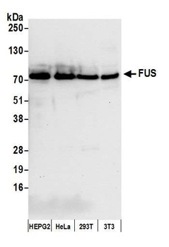
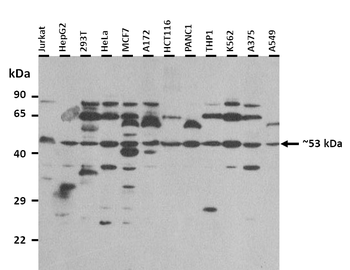
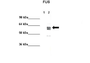
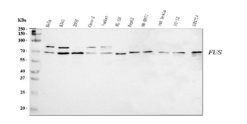
Detection of human and mouse FUS by western blot.Samples: Whole cell lysate from Hep-G2, HeLa, HEK293T, and mouse NIH 3T3 cells. Antibody: Affinity purified rabbit anti-FUS antibody orb1530387 at 0.04 µg/ml.
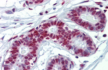
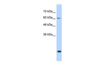
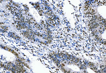
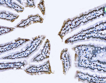
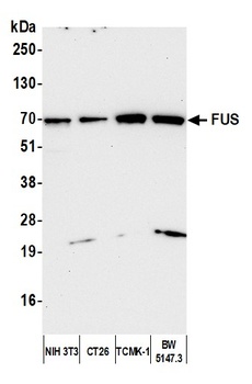
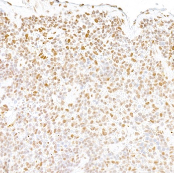
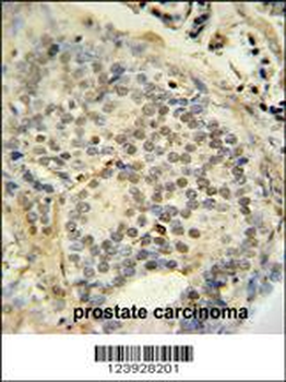
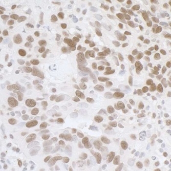
Detection of mouse FUS by immunohistochemistry. Sample: FFPE section of mouse renal cells carcinoma. Antibody: Affinity purified rabbit anti-FUS used at a dilution of 1:1, 000 (1µg/ml).
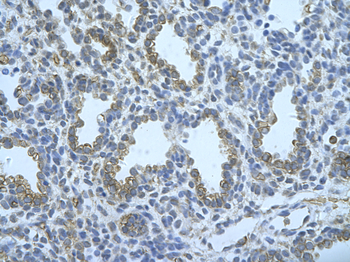
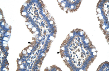
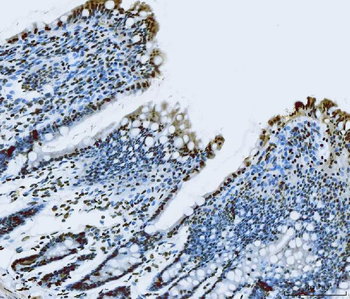
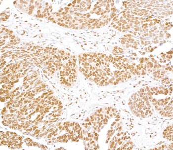
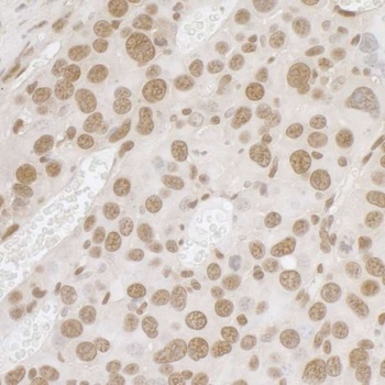
Detection of human FUS by immunohistochemistry. Sample: FFPE section of human ovarian carcinoma. Antibody: Affinity purified rabbit anti-FUS used at a dilution of 1:1, 000 (1µg/ml).
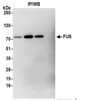
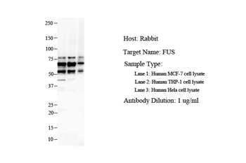
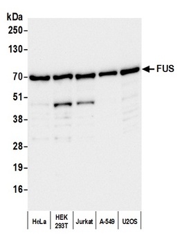
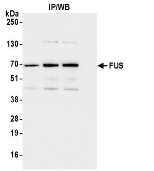
Detection of human FUS by western blot of immunoprecipitates.Samples: Whole cell lysate from HEK293T cells. Antibodies: Affinity purified rabbit anti-FUS antibody.
Quick Database Links
UniProt Details
− No UniProt data available
NCBI Reference Sequences
−Associated Accession Numbers
Curated reference sequences for the gene transcript and protein product| Protein | NP_004951.1 |
|---|
Documents Download
Datasheet
Product Information
Request a Document
Protocol Information
WB
Western Blot (IB, immunoblot)
IHC
Immunohistochemistry
IP
Immunoprecipitation
Rabbit FUS Antibody (orb1530387)

- 0.0
Based on 0 reviews
Participating in our Biorbyt product reviews program enables you to support fellow scientists by sharing your firsthand experience with our products.
Login to Submit a ReviewAvailable Sizes
Select a size below
Choose Conjugation or Carrier Free Version
Free Secondary Antibody (20 ul)0/0
Please add an antibody product to your cart first.