You have no items in your shopping cart.
Description
Research Area
Signal Transduction
Images & Validation
−Item 1 of 2
| Tested Applications | IF, WB |
|---|---|
| Dilution Range | IF - 1:25, WB - 1:2000 |
| Reactivity | Human |
Key Properties
−| Antibody Type | Primary Antibody |
|---|---|
| Host | Mouse |
| Clonality | Monoclonal |
| Isotype | IgG2a,k |
| Clone No. | B3873EV379X07X16 |
| Immunogen | This PLIN3 antibody is generated from a mouse immunized with a KLH conjugated synthetic peptide between 1-434 amino acids from human PLIN3. |
| Target | PLIN3 |
| Molecular Weight | 47075 Da |
| Conjugation | Unconjugated |
Storage & Handling
−| Storage | Maintain refrigerated at 2-8°C for up to 2 weeks. For long term storage store at -20°C in small aliquots to prevent freeze-thaw cycles |
|---|---|
| Form/Appearance | Purified monoclonal antibody supplied in PBS with 0.09% (W/V) sodium azide. This antibody is purified through a protein G column, followed by dialysis against PBS. |
| Expiration Date | 12 months from date of receipt. |
| Disclaimer | For research use only |
Alternative Names
−Perilipin-3, 47 kDa mannose 6-phosphate receptor-binding protein, 47 kDa MPR-binding protein, Cargo selection protein TIP47, Mannose-6-phosphate receptor-binding protein 1, Placental protein 17, PP17, PLIN3, M6PRBP1, TIP47
Similar Products
−- Item 1 of 9
PLIN3 Antibody [orb1240194]
ELISA, IHC-P, KO/KD Validated, WB
Porcine, Rat
Human, Mouse
Rabbit
Polyclonal
Unconjugated
0.1 mg, 0.02 mg - Item 1 of 8
PLIN3 Antibody [orb1240193]
ELISA, IF, IHC-P, KO/KD Validated, WB
Porcine
Human, Mouse, Rat
Rabbit
Polyclonal
Unconjugated
0.1 mg, 0.02 mg - Item 1 of 1

PLIN3 Antibody [orb3160508]
ELISA, IHC, WB
Human, Mouse, Rat
Rabbit
Polyclonal
Unconjugated
50 μl, 100 μl


Quality Guarantee
Explore bioreagents carefree to elevate your research. All our products are rigorously tested for performance. If a product does not perform as described on its datasheet, our scientific support team will provide expert troubleshooting, a prompt replacement, or a refund. For full details, please see our Terms & Conditions and Buying Guide. Contact us at [email protected].

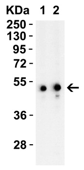
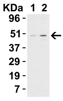
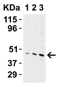
All lanes: Anti-PLIN3 Antibody at 1:2000 dilution. Lane 1: Hela whole cell lysate. Lane 2: K562 whole cell lysate. Lane 3: Daudi whole cell lysate. Lysates/proteins at 20 µg per lane. Secondary Goat Anti-mouse IgG, (H+L), Peroxidase conjugated at 1/10000 dilution. Predicted band size: 47 kDa. Blocking/Dilution buffer: 5% NFDM/TBST.
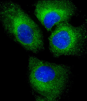
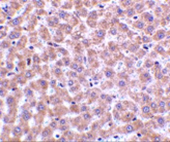
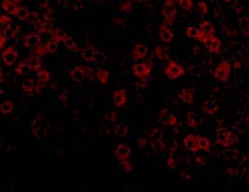
Immunofluorescent analysis of 4% paraformaldehyde-fixed, 0.1% Triton X-100 permeabilized U-2 OS (human osteosarcoma cell line) cells labeling PLIN3 at 1/25 dilution, followed by Dylight 488-conjugated goat anti-mouse IgG secondary antibody at 1/200 dilution (green). Immunofluorescence image showing cytoplasm staining on U-2 OS cell line. The nuclear counter stain is DAPI (blue).
Quick Database Links
Gene Symbol
PLIN3
UniProt
UniProt Details
− No UniProt data available
Documents Download
Datasheet
Product Information
Request a Document
Protocol Information
WB
Western Blot (IB, immunoblot)
IF
Immunofluorescence
PLIN3 Antibody (orb1925838)

- 0.0
Based on 0 reviews
Participating in our Biorbyt product reviews program enables you to support fellow scientists by sharing your firsthand experience with our products.
Login to Submit a ReviewAvailable Sizes
Select a size below
Choose Conjugation or Carrier Free Version
Free Secondary Antibody (20 ul)0/0
Please add an antibody product to your cart first.