You have no items in your shopping cart.
Description
Research Area
Metabolism Research
Images & Validation
−Item 1 of 5
| Tested Applications | FC, IF, IHC-P, WB |
|---|---|
| Dilution Range | IF - 1:10-50, WB - 1:1000, IHC-P - 1:100, FC - 1:10-50 |
| Reactivity | Human |
| Predicted Reactivity | Equine, Mouse, Rat |
Key Properties
−| Host | Rabbit |
|---|---|
| Clonality | Polyclonal |
| Isotype | Rabbit IgG |
| Immunogen | This PGK1 antibody is generated from rabbits immunized with a KLH conjugated synthetic peptide between 117-145 amino acids from the Central region of human PGK1. Antigen Region: 117-145 aa. |
| Target | PGK1 |
| Molecular Weight | 44615 Da |
| Conjugation | Unconjugated |
Storage & Handling
−| Storage | Maintain refrigerated at 2-8°C for up to 2 weeks. For long term storage store at -20°C in small aliquots to prevent freeze-thaw cycles |
|---|---|
| Form/Appearance | Purified polyclonal antibody supplied in PBS with 0.09% (W/V) sodium azide. This antibody is prepared by Saturated Ammonium Sulfate (SAS) precipitation followed by dialysis against PBS. |
| Expiration Date | 12 months from date of receipt. |
| Disclaimer | For research use only |
Alternative Names
−Phosphoglycerate kinase 1, Cell migration-inducing gene 10 protein, Primer recognition protein 2, PRP 2, PGK1, PGKA
Similar Products
−- Item 1 of 2
PGK1 Antibody (Center S320) [orb1939493]
IHC-P, WB
Monkey
Human
Rabbit
Polyclonal
Unconjugated
50 μl, 100 μl - Item 1 of 3
PGK1 Mouse Monoclonal Antibody [orb1474128]
FC, IH, KO/KD Validated, WB
Human, Mouse
Mouse
Monoclonal
Unconjugated
200 μl, 50 μl, 100 μl, 30 μl - Item 1 of 2
PGK1 Antibody (Center S320) [orb1788322]
IHC-P, WB
Human, Mouse
Rabbit
Polyclonal
Unconjugated

PGK1/2 Rabbit Polyclonal Antibody [orb2988704]
WB
Human, Mouse, Rat
Rabbit
Polyclonal
Unconjugated
200 μl, 100 μl, 50 μl, 30 μl

Quality Guarantee
Explore bioreagents carefree to elevate your research. All our products are rigorously tested for performance. If a product does not perform as described on its datasheet, our scientific support team will provide expert troubleshooting, a prompt replacement, or a refund. For full details, please see our Terms & Conditions and Buying Guide. Contact us at [email protected].
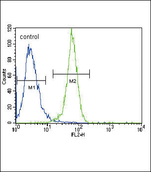
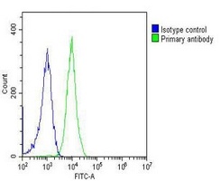
PGK1 Antibody (Center) flow cytometric analysis of Hela cells (right histogram) compared to a negative control cell (left histogram). FITC-conjugated goat-anti-rabbit secondary antibodies were used for the analysis.
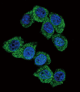
Confocal immunofluorescent analysis of PGK1 Antibody (Center) with Hela cell followed by Alexa Fluor 488-conjugated goat anti-rabbit lgG (green). DAPI was used to stain the cell nuclear (blue).
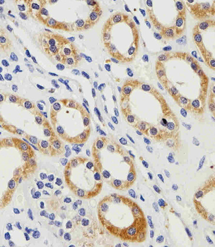
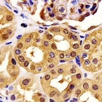
Immunohistochemical analysis of paraffin-embedded H. kidney section using PGK1 Antibody (Center). Diluted at 1:100 dilution. A peroxidase-conjugated goat anti-rabbit IgG at 1:400 dilution was used as the secondary antibody, followed by DAB staining.
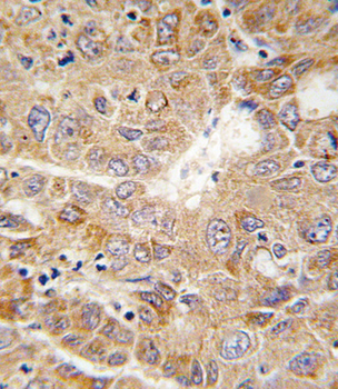
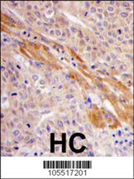
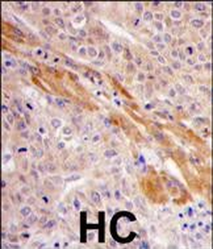
Formalin-fixed and paraffin-embedded human hepatocarcinoma tissue reacted with PGK1 antibody (Center), which was peroxidase-conjugated to the secondary antibody, followed by DAB staining. This data demonstrates the use of this antibody for immunohistochemistry; clinical relevance has not been evaluated.
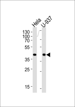
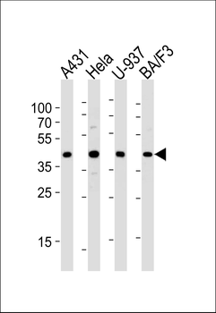
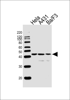
Western blot analysis of lysates from Hela, U-937 cell line (from left to right), using PGK1 Antibody (G132). Diluted at 1:1000 at each lane. A goat anti-rabbit IgG H&L (HRP) at 1:5000 dilution was used as the secondary antibody. Lysates at 35 ug per lane.
Quick Database Links
UniProt Details
− No UniProt data available
NCBI Reference Sequences
−Associated Accession Numbers
Curated reference sequences for the gene transcript and protein product| Protein | NP_000282.1 |
|---|
Documents Download
Datasheet
Product Information
Request a Document
Protocol Information
WB
Western Blot (IB, immunoblot)
IHC-P
Immunohistochemistry Paraffin
FC
Flow Cytometry
IF
Immunofluorescence
PGK1 Antibody (Center) (orb1929564)

- 0.0
Based on 0 reviews
Participating in our Biorbyt product reviews program enables you to support fellow scientists by sharing your firsthand experience with our products.
Login to Submit a ReviewAvailable Sizes
Select a size below
Choose Conjugation or Carrier Free Version
Free Secondary Antibody (20 ul)0/0
Please add an antibody product to your cart first.