You have no items in your shopping cart.
Featured
KO/KD
Validated
Validated
Description
Research Area
Metabolism Research
Images & Validation
−Item 1 of 3
| Tested Applications | FC, IH, KO/KD Validated, WB |
|---|---|
| Dilution Range | WB (1/500 - 1/2000), IH (1/50 - 1/100), FC (1/50 - 1/100) |
| Reactivity | Human, Mouse |
Key Properties
−| Antibody Type | Primary Antibody |
|---|---|
| Host | Mouse |
| Clonality | Monoclonal |
| Immunogen | KLH-conjugated synthetic peptide encompassing a sequence within the center region of human PGK1. The exact sequence is proprietary. |
| Target | PGK1 |
| Purification | The antibody was purified by immunogen affinity chromatography. |
| Conjugation | Unconjugated |
Storage & Handling
−| Storage | Maintain refrigerated at 2-8°C for up to 2 weeks. For long term storage store at -20°C in small aliquots to prevent freeze-thaw cycles. |
|---|---|
| Form/Appearance | Liquid |
| Buffer/Preservatives | 0.42% Potassium phosphate, 0.87% Sodium chloride, pH 7.3, 30% glycerol, and 0.01% sodium azide. |
| Expiration Date | 12 months from date of receipt. |
| Disclaimer | For research use only |
Alternative Names
−PGKA; Phosphoglycerate kinase 1; Cell migration-inducing gene 10 protein; Primer recognition protein 2; PRP 2
Similar Products
−- Item 1 of 4
- Item 1 of 1
PGK1 Mouse Monoclonal Antibody [orb783125]
IF, IHC-Fr, IHC-P, IP, WB
Mouse
Human, Mouse
Mouse
Monoclonal
Unconjugated
50 μl, 100 μl 

Quality Guarantee
Explore bioreagents carefree to elevate your research. All our products are rigorously tested for performance. If a product does not perform as described on its datasheet, our scientific support team will provide expert troubleshooting, a prompt replacement, or a refund. For full details, please see our Terms & Conditions and Buying Guide. Contact us at [email protected].

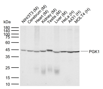
Western blot analysis of PGK1 expression in HEK293T (A), A431 (B), mouse brain (C) whole cell lysates. (Predicted band size: 44 kD; Observed band size: 44 kD)
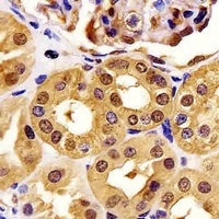
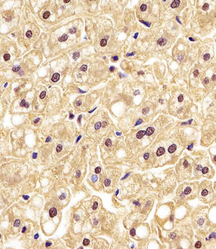
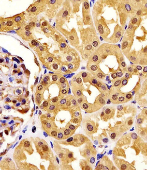
Immunohistochemical analysis of PGK1 staining in human kidney formalin fixed paraffin embedded tissue section. The section was pre-treated using heat mediated antigen retrieval with sodium citrate buffer (pH 6.0). The section was then incubated with the antibody at room temperature and detected using an HRP conjugated compact polymer system. DAB was used as the chromogen. The section was then counterstained with haematoxylin and mounted with DPX.
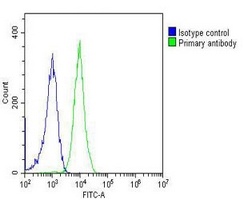
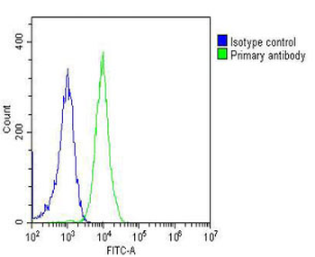
Overlay histogram showing Jurkat cells stained with Anti-PGK1 Antibody (green line). The cells were fixed with 2% paraformaldehyde (10 min) and then permeabilized with 90% methanol for 10 min. The cells were then icubated in 2% bovine serum albumin to block non-specific protein-protein interactions followed by Anti-PGK1 Antibody for 60 min at 37 °C. The secondary antibody used was Goat Anti-Mouse IgG (H&L) - AF488 at 1/200 dilution for 40 min at 37 °C. Isotype control antibody (blue line) was mouse IgG2a (1μg/1x10^6 cells) used under the same conditions. Acquisition of > 10000 events was performed.
Quick Database Links
UniProt Details
− No UniProt data available
NCBI Gene Details
− No NCBI Gene data available
Documents Download
Datasheet
Product Information
Request a Document
Protocol Information
WB
Western Blot (IB, immunoblot)
FC
Flow Cytometry
PGK1 Mouse Monoclonal Antibody (orb1474128)

- 0.0
Based on 0 reviews
Participating in our Biorbyt product reviews program enables you to support fellow scientists by sharing your firsthand experience with our products.
Login to Submit a ReviewAvailable Sizes
Select a size below
Choose Conjugation or Carrier Free Version
Free Secondary Antibody (20 ul)0/0
Please add an antibody product to your cart first.