You have no items in your shopping cart.
Description
Images & Validation
−Item 1 of 7
| Tested Applications | ELISA, FC, IHC, WB |
|---|---|
| Dilution Range | ELISA: 1:10,000 - 1:50,000, FC: 1:800, IHC: 1:100, WB: 1:1,000 |
| Reactivity | Human |
| Application Notes |
Key Properties
−| Antibody Type | Primary Antibody |
|---|---|
| Host | Mouse |
| Clonality | Monoclonal |
| Isotype | IgG2a |
| Clone No. | MN-1 |
| Immunogen | This antibody was produced in mesothelin-deficient mice by immunizations with plasmid cDNA encoding human MSLN full length protein followed by a single boost of a recombinant human mesothelin-Fc fusion protein. |
| Target | MSLN |
| Purity | This antibody is directed against human mesothelin protein. This product was purified from tissue culture supernatant fluid by Protein A chromatography. Cross reactivity with homologues from other sources has not been tested. |
| Conjugation | Unconjugated |
Storage & Handling
−| Storage | Store vial at -20° C prior to opening. Aliquot contents and freeze at -20° C or below for extended storage. Avoid cycles of freezing and thawing. Centrifuge product if not completely clear after standing at room temperature. This product is stable for several weeks at 4° C as an undiluted liquid. Dilute only prior to immediate use. |
|---|---|
| Form/Appearance | Liquid (sterile filtered) |
| Buffer/Preservatives | Preservative: 0.01% (w/v) Sodium Azide. Stabilizer: None; Buffer: 0.02 M Potassium Phosphate, 0.15 M Sodium Chloride, pH 7.2 |
| Concentration | 1.0 mg/mL |
| Expiration Date | 12 months from date of receipt. |
| Dry Ice Shipping | Please note: This product requires shipment on dry ice. A dry ice surcharge will apply. |
| Disclaimer | For research use only |
Alternative Names
−mouse anti-Mesothelin Antibody, Mesothelian, MN, MB, Pre-pro-megakaryocyte-potentiating factor, CAK1 antigen
Similar Products
−- Item 1 of 7
Mesothelin Antibody / MSLN [orb606569]
IHC-P, WB
Human, Mouse, Rat
Mouse
Monoclonal
Unconjugated
100 μg, 20 μg - Item 1 of 7
- Item 1 of 6
Mesothelin Antibody [orb757394]
ELISA, FC, IHC
Human
Rabbit
Monoclonal
Unconjugated
500 μg, 50 μg, 10 μg, 100 μg - Item 1 of 6
Mesothelin Antibody [orb757396]
ELISA, FC, WB
Human
Rabbit
Monoclonal
Unconjugated
50 μg, 10 μg, 100 μg, 500 μg - Item 1 of 7
Mesothelin Antibody / MSLN [orb2641183]
IHC-P, WB
Human, Mouse, Rat
Mouse
Monoclonal
Unconjugated
100 μg


Quality Guarantee
Explore bioreagents carefree to elevate your research. All our products are rigorously tested for performance. If a product does not perform as described on its datasheet, our scientific support team will provide expert troubleshooting, a prompt replacement, or a refund. For full details, please see our Terms & Conditions and Buying Guide. Contact us at [email protected].
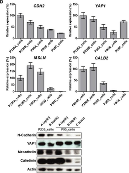
(A) Tumor processing flow-chart of 235 samples, which were processed for RNA extraction, cell culture and embedding either in PFA or OCT. Samples showing attributes which are grayed in the chart, were not used in the live cell biobank. (B) Circos whole genome copy number variations (CNVs) view of tumor and primary cell culture in patients malignant pleural mesothelioma (MPM) 236 and MPM95. The quilt plot highlights the CNV and SNVs in genes that are part of MPM landscape (2). (C) Immunofluorescence analysis of selected markers in primary culture from patient MPM236. Scale bar 200 µm. (D) Selected genes expression analysis at mRNA (upper panel) and protein (lower panel) level in primary cultures derived from samples from two patients, MPM236 and MPM95. The latter one underwent EMT during disease progression. (E) Selected genes expression analysis at mRNA in tumor samples from patients MPM236 and MPM95. (F) Significant correlation between gene expression changes in tumor and primary culture from patient MPM236 at passage 3.
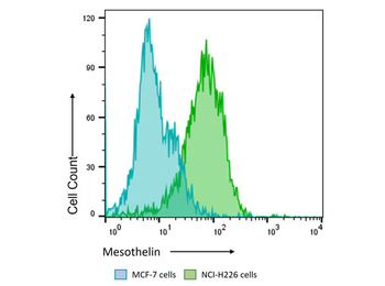
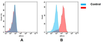
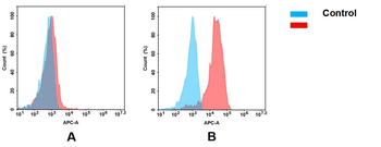
Flow Cytometry Results of Anti-Mesothelin (MOUSE) Monoclonal Antibody. The green histogram shows NCI-H226 cells and blue histogram shows MCF-7 cells. Both cell lines are stained with a 1:800 dilution Anti-Mesothelin (MOUSE) Monoclonal Antibody. The secondary antibody use was Anti-Mouse IgG (H&L) (GOAT) Antibody DyLight™ 488 Conjugated at the 1:400 dilution.

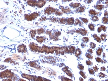
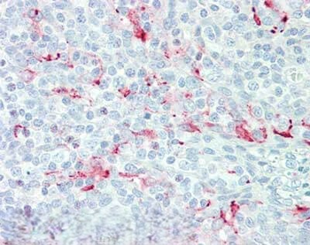
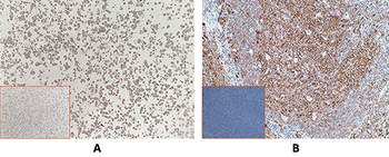
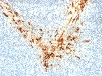
Immunohistochemistry of Mouse anti-Mesothelin antibody. Tissue: human tonsil. Fixation: formalin fixed paraffin embedded. Antigen retrieval: not required. Primary antibody: anti-Mesothelin antibody at 15 µg/ml for 1 h at RT. Secondary antibody: Peroxidase mouse secondary antibody at 1:10000 for 45 min at RT. Staining: Mesothelin as precipitated red signal with hematoxylin purple nuclear counterstain.

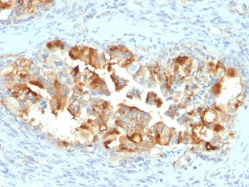
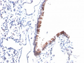
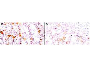
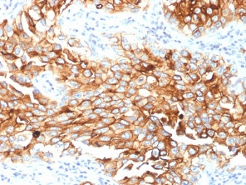
Immunohistochemistry using Biorbyt's anti-mesothelin antibody to react with two epitopes on mesothelin in PEFF human mesothelioma tissue sections treated by antigen retrieval methods. Anti-mesothelin primary antibodies were used at 10 µg/ml to label these sections as follows: C, MAb MB; and D, MAb MN followed by goat anti-mouse IgG conjugated to horseradish peroxidase at 25 µg/ml in 1% BSA/PBS for 30 minutes. (magnification, ×200; bar, 50 µm).
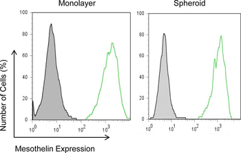
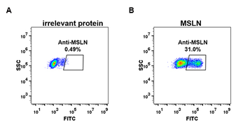
Mesothelin expression in mesothelioma monolayers and spheroids.NCI-H226 cells incubated with an anti-mesothelin mAb (MN) and detected with goat anti-mouse IgG conjugated with Alexa488 by flow cytometry.

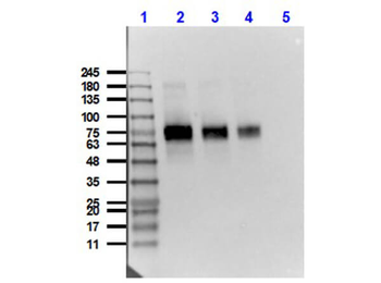
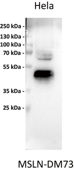
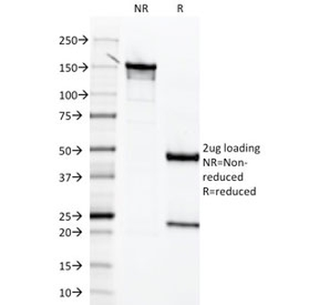
Western Blot of Mouse Anti-Mesothelin Antibody. Lane 1: Opal Prestained Molecular Weight Marker. Lane 2: HeLa [10 µg] + Mesothelin-Fc [0.1 µg]. Lane 3: HeLa [10 µg] + Mesothelin-Fc [0.05 µg]. Lane 4: HeLa [10 µg] + Mesothelin-Fc [0.02 µg]. Lane 5: HeLa Whole Cell Lysate (p/n orb348668). Primary Antibody: Anti-Mesothelin at 1 µg/ml overnight at 2-8°C. Secondary Antibody: Rabbit Anti-Mouse IgG HRP conjugated (p/n orb347544) at 1:40000 for 30 mins at RT. Block: BlockOut Buffer (p/n orb348644) 30 mins at RT. Exposure: 15 sec. Predicted MW: 40 kDa Mesothelin + Fc region 30kDa. Observed MW: ~70-75kDa.
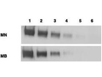
Western blotting using Biorbyt's anti-mesothelin antibodies to detect mesothelin-Fc at 100 ng (lane 1), 25 ng (lane 2), 6 ng (lane 3), 2 ng (lane 4) and 0.4 ng (lane 5). Lane 6 contains 50 ng of CDC25-Fc. Proteins were separated on 4-20% gradient gel by SDS-PAGE followed by transfer to PVDF membrane. Primary antibody was used at 1 µg/ml followed by reaction with ALP goat anti-mouse IgG and BCIP/NBT substrate.
Documents Download
Datasheet
Product Information
Request a Document
Protocol Information
WB
Western Blot (IB, immunoblot)
IHC
Immunohistochemistry
FC
Flow Cytometry
ELISA
Enzyme-linked Immunosorbent Assay (EIA)
Khanna, Swati et al. Malignant Mesothelioma Effusions Are Infiltrated by CD3+ T Cells Highly Expressing PD-L1 and the PD-L1+ Tumor Cells within These Effusions Are Susceptible to ADCC by the Anti-PD-L1 Antibody Avelumab J Thorac Oncol, 11, 1993-2005 (2016)
MSLN Antibody (orb344444)

- 0.0
Based on 0 reviews
Participating in our Biorbyt product reviews program enables you to support fellow scientists by sharing your firsthand experience with our products.
Login to Submit a ReviewAvailable Sizes
Select a size below
Choose Conjugation or Carrier Free Version
Free Secondary Antibody (20 ul)0/0
Please add an antibody product to your cart first.