You have no items in your shopping cart.
Featured
Description
Research Area
ATPases, Molecular Biology, Signal Transduction, Surface Molecules
Images & Validation
−Item 1 of 5
| Tested Applications | FC, ICC, WB |
|---|---|
| Dilution Range | WB=1:500-2000, ICC/IF=1:100-500, Flow-Cyt=1μg/Test |
| Reactivity | Human, Rat |
| Predicted Reactivity | Mouse |
Related Conjugates & Formulations
−Key Properties
−| Antibody Type | Primary Antibody |
|---|---|
| Host | Rabbit |
| Clonality | Polyclonal |
| Isotype | IgG |
| Immunogen | KLH conjugated synthetic peptide derived from human MDR1 (1051-1280/1280aa) |
| Target | ABCB1 |
| Molecular Weight | 141 kDa |
| Purification | Affinity purified by Protein A |
| Conjugation | Unconjugated |
Storage & Handling
−| Storage | Maintain refrigerated at 2-8°C for up to 2 weeks. For long term storage store at -20°C in small aliquots to prevent freeze-thaw cycles. |
|---|---|
| Form/Appearance | Liquid |
| Buffer/Preservatives | 0.01M TBS (pH7.4) with 1% rAlbumin, 0.02% Proclin300 and 50% Glycerol. |
| Concentration | 1mg/ml |
| Expiration Date | 12 months from date of receipt. |
| Disclaimer | For research use only |
Alternative Names
−ABC20; CD243; CLCS; ENPAT; GP170; MDR1; P-GP; PGY1; p-170; Abcb1; Mdr1b; Pgy-1; mdr; MDR1_HUMAN; ATP-binding cassette sub-family B member 1; Multidrug resistance protein 1; P-glycoprotein 1; Phospholipid transporter ABCB1; MDR1B_MOUSE; Abcb1b; ATP-binding cassette sub-family B member 1B; Multidrug resistance protein 1B; Pgy1-1; ATP binding cassette subfamily B member 1; colchicin sensitivity; ATP-binding cassette, sub-family B (MDR/TAP), member 1; P-glycoprotein
Similar Products
−- Item 1 of 5
MDR1 Rabbit Polyclonal Antibody [orb221459]
IF, IHC-Fr, IHC-P, WB
Mouse, Rat
Human, Mouse, Rat
Rabbit
Polyclonal
Unconjugated
50 μl, 100 μl, 200 μl 
MDR1/P Glycoprotein Rabbit Polyclonal Antibody (PE) [orb124755]
FC, ICC
Mouse
Human, Rat
Rabbit
Polyclonal
PE
100 μlMDR1/P Glycoprotein Rabbit Polyclonal Antibody (FITC) [orb15962]
FC, ICC
Mouse
Human, Rat
Rabbit
Polyclonal
FITC
100 μlMDR1/P Glycoprotein Rabbit Polyclonal Antibody (Cy5) [orb909117]
FC, ICC
Mouse
Human, Rat
Rabbit
Polyclonal
Cy5
100 μlMDR1/P Glycoprotein Rabbit Polyclonal Antibody (PE-Cy5.5) [orb911276]
FC, ICC
Mouse
Human, Rat
Rabbit
Polyclonal
PE/Cy5.5
100 μl
Quality Guarantee
Explore bioreagents carefree to elevate your research. All our products are rigorously tested for performance. If a product does not perform as described on its datasheet, our scientific support team will provide expert troubleshooting, a prompt replacement, or a refund. For full details, please see our Terms & Conditions and Buying Guide. Contact us at [email protected].
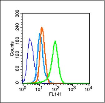
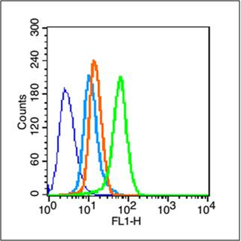
Blank control (blue line): Hela (blue). Primary Antibody (green line): Rabbit Anti-MDR1 antibody (orb11034), Dilution: 1 µg/10^6 cells, Isotype Control Antibody (orange line): Rabbit IgG. Secondary Antibody (white blue line): Goat anti-rabbit IgG-FITC, Dilution: 1 µg/Test. Protocol, The cells were fixed with 70% methanol (Overnight at -20°C) and then permeabilized with ice-cold 90% methanol for 30 min on ice. Cells stained with Primary Antibody for 30 min at room temperature. The cells were then incubated in 1 X PBS/2% BSA/10% goat serum to block non-specific protein-protein interactions followed by the antibody for 15 min at room temperature. The secondary antibody used for 40 min at room temperature. Acquisition of 20000 events was performed.
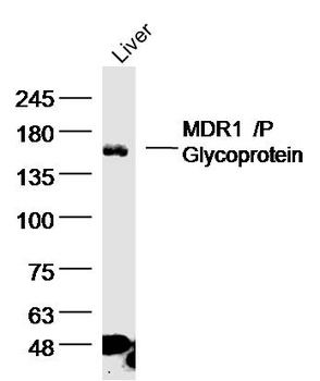
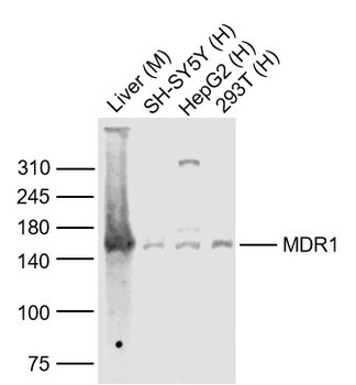
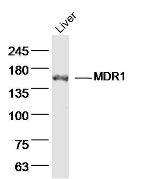
Sample: liver (Rat) Lysate at 40 ug, Primary: Anti-MDR1'P Glycoprotein (orb11034) at 1/300 dilution, Secondary: IRDye800CW Goat Anti-Rabbit IgG at 1/20000 dilution, Predicted band size: 141kD, Observed band size: 141 kD.
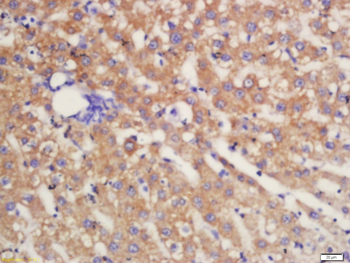
Tissue/Cell: Rat liver tissue, 4% Paraformaldehyde-fixed and paraffin-embedded, Antigen retrieval: citrate buffer (0.01M, pH6.0), Boiling bathing for 15 min, Block endogenous peroxidase by 3% Hydrogen peroxide for 30 min, Blocking buffer (normal goat serum) at 37°C for 20 min, Incubation: Anti-MDR1 / P Glycoprotein Polyclonal Antibody, Unconjugated (orb11034) 1:200, overnight at 4°C, followed by conjugation to the secondary antibody and DAB staining.
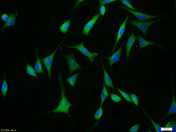
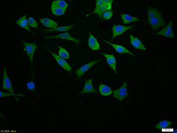
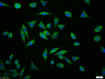
Tissue/Cell: SH-SY5Y cell, 4% Paraformaldehyde-fixed, Triton X-100 at room temperature for 20 min, Blocking buffer (normal goat serum) at 37°C for 20 min, Antibody incubation with (MDR1/P Glycoprotein) polyclonal Antibody, Unconjugated (orb11034) 1:100, 90 minutes at 37°C, followed by a FITC conjugated Goat Anti-Rabbit IgG antibody at 37°C for 90 minutes, DAPI (blue) was used to stain the cell nuclei.
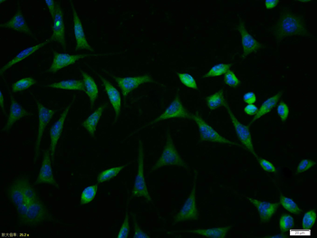
Tissue/Cell: SH-SY5Y cell, 4% Paraformaldehyde-fixed, Triton X-100 at room temperature for 20 min, Blocking buffer (normal goat serum) at 37°C for 20 min, Antibody incubation with (MDR1/P Glycoprotein) polyclonal Antibody, Unconjugated (orb11034) 1:100, 90 minutes at 37°C, followed by a FITC conjugated Goat Anti-Rabbit IgG antibody at 37°C for 90 minutes, DAPI (blue) was used to stain the cell nuclei.
Quick Database Links
Gene Symbol
ABCB1
UniProt
UniProt Details
− No UniProt data available
Documents Download
Datasheet
Product Information
Request a Document
Protocol Information
WB
Western Blot (IB, immunoblot)
FC
Flow Cytometry
ICC
Immunocytochemistry
MDR1/P Glycoprotein Rabbit Polyclonal Antibody (orb11034)

- 0.0
Based on 0 reviews
Participating in our Biorbyt product reviews program enables you to support fellow scientists by sharing your firsthand experience with our products.
Login to Submit a ReviewAvailable Sizes
Select a size below
Free Secondary Antibody (20 ul)0/0
Please add an antibody product to your cart first.