You have no items in your shopping cart.
Description
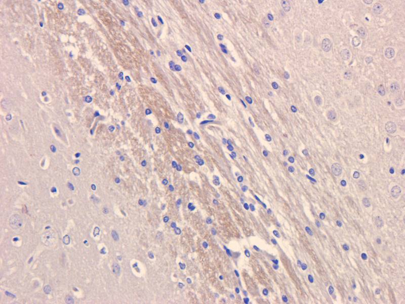
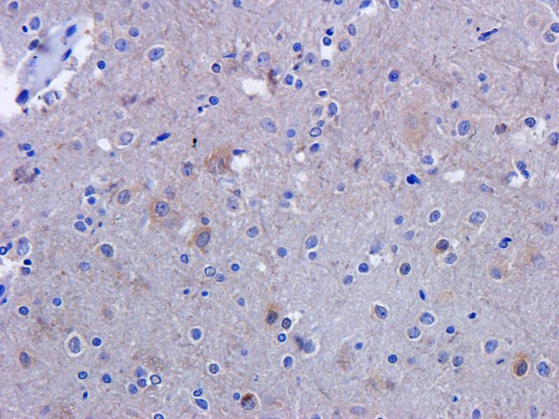
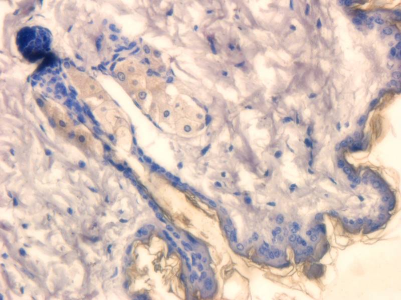
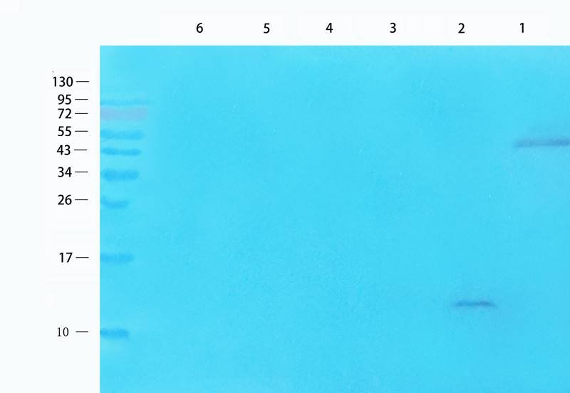
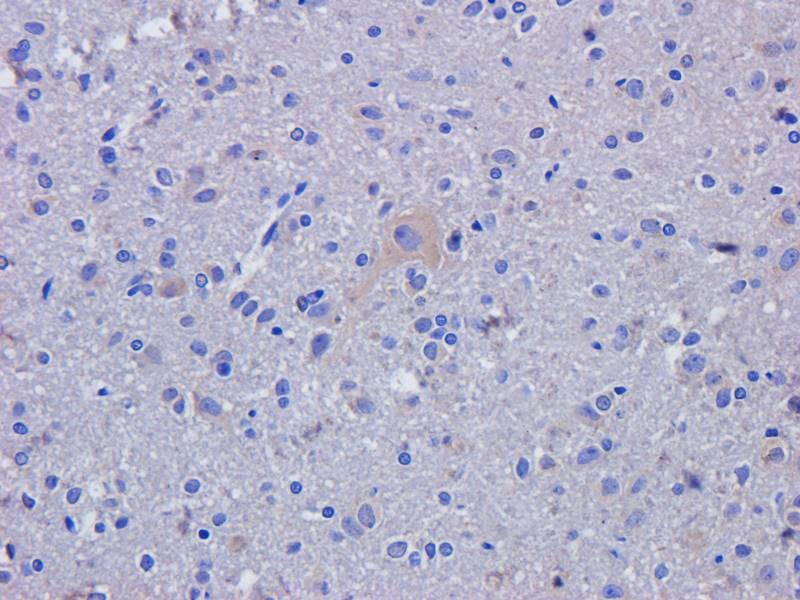
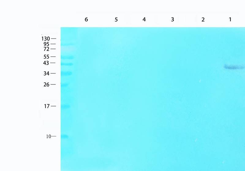
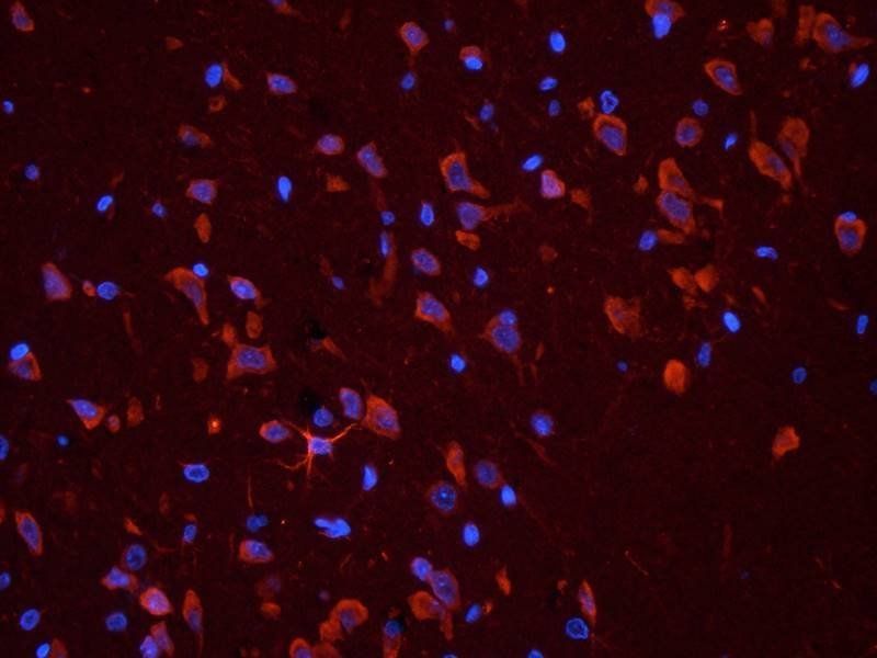
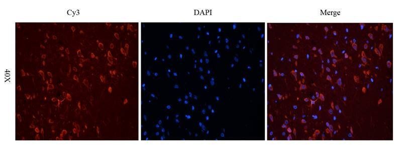
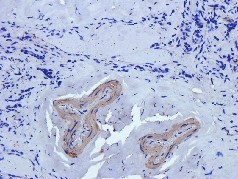
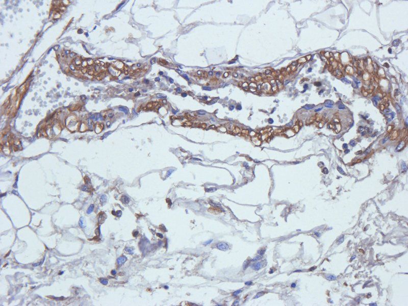
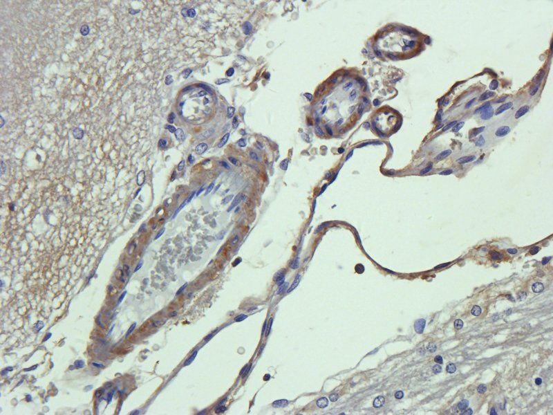
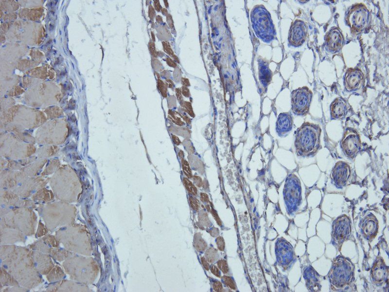
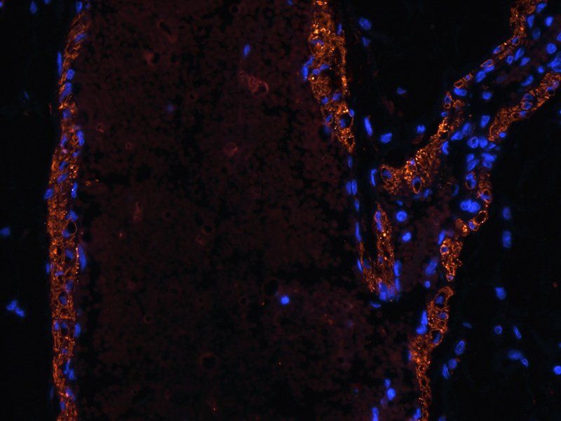
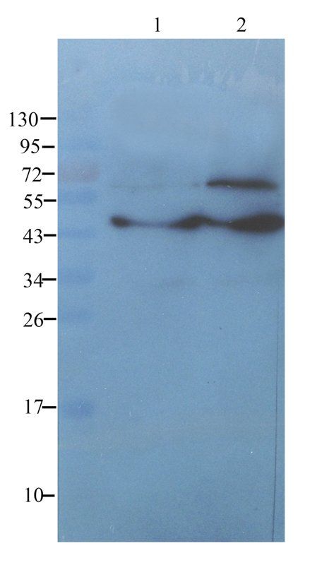
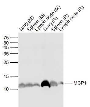
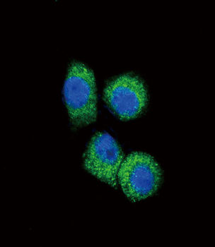
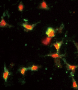
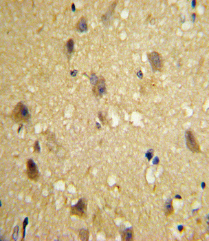
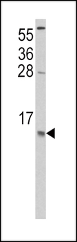
Images & Validation
−| Application Notes |
|---|
Key Properties
−| Source | Escherichia Coli |
|---|---|
| Biological Activity | ED50 =1-10ng/ml corresponding to a Specific Activity of 100,000-1,000,000IU/mg. The biological activity was determined by measuring the dose dependent chemotaxis with human THP-1 cells. The optimal concentration should be determined for each specific application by an initial dose-response assay. |
| Purity | Greater than 98.0% as determined by SDS-PAGE. |
| Protein Sequence | QPDAVNAPLT CCYSFTGKMI PMSRLENYKR ITSSRCPKEA VVFVTKLKRE ICADPNKEWV QKYIRKLDQN QVRSETTVFY KIASTLRTSA PLNVNLTHKS EANASTLFST TTSSTSVEVT SMTEN |
Storage & Handling
−| Storage | Stability: Lyophilized MCP-1 although stable at room temperature for 3 weeks, should be stored desiccated below -18°C. Upon reconstitution CCL2 should be stored at 4°C between 2-7 days and for future use below -18°C.For long term storage it is recommended to add a carrier protein (0.1% HSA or BSA).Please prevent freeze-thaw cycles |
|---|---|
| Form/Appearance | Sterile Filtered White lyophilized (freeze-dried) powder. |
| Buffer/Preservatives | The protein was lyophilized from a concentrated (1mg/ml) sterile solution containing no additives. |
| Expiration Date | 6 months from date of receipt. |
| Disclaimer | For research use only |
Alternative Names
−Small inducible cytokine A2, CCL2, Monocyte chemotactic protein 1, MCP-1, Monocyte chemoattractant protein 1, Monocyte chemotactic and activating factor, MCAF, Monocyte secretory protein JE, HC11, chemokine (C-C motif) ligand 2, MCP1, SCYA2, GDCF-2, SMC-CF, HSMCR30, MGC9434, GDCF-2 HC11, Immediate-early serum-responsive JE protein.
Similar Products
−- Item 1 of 8
- Item 1 of 7
MCP1 Rabbit Polyclonal Antibody [orb323291]
ELISA, ICC, IF, IHC-P, WB
Human, Mouse, Porcine, Rat
Rabbit
Polyclonal
Unconjugated
100 μg - Item 1 of 1
MCP1 Rabbit Polyclonal Antibody [orb13563]
ELISA, WB
Human
Mouse, Rat
Rabbit
Polyclonal
Unconjugated
50 μl, 100 μl, 200 μl - Item 1 of 7
ZC3H12A Antibody [orb1672348]
ELISA, IF, IHC, WB
Gallus, Monkey, Porcine, Primate
Human, Mouse, Rat
Rabbit
Polyclonal
Unconjugated
0.1 mg, 0.02 mg - Item 1 of 5
CCL2 Antibody (C-term) [orb1929874]
FC, IF, IHC-P, WB
Human
Rabbit
Polyclonal
Unconjugated
50 μl, 100 μl
Quality Guarantee
Explore bioreagents carefree to elevate your research. All our products are rigorously tested for performance. If a product does not perform as described on its datasheet, our scientific support team will provide expert troubleshooting, a prompt replacement, or a refund. For full details, please see our Terms & Conditions and Buying Guide. Contact us at [email protected].
Documents Download
Datasheet
Product Information
Request a Document
Protocol Information
Protein Handling and Storage Guide
Protein Handling Guide
MCP 1 Protein (orb429440)

- 0.0
Based on 0 reviews
Participating in our Biorbyt product reviews program enables you to support fellow scientists by sharing your firsthand experience with our products.
Login to Submit a ReviewAvailable Sizes
Select a size below