You have no items in your shopping cart.
Featured
Description
Research Area
Immunology & Inflammation
Images & Validation
−Item 1 of 8
| Tested Applications | ICC, IF, IHC-P, WB |
|---|---|
| Dilution Range | IF/ICC: 1:50-500, IHC-P: 1:50-300, WB: 1:200-1000 |
Key Properties
−| Host | Rabbit |
|---|---|
| Clonality | Polyclonal |
| Isotype | IgG |
| Immunogen | KLH conjugated synthetic peptide derived from human MCP1. Please contact us for the exact immunogen sequence. The peptide is available as orb374822. |
| Target | MCP1 |
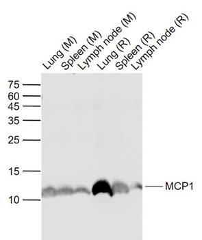
| Molecular Weight | 11 kDa |
| Purity | Polyclonal antibodies are purified by peptide affinity chromatography |
| Conjugation | Unconjugated |
Storage & Handling
−| Storage | Maintain refrigerated at 2-8°C for up to 2 weeks. For long term storage store at -20°C in small aliquots to prevent freeze-thaw cycles. |
|---|---|
| Form/Appearance | 10 mM PBS, 0.02% sodium azide |
| Concentration | - 100 μg (in 200 μl): 0.5 mg/ml- 200 μg (in 400 μl): 0.5 mg/ml |
| Expiration Date | 12 months from date of receipt. |
| Disclaimer | For research use only |
Alternative Names
−anti-C-C motif chemokine 2 antibody, anti-CCL2 antibody, anti-CCL2_HUMAN antibody, anti-GDCF 2 antibody, anti-GDCF-2 antibody, anti-GDCF2 antibody, anti-HC11 antibody, anti-MCAF antibody, anti-MCP 1 antibody, anti-MCP-1 antibody, anti-MCP1 antibody, anti-MGC9434 antibody, anti-Monocyte chemoattractant protein 1 antibody, anti-Monocyte chemotactic protein 1 antibody, anti-Monocyte secretory protein JE antibody, anti-SCYA2 antibody, anti-Small inducible cytokine A2 antibody, anti-SMC CF antibody, anti-SMC-CF antibody, anti-SMCCF antibody
Similar Products
−- Item 1 of 7
MCP1 Rabbit Polyclonal Antibody [orb323291]
ELISA, ICC, IF, IHC-P, WB
Human, Mouse, Porcine, Rat
Rabbit
Polyclonal
Unconjugated
100 μg - Item 1 of 1
MCP1 Rabbit Polyclonal Antibody [orb13563]
ELISA, WB
Human
Mouse, Rat
Rabbit
Polyclonal
Unconjugated
50 μl, 100 μl, 200 μl - Item 1 of 5
CCL2 Antibody (C-term) [orb1929874]
FC, IF, IHC-P, WB
Human
Rabbit
Polyclonal
Unconjugated
50 μl, 100 μl - Item 1 of 3
MCP1 Rabbit Polyclonal Antibody [orb181787]
ELISA, ICC, IF
Human, Mouse, Porcine, Rat
Rabbit
Polyclonal
Unconjugated
100 μg - Item 1 of 3
MCP1/CCL2 Rabbit Polyclonal Antibody [orb312096]
ELISA, IHC, WB
Human
Rabbit
Polyclonal
Unconjugated
100 μg
Quality Guarantee
Explore bioreagents carefree to elevate your research. All our products are rigorously tested for performance. If a product does not perform as described on its datasheet, our scientific support team will provide expert troubleshooting, a prompt replacement, or a refund. For full details, please see our Terms & Conditions and Buying Guide. Contact us at [email protected].
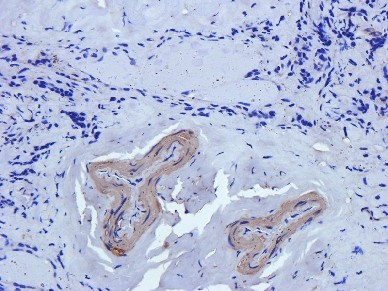
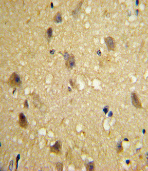
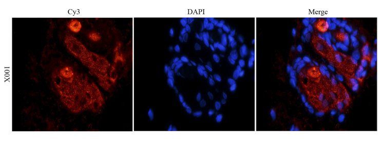
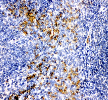
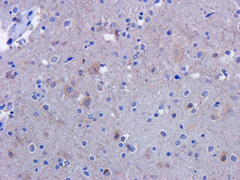
Immunohistochemical staining of rat brain tissue using anti- MCP1 (dilution of primary antibody - 2.5 ug/ml)
IHC-P staining of rat brain tissue using MCP1 antibody (dilution at 2.5 ug/ml)
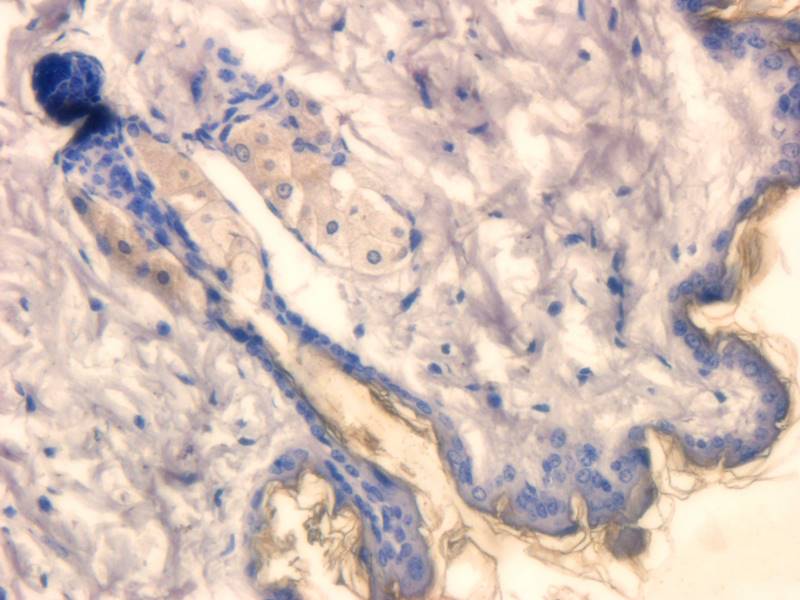
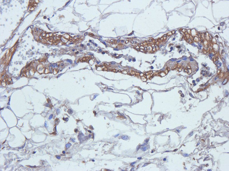
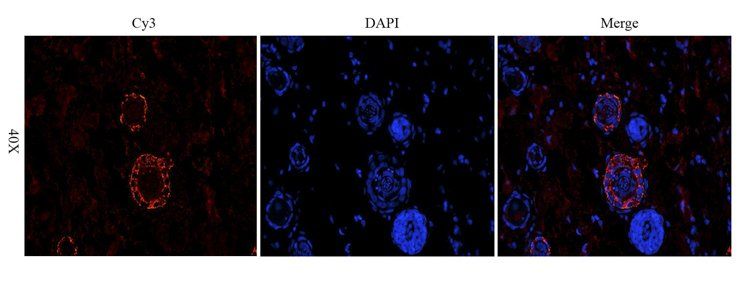
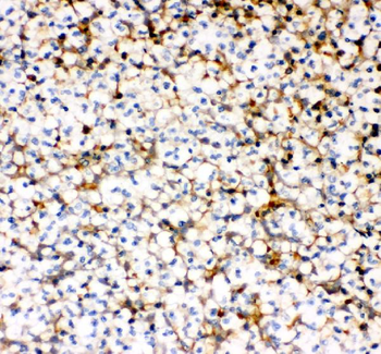
IHC-P image of rat skin tissue using MCP1 antibody (dilution of primary antibody at 2.5 ug/ml)
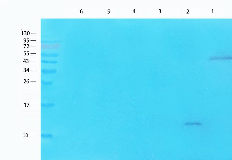
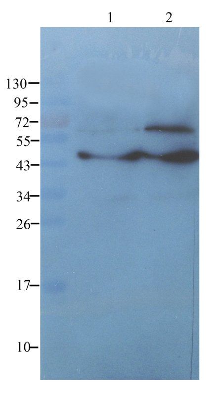
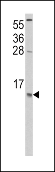
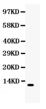
Western blot analysis of human lung cancer (lane 1), mouse brain (lane 2), rat spleen (lane 3), rat skin (lane 4), rat small intestines (lane 5), Hela cells (lane 6) using MCP1 antibody (1 ug/ml)
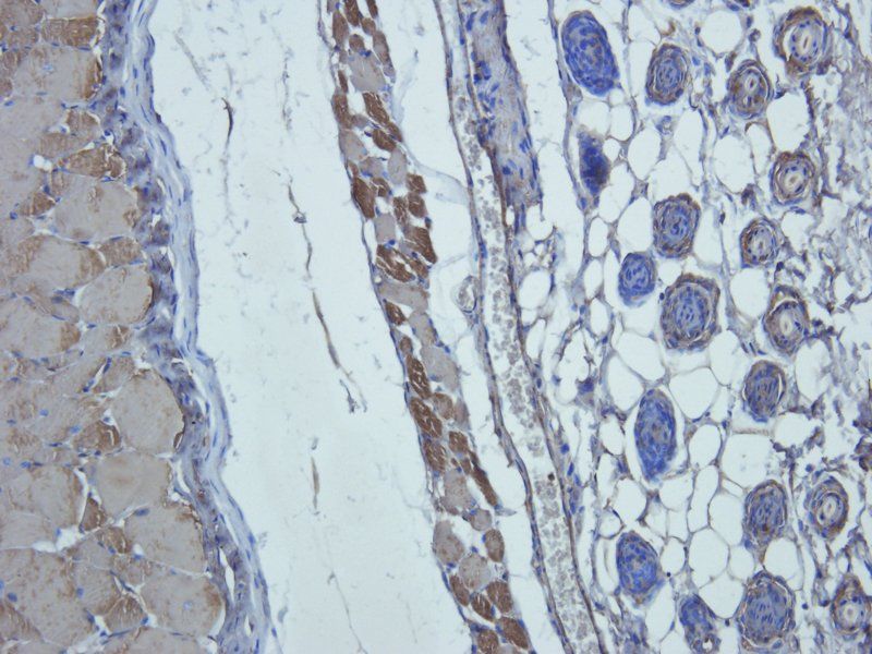
IHC-P staining of rat brain tissue using anti- MCP1 (dilution at 2.5 ug/ml)
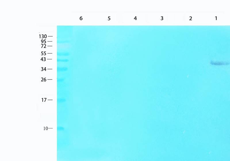
WB analysis of human lung cancer (lane 1), mouse brain (lane 2), rat spleen (lane 3), rat skin (lane 4), rat small intestines (lane 5), Hela cells (lane 6) using MCP1 antibody (1 ug/ml)
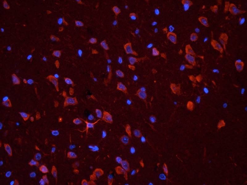
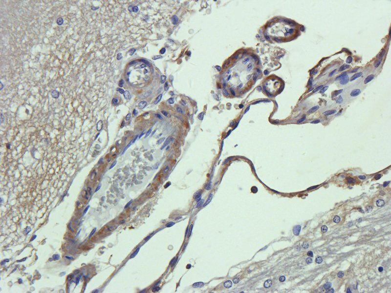
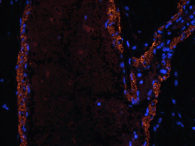
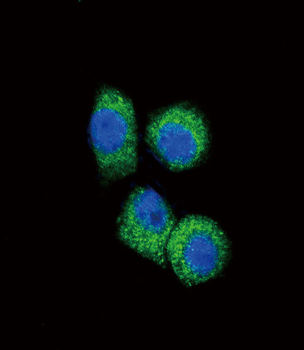
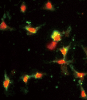
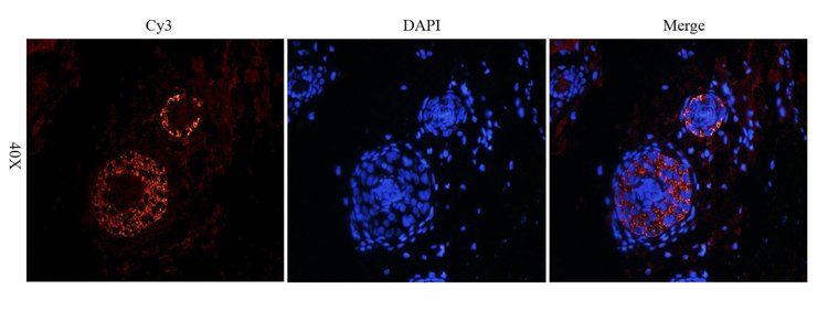
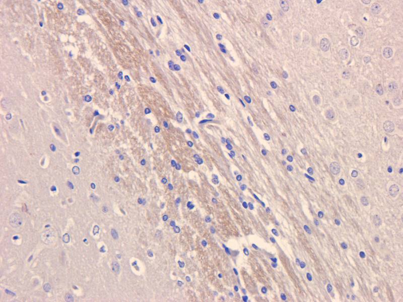
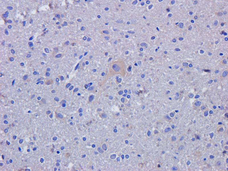
IF image of rat brain tissue using anti- MCP1 (primary antibody at 2.5 ug/ml)
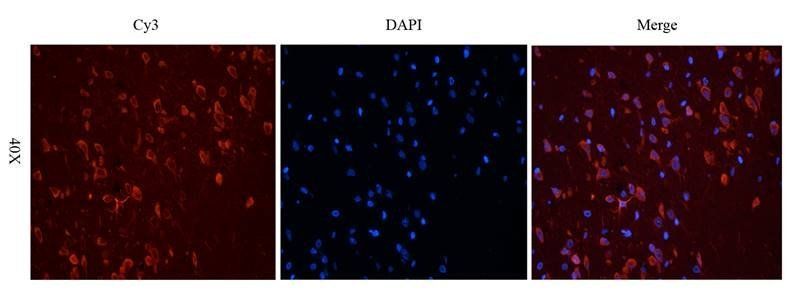
Immunofluorescence image of rat brain tissue using anti- MCP1 (dilution at 2.5 ug/ml)
Documents Download
Datasheet
Product Information
Request a Document
Protocol Information
WB
Western Blot (IB, immunoblot)
IHC-P
Immunohistochemistry Paraffin
IF
Immunofluorescence
ICC
Immunocytochemistry
Filter by Applications
Filter by Species
Jing Zhao, Katrien C. K. Poelaert, Jolien Van Cleemput, Hans J. Nauwynck CCL2 and CCL5 driven attraction of CD172a+ monocytic cells during an equine herpesvirus type 1 (EHV-1) infection in equine nasal mucosa and the impact of two migration inhibitors, rosiglitazone (RSG) and quinacrine (QC) Vet Res, 48, 1 (2017)
Applications
IF
Reactivity
Rabbit
Pierfrancesco Mirabelli, Anthony Mukwaya, Anton Lennikov, Maria Xeroudaki, Beatrice Peebo, Mira Schaupper, Neil Lagali Genome-wide expression differences in anti-Vegf and dexamethasone treatment of inflammatory angiogenesis in the rat cornea Sci Rep, 7, 7616 (2017)
Applications
IHC
Reactivity
Rat
Victoria Allahyari1, Zahra Behroozi2,3, Maziar M. Akhavan4, Aidin Shahrezaei5 and Farinaz Nasirinezhad1,6,7 Chronic exercise reduces astrocytic c-Fos and CCL2 via conditioned serum and cerebrospinal fluid Narra J, (2025)
Chang, Guo-Qing et al. CCL2/CCR2 Chemokine System in Embryonic Hypothalamus: Involvement in Sexually Dimorphic Stimulatory Effects of Prenatal Ethanol Exposure on Peptide-Expressing Neurons Neuroscience, 424, 155-171 (2020)
Applications
IF
Reactivity
Rat
Jiang, Qingkui et al. Elevated CCL2 causes Leydig cell malfunction in metabolic syndrome JCI Insight, 5, 134882 (2020)
Applications
IF, IHC
Reactivity
Mouse
MCP1 Rabbit Polyclonal Antibody (orb36895)

- 0.0
Based on 0 reviews
Participating in our Biorbyt product reviews program enables you to support fellow scientists by sharing your firsthand experience with our products.
Login to Submit a ReviewAvailable Sizes
Select a size below
Choose Conjugation or Carrier Free Version
Free Secondary Antibody (20 ul)0/0
Please add an antibody product to your cart first.