You have no items in your shopping cart.
Description
Research Area
Signal Transduction
Images & Validation
−Item 1 of 3
| Tested Applications | FC, IHC-P, WB |
|---|---|
| Dilution Range | WB - 1:1000, IHC-P - 1:10-50, FC - 1:10-50 |
| Reactivity | Human |
Key Properties
−| Host | Rabbit |
|---|---|
| Clonality | Polyclonal |
| Isotype | Rabbit IgG |
| Immunogen | This MAP4K3 antibody is generated from rabbits immunized with a recombinant protein of human MAP4K3. |
| Target | MAP4K3 (HGNC:6865) |
| Molecular Weight | 101316 Da |
| Conjugation | Unconjugated |
Storage & Handling
−| Storage | Maintain refrigerated at 2-8°C for up to 2 weeks. For long term storage store at -20°C in small aliquots to prevent freeze-thaw cycles |
|---|---|
| Form/Appearance | Purified polyclonal antibody supplied in PBS with 0.09% (W/V) sodium azide. This antibody is purified through a protein A column, followed by peptide affinity purification. |
| Expiration Date | 12 months from date of receipt. |
| Disclaimer | For research use only |
Alternative Names
−Mitogen-activated protein kinase kinase kinase kinase 3, Germinal center kinase-related protein kinase, GLK, MAPK/ERK kinase kinase kinase 3, MEK kinase kinase 3, MEKKK 3, MAP4K3, RAB8IPL1
Similar Products
−- Item 1 of 6
MAP4K3 Rabbit Polyclonal Antibody [orb1939809]
ELISA, FC, IHC, WB
Human, Mouse, Rat
Rabbit
Polyclonal
Unconjugated
100 μg - Item 1 of 4
- Item 1 of 4
Mouse Map4k3 Antibody (Center) [orb1935804]
FC, IHC-P, WB
Rat
Human, Mouse
Rabbit
Polyclonal
Unconjugated
50 μl, 100 μl 
MAP4K3 Antibody [orb3161229]
ELISA, ICC, IHC
Human, Mouse, Rat
Rabbit
Polyclonal
Unconjugated
50 μl, 100 μlMAP4K3 Antibody [orb3160561]
ELISA, ICC, IHC
Human, Mouse, Rat
Rabbit
Polyclonal
Unconjugated
50 μl, 100 μl


Quality Guarantee
Explore bioreagents carefree to elevate your research. All our products are rigorously tested for performance. If a product does not perform as described on its datasheet, our scientific support team will provide expert troubleshooting, a prompt replacement, or a refund. For full details, please see our Terms & Conditions and Buying Guide. Contact us at [email protected].

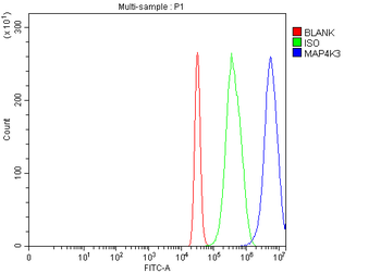
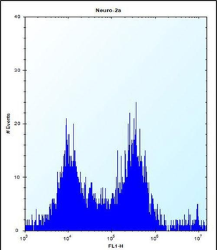
MAP4K3 Antibody flow cytometric analysis of HepG2 cells (right histogram) compared to a negative control cell (left histogram). FITC-conjugated goat-anti-rabbit secondary antibodies were used for the analysis.
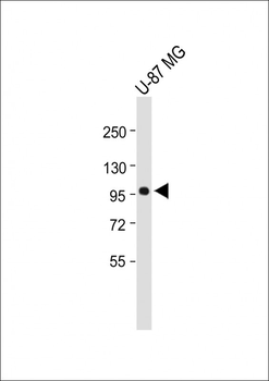
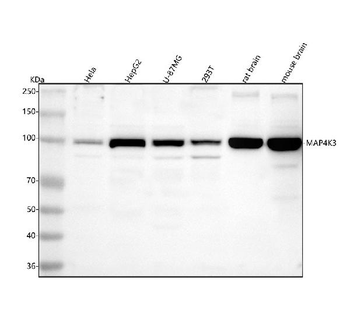
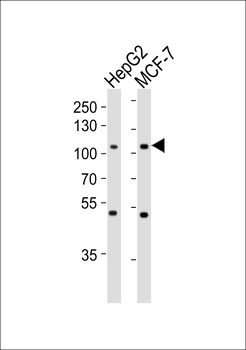
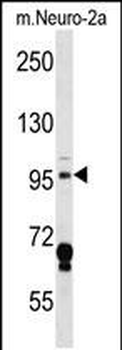
Anti-MAP4K3 Antibody at 1:1000 dilution + U-87 MG whole cell lysate. Lysates/proteins at 20 µg per lane. Secondary Goat Anti-Rabbit IgG, (H+L), Peroxidase conjugated at 1/10000 dilution. Predicted band size: 101 kDa. Blocking/Dilution buffer: 5% NFDM/TBST.
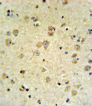
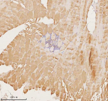
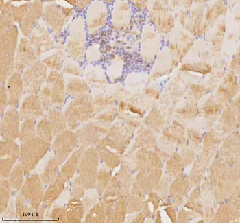
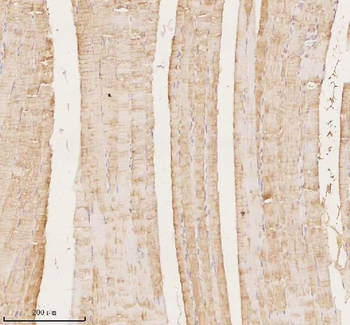
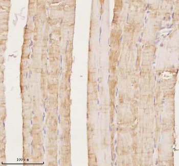
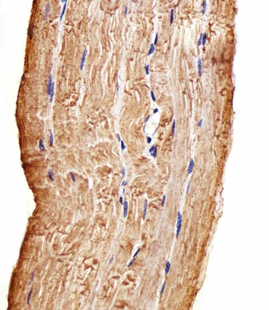
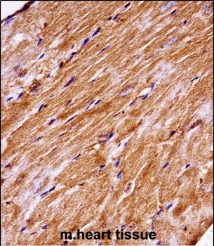
MAP4K3 Antibody IHC analysis in formalin fixed and paraffin embedded brain tissue followed by peroxidase conjugation of the secondary antibody and DAB staining. This data demonstrates the use of the MAP4K3 Antibody for immunohistochemistry. Clinical relevance has not been evaluated.
Quick Database Links
UniProt Details
− No UniProt data available
NCBI Reference Sequences
−Associated Accession Numbers
Curated reference sequences for the gene transcript and protein product| Protein | NP_001257354.1, NP_003609.2 |
|---|
Documents Download
Datasheet
Product Information
Request a Document
Protocol Information
WB
Western Blot (IB, immunoblot)
IHC-P
Immunohistochemistry Paraffin
FC
Flow Cytometry
MAP4K3 Antibody (orb1927683)

- 0.0
Based on 0 reviews
Participating in our Biorbyt product reviews program enables you to support fellow scientists by sharing your firsthand experience with our products.
Login to Submit a ReviewAvailable Sizes
Select a size below
Free Secondary Antibody (20 ul)0/0
Please add an antibody product to your cart first.