You have no items in your shopping cart.
Featured
KO/KD
Validated
Validated
Description
Research Area
Cell Biology
Images & Validation
−Item 1 of 11
| Tested Applications | ELISA, ICC, IF, IHC-P, KO/KD Validated, WB |
|---|---|
| Reactivity | Human, Mouse, Rat |
Key Properties
−| Antibody Type | Primary Antibody |
|---|---|
| Host | Rabbit |
| Clonality | Polyclonal |
| Isotype | IgG |
| Immunogen | Anti-IRE1p antibody (orb1239523) was raised against a peptide corresponding to 16 amino acids near the carboxy terminus of human IRE1P. The immunogen is located within the last 50 amino acids of IRE1p. |
| Target | ERN1 |
| Molecular Weight | Predicted: 110kDObserved: 110kD |
| Purification | IRE1p Antibody is affinity chromatography purified via peptide column. |
| Conjugation | Unconjugated |
Storage & Handling
−| Storage | Maintain refrigerated at 2-8°C for up to 2 weeks. For long term storage store at -20°C in small aliquots to prevent freeze-thaw cycles. |
|---|---|
| Form/Appearance | Liquid |
| Buffer/Preservatives | IRE1p Antibody is supplied in PBS containing 0.02% sodium azide. |
| Concentration | 1 mg/mL |
| Expiration Date | 12 months from date of receipt. |
| Disclaimer | For research use only |
Alternative Names
−IRE1p Antibody: IRE1, IRE1P, IRE1a, hIRE1p, IRE1, Endoplasmic reticulum-to-nucleus signaling 1
Similar Products
−- Item 1 of 2
Phospho-IRE1a (Ser 726) Rabbit Polyclonal Antibody [orb157704]
FC, IF, IHC-Fr, IHC-P
Bovine, Canine, Equine, Mouse, Porcine, Rabbit
Human, Rat
Rabbit
Polyclonal
Unconjugated
50 μl, 100 μl, 200 μl - Item 1 of 3
IRE1 Rabbit Polyclonal Antibody [orb157705]
IF, IHC-Fr, IHC-P
Bovine, Canine, Equine, Porcine, Rabbit
Human, Mouse, Rat
Rabbit
Polyclonal
Unconjugated
50 μl, 100 μl, 200 μl - Item 1 of 1
Human Endoplasmic Reticulum to Nucleus Signalling 1 (ERN1) ELISA Kit [orb778257]
Human
0.16-10 ng/mL
0.061 ng/mL
48 T, 96 T - Item 1 of 3
IRE1 Rabbit Polyclonal Antibody [orb526774]
FC, WB
Human, Rat
Human, Porcine
Rabbit
Polyclonal
Unconjugated
50 μl, 100 μl, 200 μl 

Quality Guarantee
Explore bioreagents carefree to elevate your research. All our products are rigorously tested for performance. If a product does not perform as described on its datasheet, our scientific support team will provide expert troubleshooting, a prompt replacement, or a refund. For full details, please see our Terms & Conditions and Buying Guide. Contact us at [email protected].
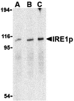
Western Blot Validation in Mouse A20 Cell Lysate. Loading: 15 µg of lysates per lane. Antibodies: IRE1p orb1239523 (A: 0.5 µg/mL, B: 1 µg/mL, C: 2 µg/mL), 1h incubation at RT in 5% NFDM/TBST. Secondary: Goat anti-rabbit IgG HRP conjugate at 1:10000 dilution.
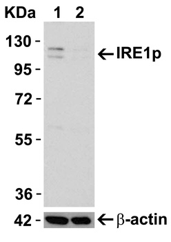
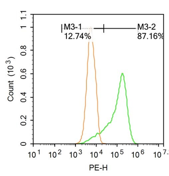
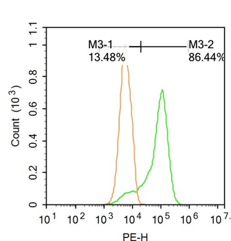
KO Validation in HeLa Cells. Loading: 10 µg of WT cell lysates (lane 1) or IRE1P KO cell lysates (lane 2). Antibodies: IRE1P orb1239523 (0.5 µg/mL) and beta-actin (1 µg/mL), 1h incubation at RT in 5% NFDM/TBST. Secondary: Goat anti-rabbit IgG HRP conjugate at 1:10000 dilution.
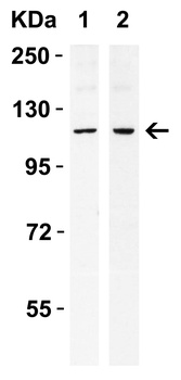
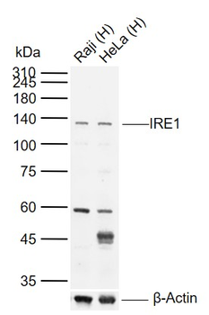
Western Blot Validation in Human Cell Lines. Loading: 15 µg of lysates per lane. Antibodies: IRE1p orb1239523 (0.4 µg/mL), 1h incubation at RT in 5% NFDM/TBST. Secondary: Goat anti-rabbit IgG HRP conjugate at 1:10000 dilution. Lane 1: Caco-2, Lane2: SK-N-SH.
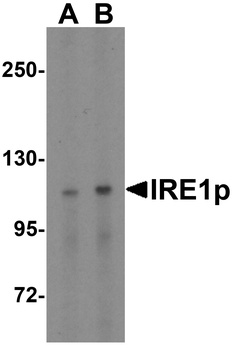
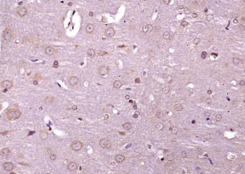
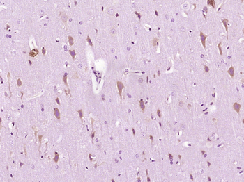
Western Blot Validation in Rat Brain Tissue Lysate. Loading: 15 µg of lysates per lane. Antibodies: IRE1p orb1239523 (A: 0.5 µg/mL, B: 1 µg/mL), 1h incubation at RT in 5% NFDM/TBST. Secondary: Goat anti-rabbit IgG HRP conjugate at 1:10000 dilution.
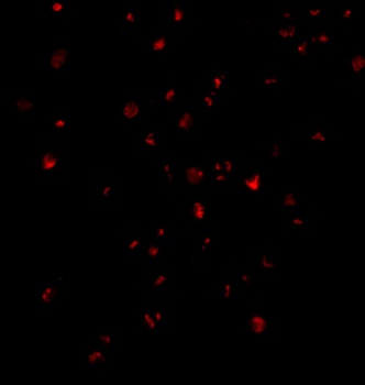
Immunofluorescence Validation of IRE1p in Mouse A20 Cells. Immunofluorescent analysis of 4% paraformaldehyde-fixed A20 Cells labeling IRE1P with orb1239523 at 2 µg/mL, followed by goat anti-rabbit IgG secondary antibody at 1/500 dilution (red).
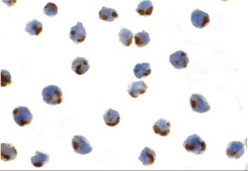
Immunocytochemistry Validation of IRE1p in Mouse A20 Cells. Immunocytochemical analysis of A20 cells using anti-IRE1p antibody (orb1239523) at 1 µg/ml. Cells was fixed with formaldehyde and blocked with 10% serum for 1 h at RT; antigen retrieval was by heat mediation with a citrate buffer (pH6). Samples were incubated with primary antibody overnight at 4°C. A goat anti-rabbit IgG H&L (HRP) at 1/250 was used as secondary. Counter stained with Hematoxylin.
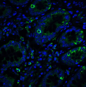
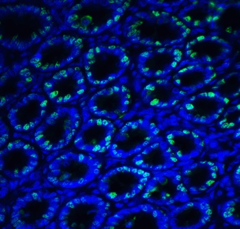
Immunofluorescence Validation of IRE1p in Human Small Intestine Tissue. Immunofluorescent analysis of 4% paraformaldehyde-fixed Human Small Intestine Tissue labeling IRE1p with orb1239523 at 20 µg/mL, followed by goat anti-rabbit IgG secondary antibody at 1/500 dilution (green) and DAPI staining (blue).
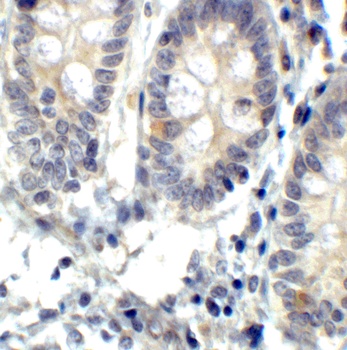
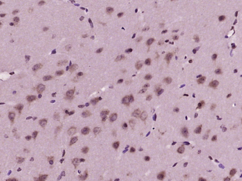
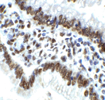
Immunohistochemistry Validation of IRE1p in Human Small Intestine Tissue. Immunohistochemical analysis of paraffin-embedded Human Small Intestine Tissue using anti-IRE1P antibody (orb1239523) at 2 µg/ml. Tissue was fixed with formaldehyde and blocked with 10% serum for 1 h at RT; antigen retrieval was by heat mediation with a citrate buffer (pH6). Samples were incubated with primary antibody overnight at 4°C. A goat anti-rabbit IgG H&L (HRP) at 1/250 was used as secondary. Counter stained with Hematoxylin.
Immunofluorescence Validation of IRE1p in Rat Small Intestine Tissue. Immunofluorescent analysis of 4% paraformaldehyde-fixed Rat Small Intestine Tissue labeling IRE1p with orb1239523 at 20 µg/mL, followed by goat anti-rabbit IgG secondary antibody at 1/500 dilution (green) and DAPI staining (blue).
Immunohistochemistry Validation of IRE1p in Rat Small Intestine Tissue. Immunohistochemical analysis of paraffin-embedded Rat Small Intestine Tissue using anti-IRE1P antibody (orb1239523) at 2 µg/ml. Tissue was fixed with formaldehyde and blocked with 10% serum for 1 h at RT; antigen retrieval was by heat mediation with a citrate buffer (pH6). Samples were incubated with primary antibody overnight at 4°C. A goat anti-rabbit IgG H&L (HRP) at 1/250 was used as secondary. Counter stained with Hematoxylin.
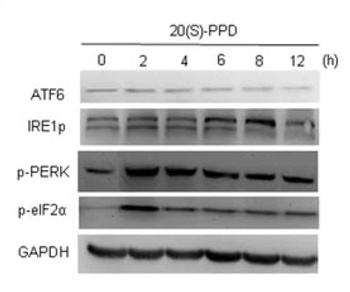
Induced Expression Validation of IRE1p in human umbilical vein endothelial cells (HUVECs) (Wang et al., 2019). IRE1p expression was examined by Western blot analysis with anti-IRE1p antibodies (orb1239523). IRE1p was increased in HUVEC cells treated with 10 µM 20 (S)‐PPD for 6 to 8 hours compared with control cells.
Documents Download
Datasheet
Product Information
Request a Document
Protocol Information
WB
Western Blot (IB, immunoblot)
IHC-P
Immunohistochemistry Paraffin
IF
Immunofluorescence
ICC
Immunocytochemistry
ELISA
Enzyme-linked Immunosorbent Assay (EIA)
ERN1 Antibody (orb1239523)

- 0.0
Based on 0 reviews
Participating in our Biorbyt product reviews program enables you to support fellow scientists by sharing your firsthand experience with our products.
Login to Submit a ReviewAvailable Sizes
Select a size below
Free Secondary Antibody (20 ul)0/0
Please add an antibody product to your cart first.