You have no items in your shopping cart.
Description
Research Area
Immunology & Inflammation
Images & Validation
−Item 1 of 5
| Tested Applications | ELISA, NeA, WB |
|---|---|
| Reactivity | Rat |
Key Properties
−| Antibody Type | Primary Antibody |
|---|---|
| Host | Goat |
| Clonality | Polyclonal |
| Immunogen | Produced from sera of goats pre-immunized with highly pure (>98%) recombinant Rat IL-1alpha (Rat IL-1alpha). |
| Target | Il1a |
| Purification | Anti-Rat IL-1alpha specific antibody was purified by affinity chromatography employing immobilized Rat IL-1alpha matrix. |
| Conjugation | Unconjugated |
Storage & Handling
−| Storage | Maintain refrigerated at 2-8°C for up to 2 weeks. For long term storage store at -20°C in small aliquots to prevent freeze-thaw cycles. |
|---|---|
| Form/Appearance | Lyophilized |
| Concentration | batch dependent |
| Expiration Date | 12 months from date of receipt. |
| Disclaimer | For research use only |
Alternative Names
−IL-1 alphaInterleukin-1 alphaIL-1 alpha
Similar Products
−- Item 1 of 7
IL1 alpha Rabbit Polyclonal Antibody [orb308737]
ELISA, ICC, IF, IHC-P, WB
Human, Mouse, Rat
Rabbit
Polyclonal
Unconjugated
100 μg - Item 1 of 6
- Item 1 of 3
IL-1 Alpha Rabbit Polyclonal Antibody [orb157678]
FC, WB
Rat
Human, Mouse
Rabbit
Polyclonal
Unconjugated
50 μl, 100 μl, 200 μl - Item 1 of 1
- Item 1 of 1

Quality Guarantee
Explore bioreagents carefree to elevate your research. All our products are rigorously tested for performance. If a product does not perform as described on its datasheet, our scientific support team will provide expert troubleshooting, a prompt replacement, or a refund. For full details, please see our Terms & Conditions and Buying Guide. Contact us at [email protected].
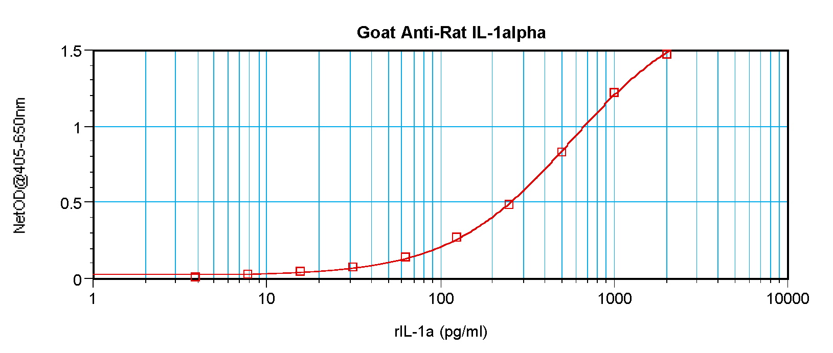
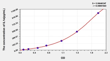
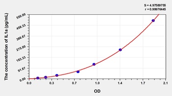
To detect Rat IL-1-alpha by sandwich ELISA (using 100 ul/well antibody solution) a concentration of 0.5 - 2.0 ug/ml of this antibody is required. This antigen affinity purified antibody, in conjunction with Anti-Rat IL-1-alpha (orb1272470) as a detection antibody, allows the detection of at least 0.2 - 0.4 ng/well of recombinant Rat IL-1-alpha.
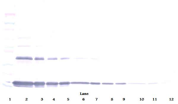
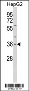
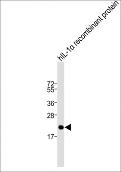
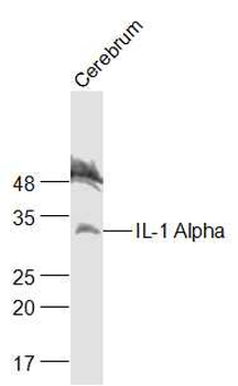
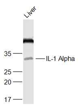
To detect Rat IL-1-alpha by Western Blot analysis this antibody can be used at a concentration of 0.1- 0.2 ug/ml. Used in conjunction with compatible secondary reagents the detection limit for recombinant Rat IL-1-alpha is 1.5-3.0 ng/lane, under either reducing or non-reducing conditions.
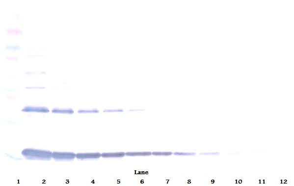
To detect Rat IL-1-alpha by Western Blot analysis this antibody can be used at a concentration of 0.1- 0.2 ug/ml. Used in conjunction with compatible secondary reagents the detection limit for recombinant Rat IL-1-alpha is 1.5-3.0 ng/lane, under either reducing or non-reducing conditions.
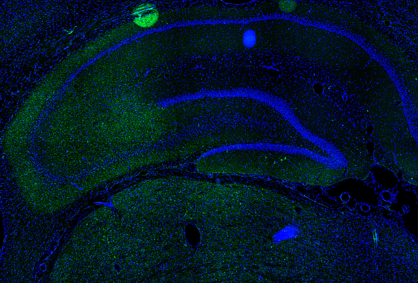
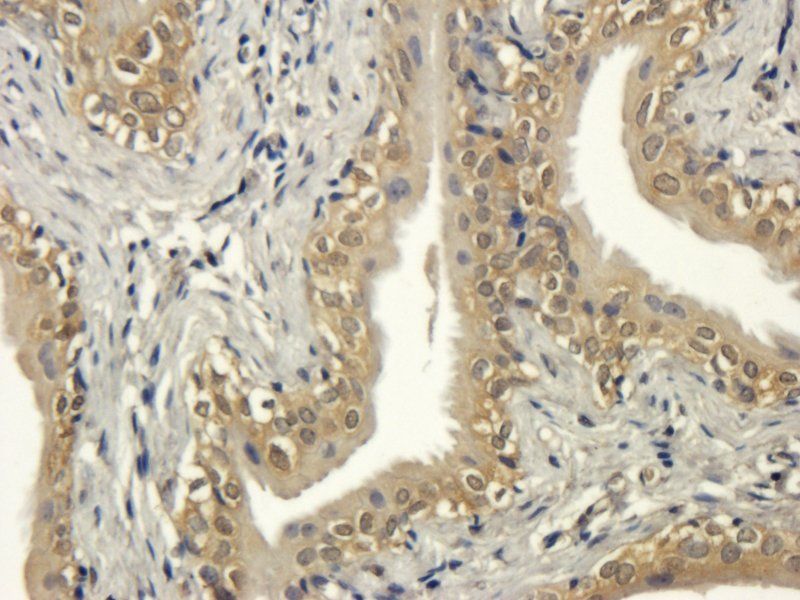
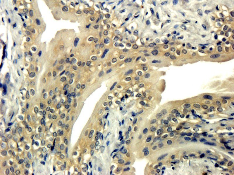
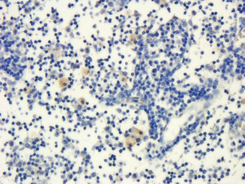
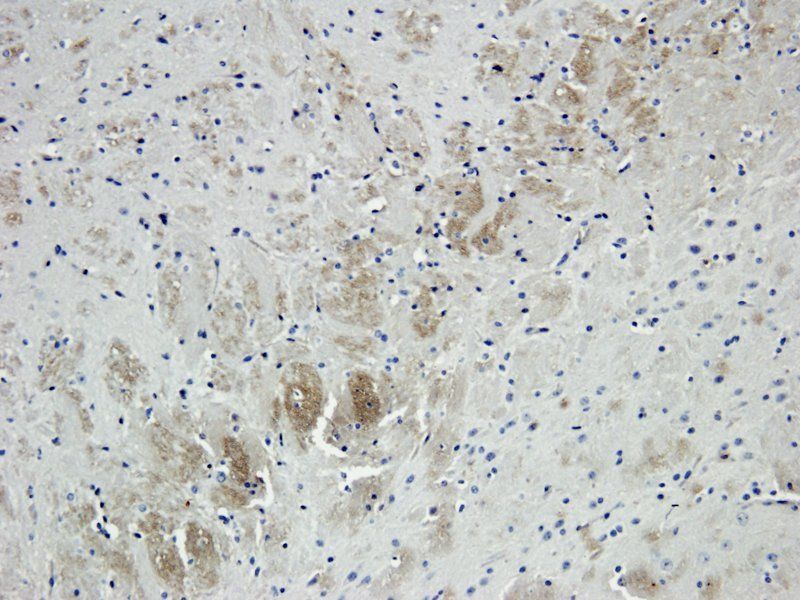
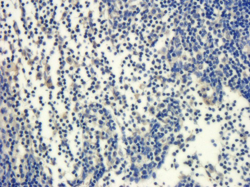
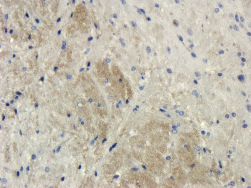
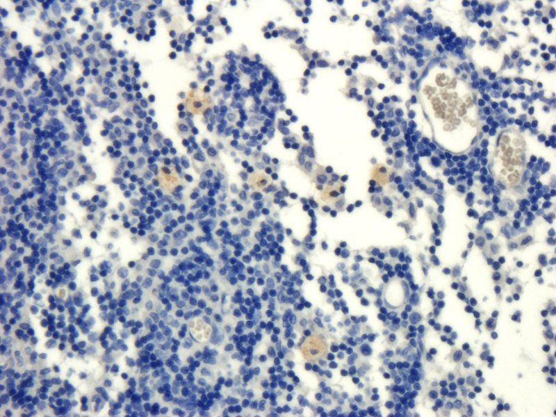
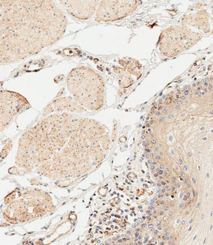
This antibody stained colchicine injected rat brain (including the ventricles and the CA3 region of the hippocampus) tissue. The primary antibody was incubated at 1.0 ug/ml overnight at 4°C. This was followed by a peroxidase conjugated secondary antibody and then a fluorescein Tyramide Signal Amplification (TSA) reagent. Optimal concentrations and conditions may vary.
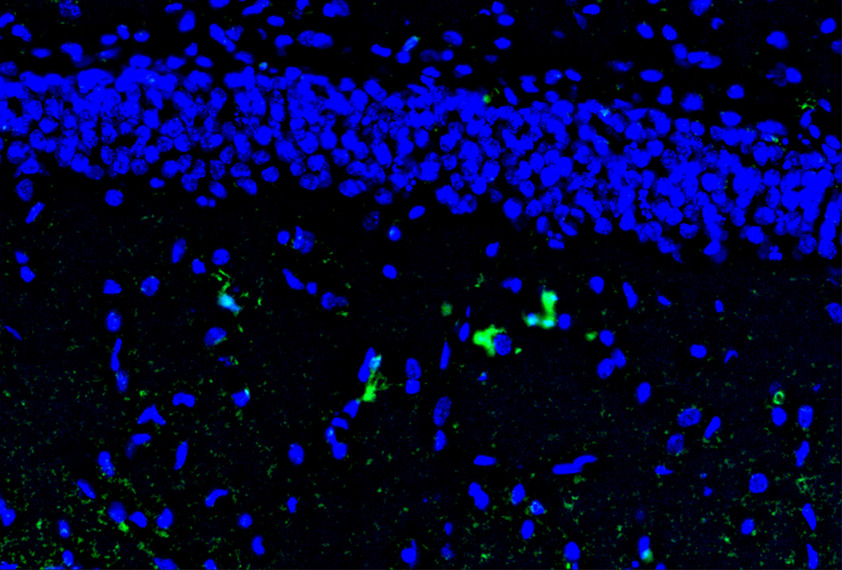
This antibody stained colchicine injected rat brain (including the ventricles and the CA3 region of the hippocampus) tissue. The primary antibody was incubated at 1.0 ug/ml overnight at 4°C. This was followed by a peroxidase conjugated secondary antibody and then a fluorescein Tyramide Signal Amplification (TSA) reagent. Optimal concentrations and conditions may vary.
Documents Download
Datasheet
Product Information
Request a Document
Protocol Information
WB
Western Blot (IB, immunoblot)
ELISA
Enzyme-linked Immunosorbent Assay (EIA)
Il1a Antibody (orb1272471)

- 0.0
Based on 0 reviews
Participating in our Biorbyt product reviews program enables you to support fellow scientists by sharing your firsthand experience with our products.
Login to Submit a ReviewAvailable Sizes
Select a size below
Free Secondary Antibody (20 ul)0/0
Please add an antibody product to your cart first.