You have no items in your shopping cart.
Description
Research Area
Neuroscience
Images & Validation
−Item 1 of 6
| Tested Applications | FC, IF, IHC, WB |
|---|---|
| Reactivity | Bovine, Canine, Drosophila, Frog, Gallus, Hamster, Human, Monkey, Mouse, Porcine, Rabbit, Rat, Sheep |
| Application Notes |
Key Properties
−| Antibody Type | Primary Antibody |
|---|---|
| Host | Mouse |
| Clonality | Monoclonal |
| Isotype | IgG1, kappa |
| Clone No. | LK1 |
| Immunogen | Recombinant human HSP60 was used as the immunogen for this antibody. |
| Target | HSPD1 |
| Purification | Protein G affinity chromatography |
| Conjugation | Unconjugated |
Storage & Handling
−| Storage | Maintain refrigerated at 2-8°C for up to 2 weeks. For long term storage store at -20°C in small aliquots to prevent freeze-thaw cycles. |
|---|---|
| Form/Appearance | Liquid |
| Buffer/Preservatives | PBS with 0.1 mg/ml rAlbumin and 0.05% sodium azide |
| Concentration | 0.2 mg/mL |
| Expiration Date | 12 months from date of receipt. |
| Disclaimer | For research use only |
Alternative Names
−60 kDa heat shock protein, mitochondrial, 60 kDa chaperonin, Chaperonin 60, CPN60, Heat shock protein 60, HSP-60, Hsp60, HuCHA60, Mitochondrial matrix protein P1, P60 lymphocyte protein, HSPD1, HSP60
Similar Products
−- Item 1 of 9
Hsp60/HSPD1 Mouse Monoclonal Antibody [orb570314]
FC, ICC, IF, IHC, WB
Human, Mouse, Rat
Mouse
Monoclonal
Unconjugated
100 μg - Item 1 of 5
HSP60 Rabbit Polyclonal Antibody [orb10846]
ICC, IF, IHC-Fr, IHC-P, WB
Bovine, Canine, Equine, Rabbit
Human, Mouse, Rat
Rabbit
Polyclonal
Unconjugated
50 μl, 100 μl, 200 μl - Item 1 of 8
- Item 1 of 8
- Item 1 of 6
Hsp60/HSPD1 Rabbit Polyclonal Antibody [orb251520]
ICC, IF, IHC, WB
Human, Mouse, Rat
Rabbit
Polyclonal
Unconjugated
100 μg


Quality Guarantee
Explore bioreagents carefree to elevate your research. All our products are rigorously tested for performance. If a product does not perform as described on its datasheet, our scientific support team will provide expert troubleshooting, a prompt replacement, or a refund. For full details, please see our Terms & Conditions and Buying Guide. Contact us at [email protected].
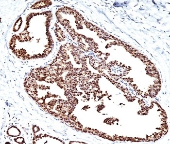
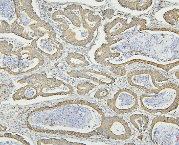
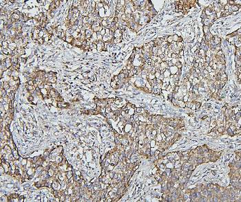
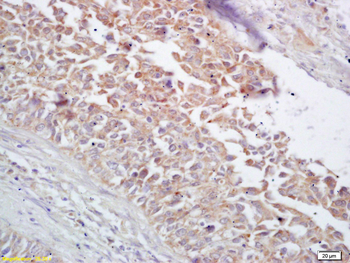
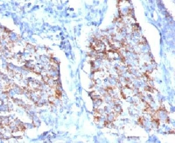
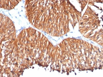
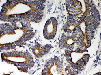
Formalin/paraffin human breast carcinoma stained with HSP60 antibody.
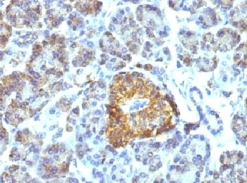
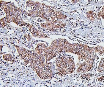
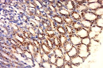
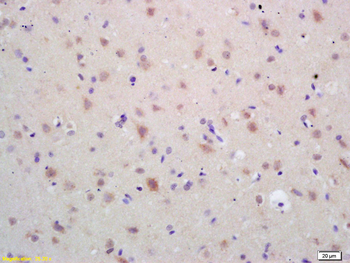
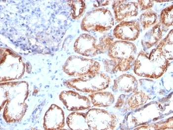
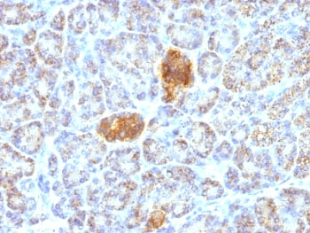
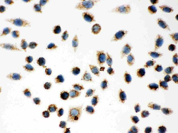
IHC testing of FFPE pancreas tissue with HSP60 antibody.
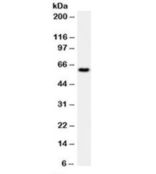
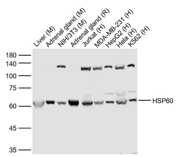
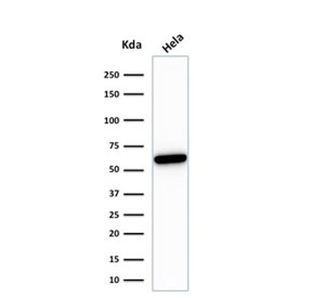
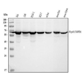
Western blot testing of HeLa cell lysate with HSP60 antibody (clone LK1).
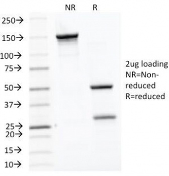
SDS-PAGE Analysis of Purified, BSA-Free HSP60 Antibody (clone LK1). Confirmation of Integrity and Purity of the Antibody.
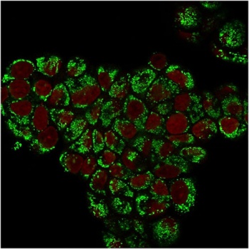
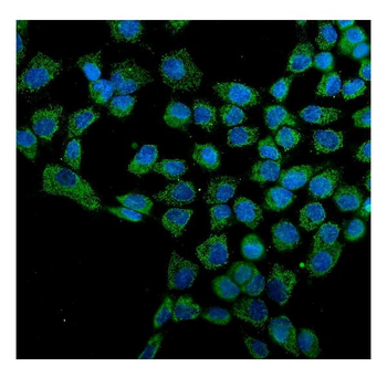
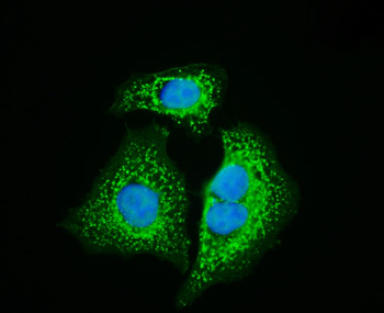
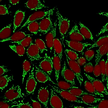
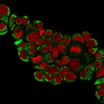
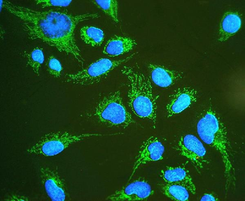
Immunofluorescent staining of PFA-fixed human MCF7 cells with HSP60 antibody (green, clone LK1) and Reddot nuclear stain (red).
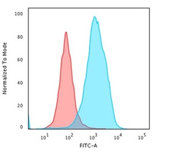
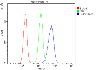
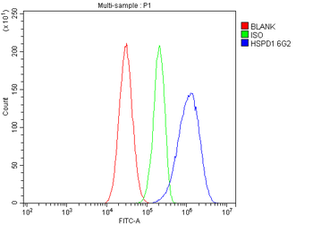
Flow cytometry testing of PFA-fixed human HeLa cells with HSP60 antibody (clone LK1); Red = isotype control, Blue = HSP60 antibody.
Quick Database Links
Gene Symbol
HSPD1
UniProt
UniProt Details
− No UniProt data available
Documents Download
Datasheet
Product Information
Request a Document
Protocol Information
WB
Western Blot (IB, immunoblot)
IHC
Immunohistochemistry
FC
Flow Cytometry
IF
Immunofluorescence
HSPD1 Antibody (orb1252759)

- 0.0
Based on 0 reviews
Participating in our Biorbyt product reviews program enables you to support fellow scientists by sharing your firsthand experience with our products.
Login to Submit a ReviewAvailable Sizes
Select a size below
Choose Conjugation or Carrier Free Version
Free Secondary Antibody (20 ul)0/0
Please add an antibody product to your cart first.