You have no items in your shopping cart.
Description
Research Area
Cancer Biology
Images & Validation
−Item 1 of 5
| Tested Applications | FC, IHC-P, WB |
|---|---|
| Dilution Range | WB - 1:1000, IHC-P - 1:25, FC - 1:10-50 |
| Reactivity | Human |
Key Properties
−| Antibody Type | Primary Antibody |
|---|---|
| Host | Rabbit |
| Clonality | Polyclonal |
| Isotype | Rabbit IgG |
| Immunogen | This HIF1A antibody is generated from rabbits immunized with HIF1A recombinant protein. |
| Target | HIF1A {ECO:0000303|PubMed:7539918, ECO:0000312|HGNC:HGNC:4910} |
| Molecular Weight | 92670 Da |
| Conjugation | Unconjugated |
Storage & Handling
−| Storage | Maintain refrigerated at 2-8°C for up to 2 weeks. For long term storage store at -20°C in small aliquots to prevent freeze-thaw cycles |
|---|---|
| Form/Appearance | Purified polyclonal antibody supplied in PBS with 0.09% (W/V) sodium azide. This antibody is purified through a protein A column, followed by peptide affinity purification. |
| Expiration Date | 12 months from date of receipt. |
| Disclaimer | For research use only |
Alternative Names
−Hypoxia-inducible factor 1-alpha, HIF-1-alpha, HIF1-alpha, ARNT-interacting protein, Basic-helix-loop-helix-PAS protein MOP1, Class E basic helix-loop-helix protein 78, bHLHe78, Member of PAS protein 1, PAS domain-containing protein 8, HIF1A, BHLHE78, MOP1, PASD8
Similar Products
−- Item 1 of 8
HIF-1 Alpha Rabbit Polyclonal Antibody [orb500810]
FC, ICC
Gallus, Porcine
Human
Rabbit
Polyclonal
Unconjugated
50 μl, 100 μl, 200 μl - Item 1 of 8
HIF-1 Alpha Rabbit Polyclonal Antibody [orb500743]
FC, WB
Gallus, Mouse, Rat
Human
Rabbit
Polyclonal
Unconjugated
50 μl, 100 μl, 200 μl - Item 1 of 5
HIF1Alpha Antibody (Center) [orb1928917]
FC, IF, IHC-P, WB
Rat
Human, Mouse
Rabbit
Polyclonal
Unconjugated
400 μl - Item 1 of 7
HIF-1α Polyclonal Antibody [orb1413487]
IF, IHC-P, WB
Human, Mouse, Rat
Rabbit
Polyclonal
Unconjugated
100 μl - Item 1 of 6
HIF-1 alpha/HIF1A/HIF Rabbit Polyclonal Antibody [orb669094]
ELISA, FC, IHC, WB
Human, Mouse, Rat
Rabbit
Polyclonal
Unconjugated
100 μg


Quality Guarantee
Explore bioreagents carefree to elevate your research. All our products are rigorously tested for performance. If a product does not perform as described on its datasheet, our scientific support team will provide expert troubleshooting, a prompt replacement, or a refund. For full details, please see our Terms & Conditions and Buying Guide. Contact us at [email protected].
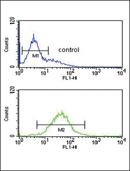
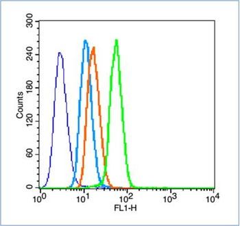
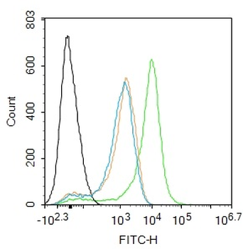
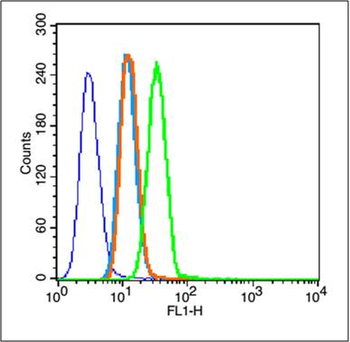
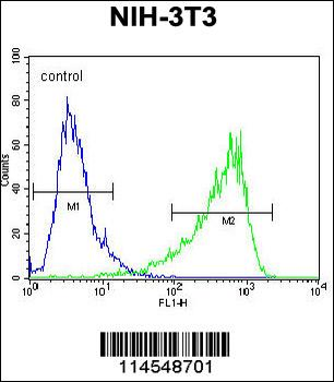
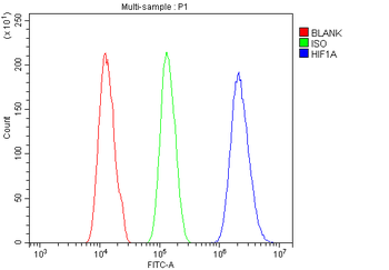
HIF1A Antibody flow cytometric analysis of K562 cells (bottom histogram) compared to a negative control cell (top histogram). FITC-conjugated goat-anti-rabbit secondary antibodies were used for the analysis.
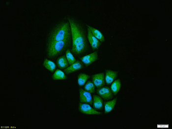
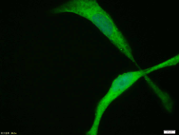
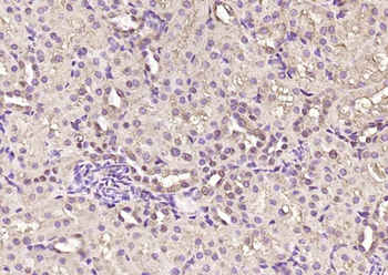
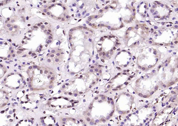
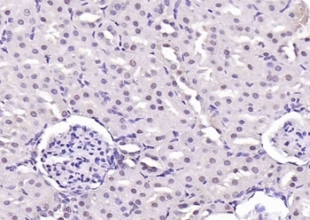
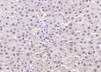
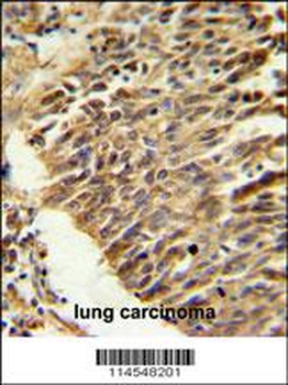
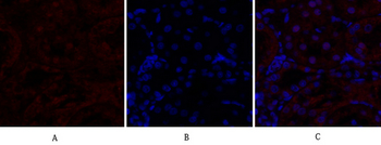
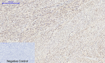
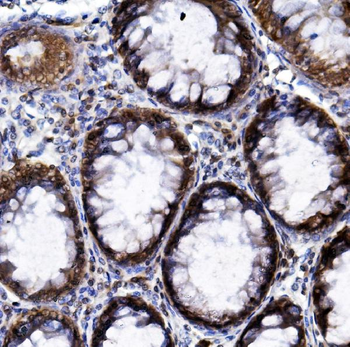
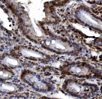
Staining HIF1A in human kidney tissue sections by Immunohistochemistry (IHC-P - paraformaldehyde-fixed, paraffin-embedded sections). Tissue was fixed with formaldehyde and blocked with 3% BSA for 0.5 hour at room temperature; antigen retrieval was by heat mediation with a citrate buffer (pH6). Samples were incubated with primary antibody (1/25) for 1 hours at 37°C. A undiluted biotinylated goat polyvalent antibody was used as the secondary antibody.
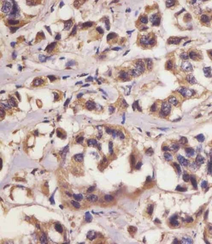
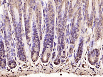
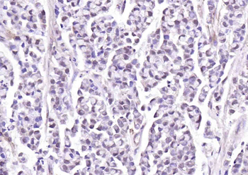
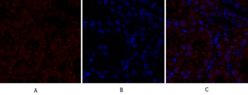
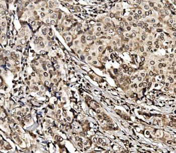
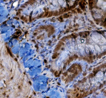
Staining HIF1A in human breast carcinoma sections by Immunohistochemistry (IHC-P - paraformaldehyde-fixed, paraffin-embedded sections). Tissue was fixed with formaldehyde and blocked with 3% BSA for 0.5 hour at room temperature; antigen retrieval was by heat mediation with a citrate buffer (pH6). Samples were incubated with primary antibody (1/25) for 1 hours at 37°C. A undiluted biotinylated goat polyvalent antibody was used as the secondary antibody.
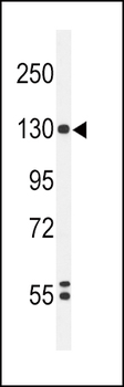
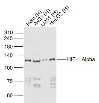
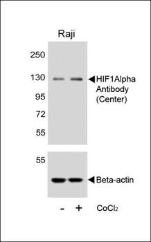
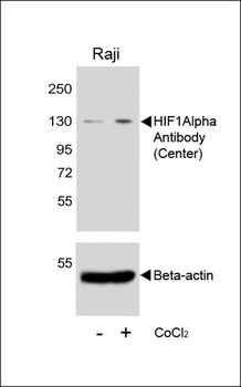
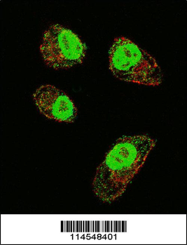
Western blot analysis of HIF1A Antibody in K562 cell line lysates (35 ug/lane). HIF1A (arrow) was detected using the purified Pab.
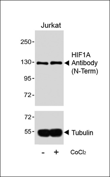
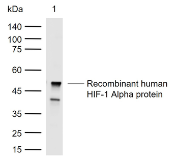
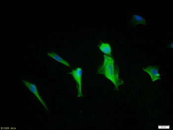
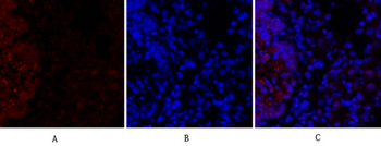
WB analysis of orb1930780.
Quick Database Links
Gene Symbol
HIF1A {ECO:0000303|PubMed:7539918, ECO:0000312|HGNC:HGNC:4910}
UniProt
RefSeq (Protein):NP_001230013.1, NP_001521.1, NP_851397.1
UniProt Details
− No UniProt data available
NCBI Reference Sequences
−Associated Accession Numbers
Curated reference sequences for the gene transcript and protein product| Protein | NP_001230013.1, NP_001521.1, NP_851397.1 |
|---|
Documents Download
Datasheet
Product Information
Request a Document
Protocol Information
WB
Western Blot (IB, immunoblot)
IHC-P
Immunohistochemistry Paraffin
FC
Flow Cytometry
HIF1A Antibody (orb1930780)

- 0.0
Based on 0 reviews
Participating in our Biorbyt product reviews program enables you to support fellow scientists by sharing your firsthand experience with our products.
Login to Submit a ReviewAvailable Sizes
Select a size below
Choose Conjugation or Carrier Free Version
Free Secondary Antibody (20 ul)0/0
Please add an antibody product to your cart first.