You have no items in your shopping cart.
Featured
Description
Research Area
Neuroscience
Images & Validation
−Item 1 of 13
| Tested Applications | ICC, IF, IHC, WB |
|---|---|
| Dilution Range | WB: 1:500-1:3000 , ICC/IF: 1:100-1:1000 , IHC-P: 1:100-1:1000 , IHC-Fr: 1:100-1:1000 |
| Reactivity | Human, Monkey, Mouse, Rat, Zebrafish |
| Application Notes |
Key Properties
−| Antibody Type | Primary Antibody |
|---|---|
| Host | Rabbit |
| Clonality | Polyclonal |
| Isotype | IgG |
| Immunogen | Recombinant protein encompassing a sequence within the N-terminus region of human GAD67 |
| Target | This antibody is specific for GAD67 protein, and it does not cross-react with GAD65 protein |
| Molecular Weight | 67 kDa |
| Purity | Purified by antigen-affinity chromatography |
| Purification | Purified by antigen-affinity chromatography. |
| Conjugation | Unconjugated |
Storage & Handling
−| Storage | Store as concentrated solution. Centrifuge briefly prior to opening vial. For short-term storage (1-2 weeks), store at 4°C. For long-term storage, aliquot and store at -20°C or below. Avoid multiple freeze-thaw cycles |
|---|---|
| Form/Appearance | PBS, 1% rAlbumin, 20% Glycerol, 0.025% ProClin 300 |
| Concentration | Batch specific - Reach out to find out the current concentration |
| Expiration Date | 12 months from date of receipt. |
| Disclaimer | For research use only |
Alternative Names
−glutamate decarboxylase 1 , CPSQ1 , GAD , SCP
Similar Products
−- Item 1 of 3
GAD67 Rabbit Polyclonal Antibody [orb10681]
FC, IF, IHC-Fr, IHC-P, WB
Rat
Human, Mouse
Rabbit
Polyclonal
Unconjugated
50 μl, 100 μl, 200 μl - Item 1 of 3
GAD1/2 Rabbit Polyclonal Antibody [orb213964]
IF, IHC, WB
Bovine, Canine, Human, Mouse, Porcine, Rat
Rabbit
Polyclonal
Unconjugated
30 μl, 100 μl, 200 μl, 50 μl - Item 1 of 3
- Item 1 of 3
GAD1 Rabbit Polyclonal Antibody [orb1289742]
IF, IHC, WB
Human, Mouse, Rat
Rabbit
Polyclonal
Unconjugated
50 μl, 100 μl, 200 μl, 30 μl - Item 1 of 2
GAD67/GAD1 Rabbit Polyclonal Antibody [orb180678]
ICC, IHC, WB
Human, Mouse, Rat
Rabbit
Polyclonal
Unconjugated
100 μg


Quality Guarantee
Explore bioreagents carefree to elevate your research. All our products are rigorously tested for performance. If a product does not perform as described on its datasheet, our scientific support team will provide expert troubleshooting, a prompt replacement, or a refund. For full details, please see our Terms & Conditions and Buying Guide. Contact us at [email protected].
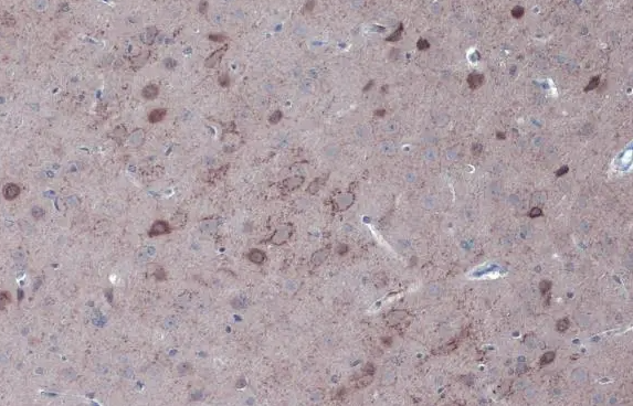
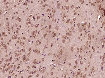
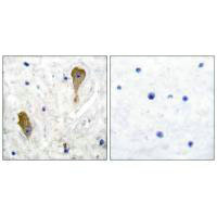
IHC-P Image - GAD67 antibody detects GAD67 protein at cytoplasm by immunohistochemical analysis. Sample: Paraffin-embedded rat brain. GAD67 stained by GAD67 antibody (orb555997) diluted at 1:500. Antigen Retrieval: Citrate buffer, pH 6.0, 15 min
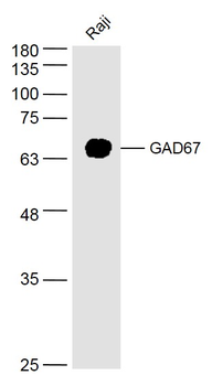
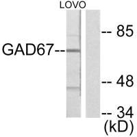
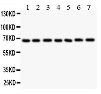
Various whole cell extracts (30 μg) were separated by 7.5% SDS-PAGE, and the membrane was blotted with GAD67 antibody diluted at 1:500. The HRP-conjugated anti-rabbit IgG antibody was used to detect the primary antibody
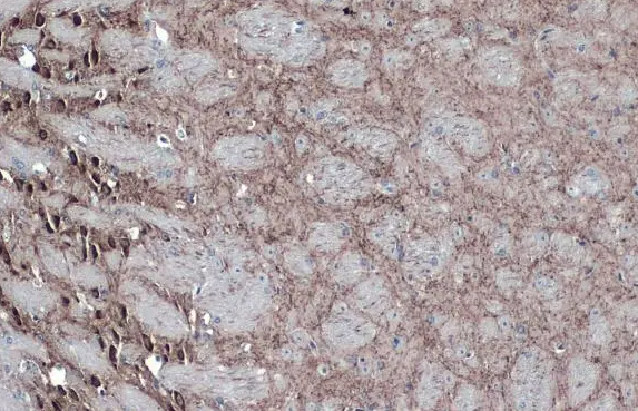
GAD67 antibody detects GAD67 protein at cytoplasm by immunohistochemical analysis. Sample: Paraffin-embedded mouse brain. GAD67 stained by GAD67 antibody diluted at 1:500. Antigen Retrieval: Citrate buffer, pH 6.0, 15 min
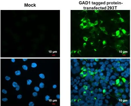
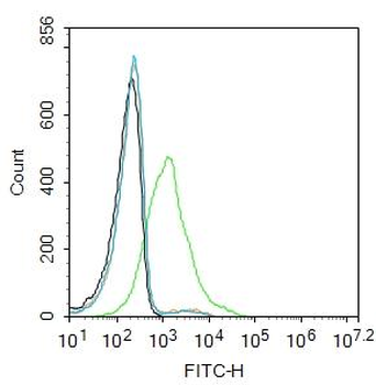
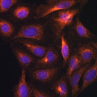
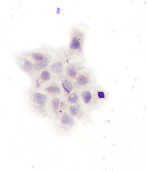
GAD67 antibody detects GAD67 protein by immunofluorescent analysis. Sample: Mock and transfected 293T cells were fixed in 4% paraformaldehyde at RT for 15 min. Green: GAD67 stained by GAD67 antibody diluted at 1:500. Blue: Fluoroshield with DAPI
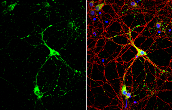
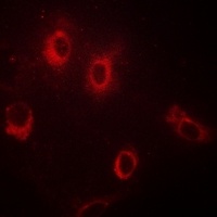
GAD67 antibody detects GAD67 protein by immunofluorescent analysis. Sample: DIV10 rat E18 primary cortical neuron cells were fixed in 4% paraformaldehyde at RT for 15 min. Green: GAD67 stained by GAD67 antibody diluted at 1:500. Red: Tau, stained by Tau antibody diluted at 1:500. Blue: Fluoroshield with DAPI
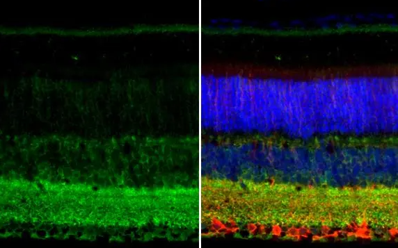
GAD67 antibody detects GAD67 protein at cell membrane and cytoplasm by immunohistochemical analysis. Sample: Paraffin-embedded mouse eye. Green: GAD67 stained by GAD67 antibody diluted at 1:250. Red: beta Tubulin 3/ Tuj1, a neural marker, stained by beta Tubulin 3/ Tuj1 antibody diluted at 1:500. Blue: Fluoroshield with DAPI Antigen Retrieval: Citrate buffer, pH 6.0, 15 min
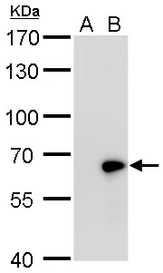
GAD67 antibody detects GAD67 protein by western blot analysis. A. 30 μg 293T whole cell lysate/extract B. 30 μg whole cell lysate/extract of human GAD1-transfected 293T cells 7.5% SDS-PAGE GAD67 antibody dilution: 1:5000. The HRP-conjugated anti-rabbit IgG antibody was used to detect the primary antibody
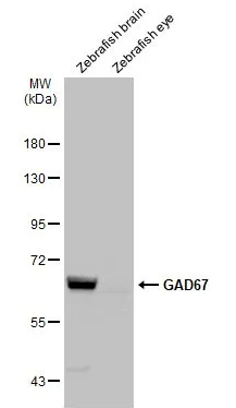
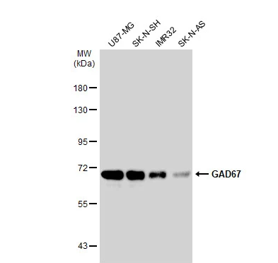
Various tissue extracts (30 μg) were separated by 7.5% SDS-PAGE, and the membrane was blotted with GAD67 antibody diluted at 1:500. The HRP-conjugated anti-rabbit IgG antibody was used to detect the primary antibody
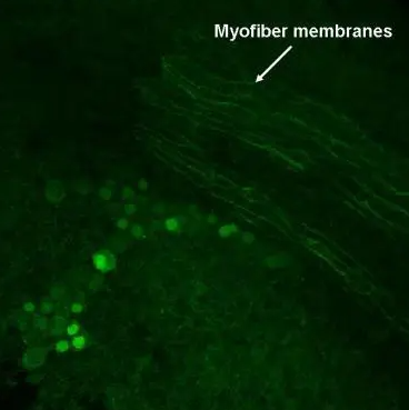
GAD67 antibody detects GAD67 protein on myofiber membranes by immunohistochemical analysis. Sample: Whole-mount zebrafish embryo GAD67 antibody dilution: 1:200
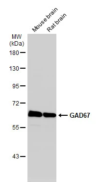
Various tissue extracts (50 μg) were separated by 7.5% SDS-PAGE, and the membrane was blotted with GAD67 antibody diluted at 1:1000. The HRP-conjugated anti-rabbit IgG antibody was used to detect the primary antibody
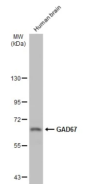
Human tissue extract (30 μg) was separated by 7.5% SDS-PAGE, and the membrane was blotted with GAD67 antibody diluted at 1:1000. The HRP-conjugated anti-rabbit IgG antibody was used to detect the primary antibody
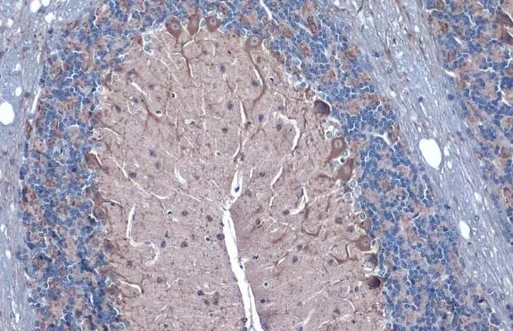
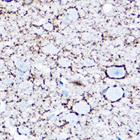
GAD67 antibody detects GAD67 protein at cell membrane and cytoplasm by immunohistochemical analysis. Sample: Paraffin-embedded mouse cerebellum. GAD67 stained by GAD67 antibody diluted at 1:1000. Antigen Retrieval: Citrate buffer, pH 6.0, 15 min
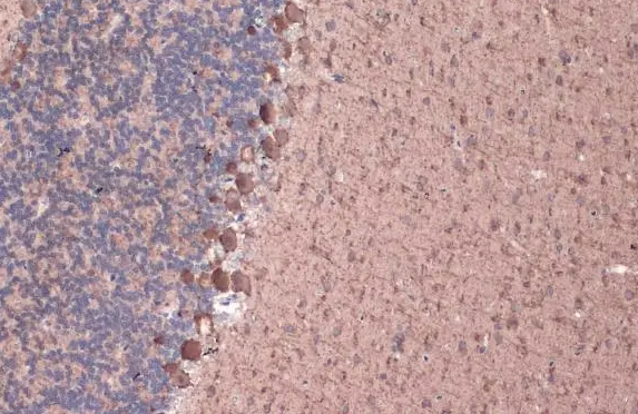
GAD67 antibody detects GAD67 protein at cell membrane and cytoplasm by immunohistochemical analysis. Sample: Paraffin-embedded rat cerebellum. GAD67 stained by GAD67 antibody diluted at 1:1000. Antigen Retrieval: Citrate buffer, pH 6.0, 15 min
Quick Database Links
Gene Symbol
This antibody is specific for GAD67 protein, and it does not cross-react with GAD65 protein
UniProt
UniProt Details
− No UniProt data available
Documents Download
Datasheet
Product Information
Request a Document
Protocol Information
WB
Western Blot (IB, immunoblot)
IHC
Immunohistochemistry
IF
Immunofluorescence
ICC
Immunocytochemistry
GAD67 Rabbit Polyclonal Antibody (orb555997)

- 0.0
Based on 0 reviews
Participating in our Biorbyt product reviews program enables you to support fellow scientists by sharing your firsthand experience with our products.
Login to Submit a ReviewAvailable Sizes
Select a size below
Choose Conjugation or Carrier Free Version
Free Secondary Antibody (20 ul)0/0
Please add an antibody product to your cart first.