You have no items in your shopping cart.
Description
Research Area
Signal Transduction
Images & Validation
−Item 1 of 7
| Tested Applications | IF, WB |
|---|---|
| Dilution Range | WB - 1:1000, IF - 1:25 |
| Reactivity | Human |
Key Properties
−| Host | Mouse |
|---|---|
| Clonality | Monoclonal |
| Isotype | IgG1,κ |
| Clone No. | B619EV37X4X7 |
| Immunogen | This FASN antibody is generated from mice immunized with a KLH conjugated synthetic peptide between 942-973 amino acids from the Central region of human FASN. Antigen Region: 942-973 aa. |
| Target | FASN |
| Molecular Weight | 273427 Da |
| Conjugation | Unconjugated |
Storage & Handling
−| Storage | Maintain refrigerated at 2-8°C for up to 2 weeks. For long term storage store at -20°C in small aliquots to prevent freeze-thaw cycles |
|---|---|
| Form/Appearance | Purified polyclonal antibody supplied in PBS with 0.09% (W/V) sodium azide. This antibody is prepared by Saturated Ammonium Sulfate (SAS) precipitation followed by dialysis against PBS. |
| Expiration Date | 12 months from date of receipt. |
| Disclaimer | For research use only |
Alternative Names
−Fatty acid synthase, [Acyl-carrier-protein] S-acetyltransferase, [Acyl-carrier-protein] S-malonyltransferase, 3-oxoacyl-[acyl-carrier-protein] synthase, 3-oxoacyl-[acyl-carrier-protein] reductase, 3-hydroxyacyl-[acyl-carrier-protein] dehydratase, Enoyl-[acyl-carrier-protein] reductase, Oleoyl-[acyl-carrier-protein] hydrolase, FASN, FAS
Similar Products
−- Item 1 of 7
- Item 1 of 2
FASN Mouse Monoclonal Antibody [orb1473885]
IF, WB
Human
Mouse
Monoclonal
Unconjugated
200 μl, 100 μl, 50 μl, 30 μl - Item 1 of 2
FASN Rabbit Polyclonal Antibody [orb213921]
IF, WB
Human
Rabbit
Polyclonal
Unconjugated
30 μl, 100 μl, 200 μl, 50 μl
Quality Guarantee
Explore bioreagents carefree to elevate your research. All our products are rigorously tested for performance. If a product does not perform as described on its datasheet, our scientific support team will provide expert troubleshooting, a prompt replacement, or a refund. For full details, please see our Terms & Conditions and Buying Guide. Contact us at [email protected].
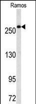
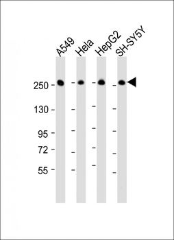
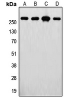
All lanes: Anti-FASN Antibody (Center) at 1:500-1:2000 dilution. Lane 1: A549 whole cell lysate. Lane 2: Hela whole cell lysate. Lane 3: 293 whole cell lysate. Lane 4: Ramos whole cell lysate. Lane 5: HepG2 whole cell lysate. Lysates/proteins at 20 µg per lane. Secondary Goat Anti-mouse IgG, (H+L), Peroxidase conjugated at 1/10000 dilution. Predicted band size: 273 kDa. Blocking/Dilution buffer: 5% NFDM/TBST.
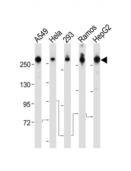
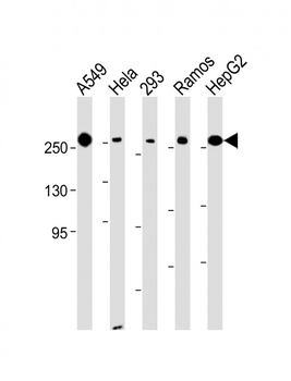
All lanes: Anti-FASN Antibody (Center) at 1:8000 dilution. Lane 1: A549 whole cell lysate. Lane 2: Hela whole cell lysate. Lane 3: 293 whole cell lysate. Lane 4: Ramos whole cell lysate. Lane 5: HepG2 whole cell lysate.Lysates/proteins at 20 µg per lane. Secondary Goat Anti-mouse IgG, (H+L), Peroxidase conjugated at 1/10000 dilution. Predicted band size: 273 kDa. Blocking/Dilution buffer: 5% NFDM/TBST.
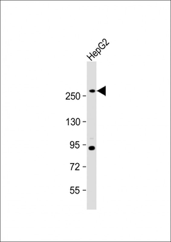
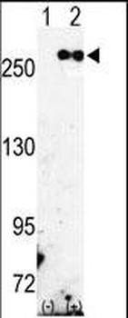
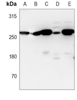
Anti- at 1:1000 dilution + HepG2 whole cell lysate.Lysates/proteins at 20 µg per lane. Secondary Goat Anti-mouse IgG, (H+L), Peroxidase conjugated at 1/10000 dilution. Predicted band size: 273 kDa. Blocking/Dilution buffer: 5% NFDM/TBST.

FASN Antibody (Center) western blot analysis in mouse brain tissue lysates (35 μg/lane). This demonstrates the FASN (Center) antibody detected the FASN (Center) protein (arrow).
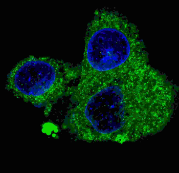
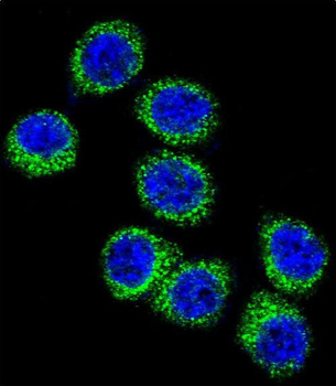
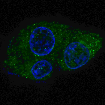
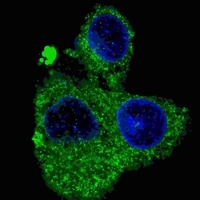
Fluorescent confocal image of HepG2 cells stained with FASN (Center) Antibody. HepG2 cells were fixed with 4% PFA (20 min), permeabilized with Triton X-100 (0.2%, 30 min). Cells were then incubated with FASN primary antibody (1:200, 2 h at room temperature). For secondary antibody, Alexa Fluor 488 conjugated donkey anti-mouse antibody (green) was used (1:1000, 1h). Nuclei were counterstained with Hoechst 33342 (blue) (10 μg/ml, 5 min).
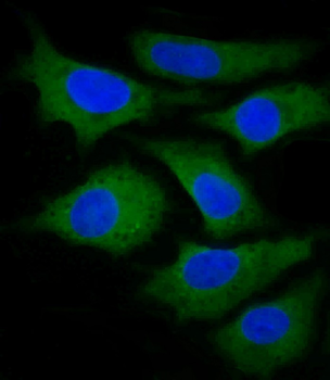
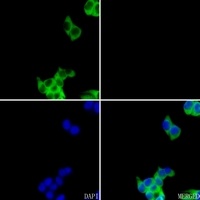
Immunofluorescent analysis of 4% paraformaldehyde-fixed, 0. 1% Triton X-100 permeabilized HepG2 (human liver hepatocellular carcinoma cell line) cells labeling FASN at 1/25 dilution, followed by Dylight 488-conjugated goat anti-mouse IgG (35503) secondary antibody at 1/200 dilution (green). Immunofluorescence image showing cytoplasm HepG2 cell line. The nuclear counter stain is DAPI (blue).
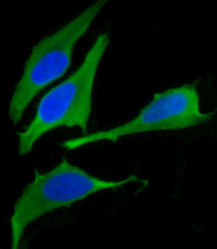
Immunofluorescent analysis of 4% paraformaldehyde-fixed, 0. 1% Triton X-100 permeabilized U-2 OS ((human cervical epithelial adenocarcinoma cell line) cells labeling FASN at 1/25 dilution, followed by Dylight 488-conjugated goat anti-mouse IgG (35503) secondary antibody at 1/200 dilution (green). Immunofluorescence image showing cytoplasm Hela cell line. The nuclear counter stain is DAPI (blue).
Quick Database Links
UniProt Details
− No UniProt data available
NCBI Reference Sequences
−Associated Accession Numbers
Curated reference sequences for the gene transcript and protein product| Protein | NP_004095.4 |
|---|
Documents Download
Datasheet
Product Information
Request a Document
Protocol Information
WB
Western Blot (IB, immunoblot)
IF
Immunofluorescence
FASN Antibody (Center) (orb1939263)

- 0.0
Based on 0 reviews
Participating in our Biorbyt product reviews program enables you to support fellow scientists by sharing your firsthand experience with our products.
Login to Submit a ReviewAvailable Sizes
Select a size below
Choose Conjugation or Carrier Free Version
Free Secondary Antibody (20 ul)0/0
Please add an antibody product to your cart first.